Photo AI
Figure 4 shows ciliated epithelial cells from the airways of a human as seen using a light microscope - Edexcel - GCSE Biology - Question 3 - 2021 - Paper 1
Question 3
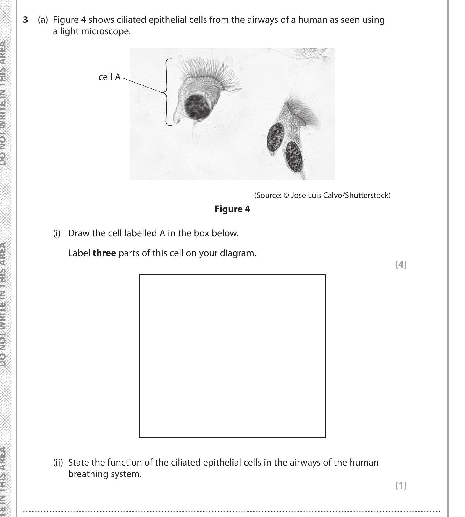
Figure 4 shows ciliated epithelial cells from the airways of a human as seen using a light microscope. (i) Draw the cell labelled A in the box below. Label three pa... show full transcript
Worked Solution & Example Answer:Figure 4 shows ciliated epithelial cells from the airways of a human as seen using a light microscope - Edexcel - GCSE Biology - Question 3 - 2021 - Paper 1
Step 1
Draw the cell labelled A in the box below. Label three parts of this cell on your diagram.

Answer
To draw the ciliated epithelial cell:
- Structure: Begin with a rectangle for the main cell body.
- Cilia: Add multiple hair-like structures at the top of the rectangle representing cilia. These protrude from the surface.
- Nucleus: Draw a circle within the rectangle, indicating the position of the nucleus.
- Cytoplasm: Shade the inside of the rectangle lightly to represent cytoplasm.
Labeling:
- Label the cilia, nucleus, and cytoplasm accordingly to ensure clarity in your diagram.
Step 2
State the function of the ciliated epithelial cells in the airways of the human breathing system.
Answer
The primary function of ciliated epithelial cells is to facilitate the movement of mucus along the airways. These cells possess cilia that beat in a coordinated manner to propel mucus, which traps dust, pathogens, and other particles, out of the respiratory system. This mechanism helps keep the airways clear and protects against infections.