Photo AI
Last Updated Sep 27, 2025
Magnetic resonance (MR) scanner Simplified Revision Notes for A-Level AQA Physics
Revision notes with simplified explanations to understand Magnetic resonance (MR) scanner quickly and effectively.
448+ students studying
10.4.3 Magnetic resonance (MR) scanner
Overview of Magnetic Resonance Imaging (MRI)
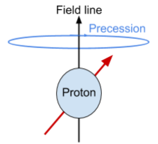
An MRI scanner uses a powerful superconducting magnet, which is cooled by liquid helium, to create a uniform magnetic field. Patients lie within this magnetic field so that an image of their internal tissues can be produced. MRI relies on properties of protons (hydrogen nuclei) within the body, which behave like tiny magnets due to their intrinsic spin.
Initially, these protons are oriented randomly. When placed in a strong magnetic field, they align either parallel or antiparallel to the field. The parallel alignment is lower in energy, so most protons align this way. These protons also undergo precession — a slight, wobbling rotation around the magnetic field lines.
Gradient Coils and Image Formation
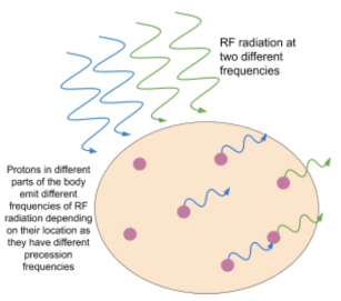
The MRI scanner contains smaller gradient coils which vary the magnetic field strength across different parts of the patient's body. This variation means that protons in different regions of the body processes at different frequencies, enabling MRI to differentiate between these areas.
To create an image, radio frequency (RF) pulses are transmitted into the body at the same frequency as the precession of protons in a specific region. These pulses temporarily change the alignment of the protons, causing them to become excited. As the protons return to their aligned state, they emit RF signals at their specific precession frequency, which can then be detected and used to form an image.
This process can be applied to produce 2D cross-sectional images or combined to create 3D images.
MRI Process: Step-by-Step
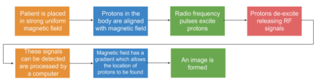
- Patient in a Strong Magnetic Field
- The patient is positioned inside a powerful, uniform magnetic field. This field aligns the protons (hydrogen nuclei) in the body along its direction.
- Alignment of Protons
- In the magnetic field, protons behave like tiny magnets due to their spin, aligning either with or against the field. Most align with the field due to lower energy requirements.
- Radiofrequency Pulses
- A radiofrequency (RF) pulse is then applied, which provides energy to the protons, temporarily exciting them and causing them to flip out of alignment with the magnetic field.
- Proton De-excitation and Signal Emission
- When the RF pulse is turned off, the protons return to their original alignment with the magnetic field, releasing the absorbed energy in the form of radiofrequency signals.
- Detection of RF Signals
- These RF signals are detected by receivers in the MRI scanner.
- Gradient Magnetic Field for Spatial Location
- To create an image, the MRI scanner applies a gradient to the magnetic field, which changes the field strength at different positions. This variation allows the scanner to locate the specific origin of the emitted signals, helping to pinpoint where in the body each signal is coming from.
- Signal Processing and Image Formation
- The detected signals are then processed by a computer to form a detailed image, highlighting different tissue types based on their hydrogen proton density and the behaviour of the protons in the magnetic field.
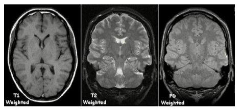
Adjusting Image Contrast
The contrast in MRI images can be adjusted by modifying the time between RF pulses. For instance:
- Tissues with large molecules (like certain brain structures) are best captured using short pulse intervals.
- Different pulse timing can enhance certain tissues, providing flexibility in examining various body structures.
Advantages of MRI
- Non-invasive: MRI does not involve radiation and is safe for repeated use.
- High-quality images of soft tissues: Unlike X-rays, MRI can capture detailed images of soft tissues (e.g., brain, muscles).
- Adjustable contrast: The contrast can be adjusted to focus on specific types of tissues or abnormalities.
- Real-time imaging: Allows observation of dynamic processes, such as blood flow or organ movement.
Disadvantages of MRI
- High cost: MRI machines are expensive to purchase and operate.
- Limited for bone imaging: MRI is not ideal for imaging bones, as they appear dark in scans.
- Noise and duration: Scanners are noisy, and scans may take a considerable time, which can be uncomfortable.
- Not suitable for patients with metal implants: Due to the magnetic field, patients with certain metallic implants (like pacemakers) cannot undergo MRI.
Example Calculation for Frequency Adjustment
- Determine the frequency for RF pulses in a specific body region by adjusting for the magnetic gradient and precession frequency.
- Adjust the pulse intervals to maximise contrast for the targeted tissue type.

500K+ Students Use These Powerful Tools to Master Magnetic resonance (MR) scanner For their A-Level Exams.
Enhance your understanding with flashcards, quizzes, and exams—designed to help you grasp key concepts, reinforce learning, and master any topic with confidence!
30 flashcards
Flashcards on Magnetic resonance (MR) scanner
Revise key concepts with interactive flashcards.
Try Physics Flashcards3 quizzes
Quizzes on Magnetic resonance (MR) scanner
Test your knowledge with fun and engaging quizzes.
Try Physics Quizzes29 questions
Exam questions on Magnetic resonance (MR) scanner
Boost your confidence with real exam questions.
Try Physics Questions27 exams created
Exam Builder on Magnetic resonance (MR) scanner
Create custom exams across topics for better practice!
Try Physics exam builder56 papers
Past Papers on Magnetic resonance (MR) scanner
Practice past papers to reinforce exam experience.
Try Physics Past PapersOther Revision Notes related to Magnetic resonance (MR) scanner you should explore
Discover More Revision Notes Related to Magnetic resonance (MR) scanner to Deepen Your Understanding and Improve Your Mastery