Photo AI
Last Updated Sep 27, 2025
Use of high energy X-rays Simplified Revision Notes for A-Level AQA Physics
Revision notes with simplified explanations to understand Use of high energy X-rays quickly and effectively.
407+ students studying
10.6.4 Use of high energy X-rays
High-energy X-rays
High-energy X-rays are commonly used in radiotherapy to target and destroy malignant (cancerous) cells. This approach is effective in either killing tumour cells or limiting their growth. Since X-rays are a form of ionising radiation, they are capable of damaging any cell they penetrate, including healthy ones. Therefore, specific strategies are employed to minimise exposure to healthy tissue while maximising the dose to the tumour.
Techniques for Targeted Use of X-rays in Cancer Treatment
-
Tumour Localisation with Scans: Before treatment, scans are conducted to accurately pinpoint the tumour's location. This helps to ensure that the high-energy X-rays are directed precisely at the tumour, reducing unnecessary exposure to surrounding healthy tissue.
-
Selecting the Correct Energy Level: The energy level of the X-rays must be carefully chosen. Higher energy levels are used to penetrate deeply situated tumours, while lower energy levels may suffice for tumours near the surface. Choosing the correct energy allows for effective tumour targeting with minimal harm to surrounding tissue.
-
Use of Shielding: To protect non-cancerous tissue from radiation, shielding materials such as lead may be placed over healthy areas. This reduces the risk of radiation damage to critical organs and tissues adjacent to the tumour site.
-
Collimated X-ray Beams: The X-ray beam is narrowed and directed through a collimator to make the photon stream more parallel. This process focuses the radiation onto the tumour and reduces scatter, thereby lowering the dose received by healthy tissues.
-
Multiple Beams Targeting the Tumour: Several beams of radiation are directed at the tumour from different angles. Each individual beam is relatively weak and thus causes minimal damage to the tissue it passes through. However, all the beams converge on the tumour, delivering a higher cumulative dose precisely at the target site.
-
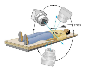
Rotating Beam (Isocentric Technique): In this method, the X-ray source rotates around the patient with the tumour located at the centre of rotation. By continuously changing the angle, the beam delivers the highest dose to the tumour while limiting exposure to surrounding tissues, as each part of the surrounding tissue only briefly intercepts the beam.
Key Points to Understand
- Ionising Radiation: X-rays have sufficient energy to ionise atoms, which can damage or kill cells. This property makes them effective in cancer treatment but also necessitates careful planning to avoid harming healthy cells.
- Targeted Therapy: The goal is to concentrate the X-ray energy on the tumour, sparing healthy tissue by adjusting beam angles, energy levels, and using protective shielding.
- Radiotherapy Planning: Treatment is carefully planned, often with the help of imaging scans (such as CT or MRI), to map the tumour's position and shape. Sophisticated software may be used to determine the optimal beam paths and intensities.
Example to Illustrate Beam Targeting in Radiotherapy
Imagine a tumour located near the lungs, surrounded by healthy tissue. In this scenario:
- Imaging scans are first performed to locate the tumour precisely and assess its size and shape.
- Shielding materials are placed over nearby healthy organs, like the heart, to protect them.
- Multiple low-dose beams are directed towards the tumour from different angles, converging to deliver a high dose precisely at the tumour site.
- A rotating beam technique might be used, where the X-ray source rotates around the patient, targeting the tumour from all angles. This minimises exposure to healthy lung tissue while focusing radiation on the tumour itself.

500K+ Students Use These Powerful Tools to Master Use of high energy X-rays For their A-Level Exams.
Enhance your understanding with flashcards, quizzes, and exams—designed to help you grasp key concepts, reinforce learning, and master any topic with confidence!
60 flashcards
Flashcards on Use of high energy X-rays
Revise key concepts with interactive flashcards.
Try Physics Flashcards6 quizzes
Quizzes on Use of high energy X-rays
Test your knowledge with fun and engaging quizzes.
Try Physics Quizzes29 questions
Exam questions on Use of high energy X-rays
Boost your confidence with real exam questions.
Try Physics Questions27 exams created
Exam Builder on Use of high energy X-rays
Create custom exams across topics for better practice!
Try Physics exam builder56 papers
Past Papers on Use of high energy X-rays
Practice past papers to reinforce exam experience.
Try Physics Past PapersOther Revision Notes related to Use of high energy X-rays you should explore
Discover More Revision Notes Related to Use of high energy X-rays to Deepen Your Understanding and Improve Your Mastery
96%
114 rated
Radionuclide Imaging and Therapy
Use of radioactive implants
205+ studying
185KViews