Photo AI
Figure 3 shows the banding pattern of a single sarcomere - AQA - A-Level Biology - Question 4 - 2020 - Paper 1
Question 4
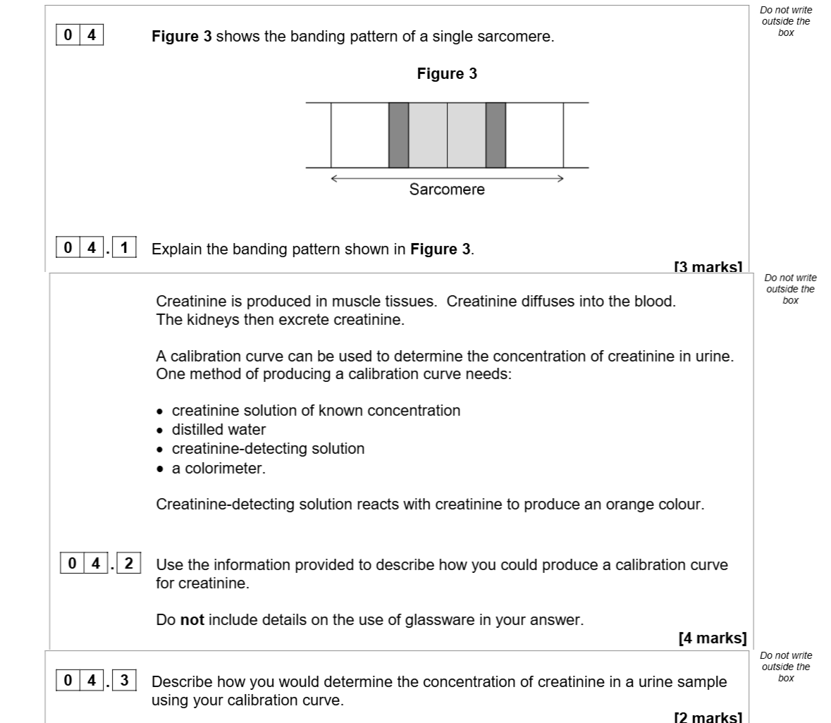
Figure 3 shows the banding pattern of a single sarcomere. 0 4 . 1 Explain the banding pattern shown in Figure 3. Creatinine is produced in muscle tissues. Creatini... show full transcript
Worked Solution & Example Answer:Figure 3 shows the banding pattern of a single sarcomere - AQA - A-Level Biology - Question 4 - 2020 - Paper 1
Step 1
Explain the banding pattern shown in Figure 3.

Answer
The banding pattern of the sarcomere, as displayed in Figure 3, consists of alternating light and dark regions. The light bands represent areas where the thin filaments, primarily composed of actin, are present. In contrast, the dark bands indicate regions where thick filaments, primarily made of myosin, overlap with the actin filaments.
Specifically, the central part of the dark band is referred to as the H zone, which contains only myosin and appears darker when visualized. The distinct appearance of these bands is crucial for muscle contraction, as it reflects the arrangement of the filaments that slide past one another.
Step 2
Use the information provided to describe how you could produce a calibration curve for creatinine.
Answer
To produce a calibration curve for creatinine:
- Use distilled water to prepare a range of creatinine solutions of known concentrations.
- Mix each creatinine solution with the creatinine-detecting solution in a suitable container.
- Measure the absorbance or transmission of each solution using a colorimeter after the reaction has occurred, noting the color change to orange.
- Plot the concentration of creatinine solutions against their corresponding absorbance/transmission values on a graph to create the calibration curve.
Step 3
Describe how you would determine the concentration of creatinine in a urine sample using your calibration curve.
Answer
To determine the concentration of creatinine in a urine sample:
- Prepare the urine sample and react it with the creatinine-detecting solution.
- Measure the absorbance or transmission of the reacted sample using the same colorimeter used for the calibration curve.
- Refer to the calibration curve plotted earlier, locating the absorbance value obtained from the urine sample on the graph.
- From the graph, read off the corresponding creatinine concentration that aligns with the measured absorbance, applying the 'line of best fit' to interpolate the result accurately.