Photo AI
Figure 3 shows the banding pattern of a single sarcomere - AQA - A-Level Biology - Question 4 - 2020 - Paper 1
Question 4
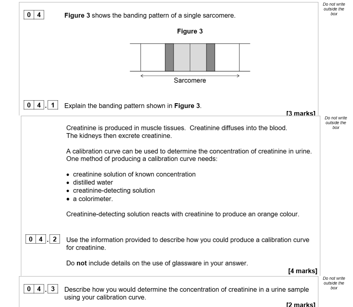
Figure 3 shows the banding pattern of a single sarcomere. 0 4 . 1 Explain the banding pattern shown in Figure 3. Creatinine is produced in muscle tissues. Creatini... show full transcript
Worked Solution & Example Answer:Figure 3 shows the banding pattern of a single sarcomere - AQA - A-Level Biology - Question 4 - 2020 - Paper 1
Step 1
Explain the banding pattern shown in Figure 3.

Answer
The banding pattern in Figure 3 illustrates distinct regions within a sarcomere. The light bands, known as I bands, consist primarily of actin filaments, providing sites for muscle contraction. The darker regions, called A bands, represent areas with overlapping actin and myosin filaments, which are essential for the muscle contraction process. The H zone, located in the center of the A band, shows only myosin filaments and appears lighter due to the absence of actin in that region.
Step 2
Use the information provided to describe how you could produce a calibration curve for creatinine.
Answer
To create a calibration curve for creatinine, start by preparing a series of known concentrations of creatinine solutions. Dilute each solution with distilled water and add the creatinine-detecting solution. Record the absorbance or transmission of each solution using a colorimeter, noting the intensity of the resulting orange color as it correlates with creatinine concentration. Finally, plot the concentration of creatinine solutions against their corresponding absorbance/transmission values on a graph to create the calibration curve.
Step 3
Describe how you would determine the concentration of creatinine in a urine sample using your calibration curve.
Answer
To determine the concentration of creatinine in a urine sample, first measure the absorbance of the urine sample using the colorimeter. Compare the obtained absorbance value against the calibration curve plotted earlier. Locate the corresponding creatinine concentration on the curve that aligns with the absorbance measurement to find the concentration of creatinine present in the urine sample.