Human Skeleton (Grade 10 NSC Matric Life Sciences): Revision Notes
Human Skeleton
The human skeleton is a remarkable living framework that provides structure and support to our bodies. Understanding its organisation and functions is essential for grasping how our bodies move and protect vital organs.
Overview of the human skeleton
Humans possess a living endoskeleton (internal skeleton) composed of bone, cartilage, and connective tissue. At birth, the human skeleton contains over 270 bones, but in adults this number reduces to 206 bones due to the fusion of smaller bones into larger structures as we grow and develop.
The reduction from 270 bones at birth to 206 bones in adults occurs because many smaller bones fuse together during growth and development, particularly in the skull, spine, and pelvis.
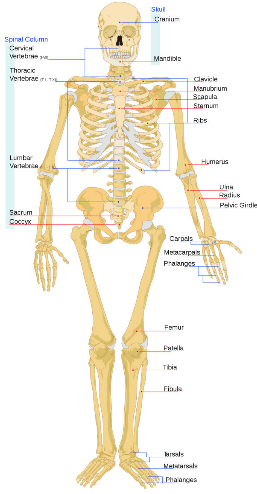
The adult human skeleton consists of two main divisions: the axial skeleton and the appendicular skeleton, each serving distinct but complementary functions in body support and movement.
Two main divisions of the skeleton
Understanding Skeletal Organization
The division of the skeleton into axial and appendicular components helps us understand how different bone groups work together to provide both stability and mobility.
The human skeletal system can be divided into two primary parts:
- Axial skeleton: Forms the central axis of the body, including the skull, vertebral column, and rib cage
- Appendicular skeleton: Consists of the limbs (arms and legs) and the structures that attach them to the axial skeleton
Axial skeleton
The axial skeleton forms the central supporting structure of the body and provides protection for vital organs. It consists of three main components: the skull, vertebral column, and rib cage with sternum.
The skull
The skull is a complex structure that houses and protects the brain whilst providing attachment points for facial muscles. It consists of two main parts:
The cranium contains eight flat bones that are joined together by immovable joints called sutures. These bones surround and protect the brain. At the base of the skull is a large opening called the foramen magnum through which the spinal cord passes. On either side of this opening are projections that connect with the first vertebra (called the atlas) to allow nodding movements of the head.
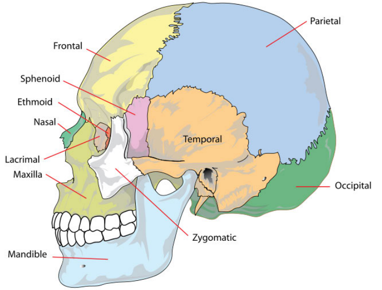
The facial bones number 15 in total and include irregular bones such as the cheek bones, nasal bones, temples, upper jaw bone (maxilla), and lower jaw bone (mandible). The mandible is the only movable bone in the skull, allowing us to speak, chew, and make facial expressions.
Dental Formula Understanding
The arrangement of teeth in humans follows a specific pattern called a dental formula: 2.1.2.3/2.1.2.3. This represents 2 incisors, 1 canine, 2 premolars, and 3 molars in each half of both the upper and lower jaws, giving us a total of 32 permanent teeth.
The vertebral column
The vertebral column, commonly called the spine or backbone, is a remarkable structure that typically consists of 24 individual (articulating) vertebrae plus 9 fused vertebrae in the sacrum and coccyx regions. Between the vertebrae are discs of fibrocartilage that act as shock absorbers during movement and prevent friction between bones.
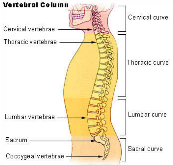
The spinal column is divided into five distinct regions:
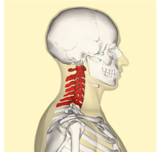
- Cervical region (neck): 7 vertebrae, including the atlas and axis
- Thoracic region (chest): 12 vertebrae, each bearing a pair of ribs
- Lumbar region (lower back): 5 vertebrae, the largest as they support body weight
- Sacral region: 5 fused vertebrae forming the sacrum
- Coccyx: 4 fused bones forming the tailbone
The vertebrae are connected by strong ligaments and muscles that stabilise the spine and control movement. Together, they form a continuous spinal canal that runs from the skull to the pelvic region, housing and protecting the spinal cord.
Functions of the vertebral column:
- Supports the skull and maintains upright posture
- Surrounds and protects the spinal cord
- Provides attachment points for ribs, muscle groups, and back muscles
- The separate vertebrae and S-shaped curves provide flexibility, allowing humans to bend forwards, backwards, and sideways
- Fibrocartilage discs act as shock absorbers during movement
The rib cage and sternum
The rib cage is a protective bony and cartilaginous structure surrounding the thoracic cavity. It consists of 24 ribs (12 pairs), the sternum (breastbone), coastal cartilages, and the 12 thoracic vertebrae.
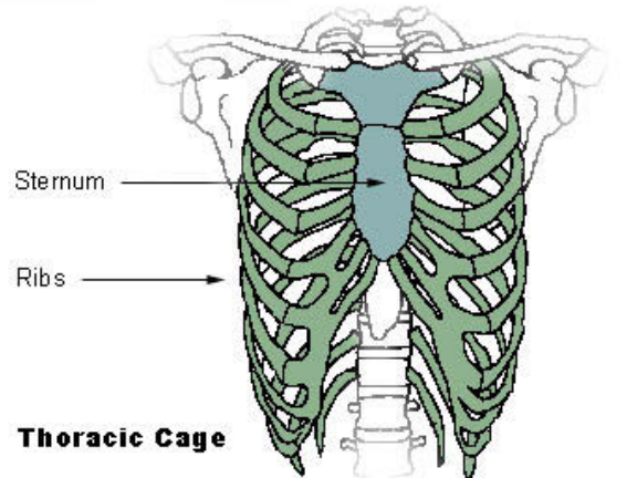
The first 7 pairs of ribs connect directly to the sternum and are called true ribs. The remaining 5 pairs do not connect directly to the sternum and are referred to as false ribs. This structure protects the heart and lungs whilst allowing the chest to expand and contract during breathing with help from the diaphragm and intercostal muscles.
Appendicular skeleton
The appendicular skeleton includes all the bones of the limbs plus the structures that attach them to the axial skeleton. This system enables locomotion and manipulation of the environment.
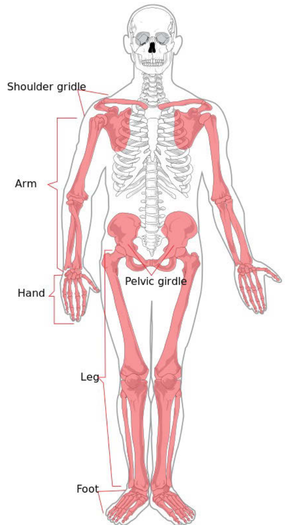
Pectoral girdle and arms
The pectoral girdle consists of 2 clavicles (collar bones) and 2 scapulae (shoulder blades). These bones are attached to the sternum at the front and connect to the back of the thorax via muscles, providing support for the shoulders whilst allowing considerable freedom of movement.
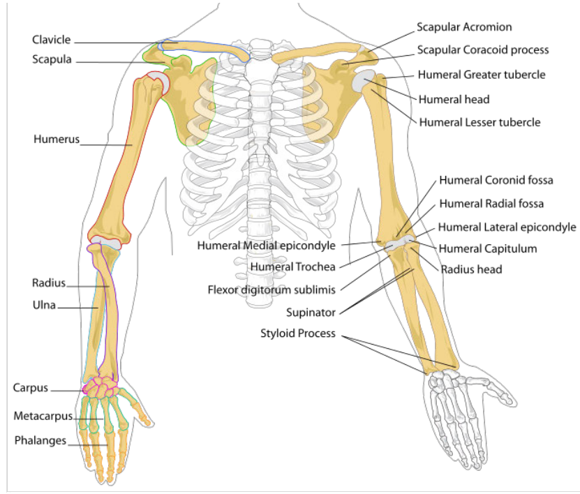
Each upper limb contains:
- Humerus: The single bone of the upper arm that fits into the shallow glenoid cavity of the scapula, forming a ball-and-socket joint
- Ulna and radius: The two bones of the forearm, with the joint at the elbow being a hinge joint
- Carpals: 8 small wrist bones arranged in two rows
- Metacarpals: 5 bones forming the palm of the hand
- Phalanges: 14 finger bones (2 in the thumb, 3 in each finger)
Functions of the pectoral girdle:
- Forms a strong support structure for arm attachment
- Provides large surface areas for muscle attachment
- Creates ball-and-socket joints allowing arms to move freely in multiple directions
Pelvic girdle and legs
The pelvic girdle consists of hip bones joined at the front by cartilage called the pubic symphysis and attached to the sacrum at the back. Each hip bone is formed from three fused bones that contribute to forming the acetabulum, a deep socket where the head of the femur (thigh bone) connects to form the hip joint.
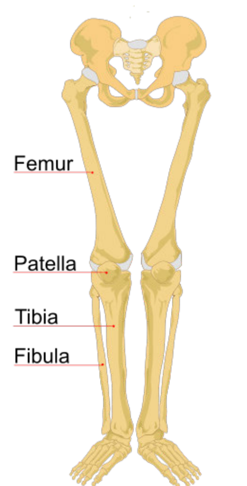
Each lower limb contains:
- Femur: The largest and strongest bone in the body, forming ball-and-socket joints with the hip and hinge joints with the tibia at the knee
- Patella: The kneecap, a flat triangular bone embedded in the tendon of the thigh muscle
- Tibia: The larger shin bone that supports most body weight
- Fibula: The smaller, thinner bone that serves mainly for muscle attachment
- Tarsals: 7 ancle bones, including the large heel bone (calcaneum)
- Metatarsals: 5 bones forming the arch and ball of the foot
- Phalanges: 14 toe bones (2 in the big toe, 3 in each other toe)

The structure of the foot is similar to the hand but is stronger and less mobile, as it must support body weight during standing, walking, and running.
Functions of the human skeleton
Essential Functions of the Skeletal System
The human skeleton is living tissue that performs many essential functions that are critical for survival and daily activities.
The human skeleton serves six main functions:
- Movement: Muscles attach to bones, enabling all body movements
- Protection: The skull protects the brain, the rib cage protects the heart and lungs, and pelvic bones protect digestive and reproductive organs
- Support: Provides shape and structural support to maintain body posture
- Storage of minerals: Bones store important minerals such as calcium and phosphate ions
- Hearing: Three tiny bones in the middle ear (hammer, anvil, and stirrup) amplify sound waves
- Red blood cell production: Long bones and flat bones contain red bone marrow that produces red blood cells
Structure of long bone
Long bones, such as the femur, humerus, tibia, and radius, have a characteristic structure that enables them to provide support whilst remaining relatively lightweight.
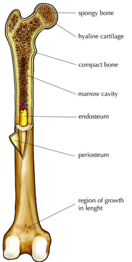
Key components of a long bone:
- Epiphysis: The enlarged ends of long bones, consisting largely of spongy bone and covered with hyaline cartilage at joint surfaces
- Spongy bone: Found in the epiphysis, contains red bone marrow where blood cells are produced
- Compact bone: Dense, hard bone forming the outer shaft (diaphysis) that provides strength and rigidity
- Marrow cavity: The hollow centre filled with yellow marrow (mostly fat) that stores energy
- Periosteum: The tough outer membrane containing blood vessels that nourish the bone and provide attachment sites for muscles via tendons and ligaments
- Endosteum: The delicate inner lining of the marrow cavity
- Trabeculae: The interconnected struts within spongy bone that transfer stress efficiently whilst maintaining light weight
This structure allows long bones to be both strong enough to support body weight and light enough to enable efficient movement.
Key Points to Remember:
- The human skeleton contains 206 bones in adults, divided into axial and appendicular skeletons
- The axial skeleton (skull, spine, rib cage) protects vital organs and forms the body's central support
- The vertebral column has five regions with natural curves that provide flexibility and shock absorption
- The appendicular skeleton (limbs and their attachments) enables movement and manipulation
- Long bones have a sophisticated structure with different tissue types serving specific functions
- The skeleton serves six main functions: movement, protection, support, mineral storage, hearing, and blood cell production