Autotroph Structure and Function (HSC SSCE Biology): Revision Notes
Autotroph Structure and Function
Introduction to nutrients in living organisms
All living organisms require specific substances to function efficiently. These essential nutrients fall into two categories:
Organic nutrients include:
- Glucose
- Amino acids
- Fatty acids
- Glycerol
- Nucleotides
- Vitamins
Inorganic nutrients include:
- Mineral ions (phosphates, sodium ions, chloride ions)
- Water
- Gases (carbon dioxide, oxygen)
These nutrients supply energy and provide raw materials for building cellular structures and living tissues.
Understanding the distinction between organic and inorganic nutrients is essential for comprehending how organisms obtain and process the materials they need to survive and grow.
Autotrophs vs. heterotrophs
Autotrophs are organisms that can produce their own organic nutrients. They only need to obtain water, mineral ions, and gases (carbon dioxide and oxygen) from external sources.
Heterotrophs must obtain all nutrients (both organic and inorganic) from external sources.
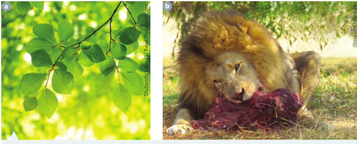
The key difference between autotrophs and heterotrophs lies in their ability to produce organic nutrients. Autotrophs are self-sufficient in organic nutrient production, while heterotrophs depend entirely on external sources for these essential compounds.
The majority of autotrophs are plants, which have evolved specialized structures to efficiently obtain and process the nutrients they need.
Vascular and non-vascular plants
Vascular plants
Most plants are vascular plants, meaning they possess a specialized transport system to move substances from one part of the plant to another. This transport system consists of two types of conducting tissue:
- Xylem: Transports water and water-soluble nutrients (mineral salts) absorbed from the soil through the root system
- Phloem: Transports sugars in the form of dissolved sucrose and other plant products from one part of the plant to another
The vascular system in plants is analogous to the circulatory system in animals, serving as a vital transportation network throughout the organism.
Non-vascular plants
A small number of plants, such as mosses and liverworts, are non-vascular plants. These plants have a very simple structure and lack specialized transport systems. All nutrients are absorbed and wastes are removed by diffusion and osmosis directly through the plant's surfaces.
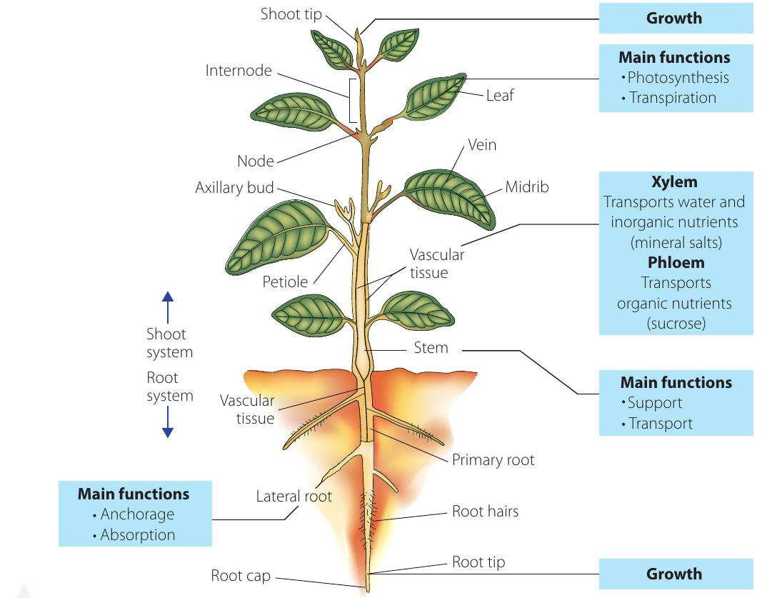
Organization of vascular plants
Vascular plants have specialized cells grouped together into tissues (such as photosynthetic tissue). These tissues combine with other tissues to form organs (such as leaves, stems, and roots) that carry out specific functions.
The three plant body systems
Vascular plants contain three main "body" systems that work together:
- Root system - anchors the plant and absorbs water and minerals
- Shoot system - includes stems and leaves; supports the plant and carries out photosynthesis
- Vascular system - transports substances throughout the plant using xylem and phloem tissues
The Three Plant Systems:
- Root system provides anchorage and absorption
- Shoot system supports the plant and enables photosynthesis
- Vascular system connects all parts through transport pathways
The root system
The root system has two main functions:
- Anchoring the plant in the soil
- Absorbing water and inorganic nutrients (mineral ions) from the soil
The root system is usually located underground.
Maximizing surface area for absorption
Roots have evolved several adaptations to maximize their surface area for efficient absorption:
1. Extensive branching
- Mature root systems branch extensively
- This increases the total surface area available for absorption
- Branching also provides better anchorage for the plant
2. Root hairs
- Located in the younger part of each root, near the tip
- Specialized epidermal cells (outer layer cells) extend outwards into the soil as microscopic projections called root hairs
- Root hairs can increase the surface area of a root up to 12 times
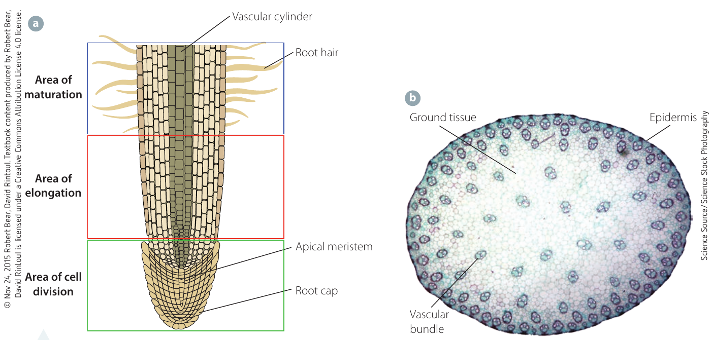
Root hairs are crucial for plant survival. By increasing surface area up to 12 times, they dramatically enhance the plant's ability to absorb water and minerals from the soil. These microscopic structures represent one of nature's most efficient solutions to the challenge of nutrient absorption.
3. Flattened epidermal cells
- Water enters the root through epidermal cells across the entire root surface
- These cells have a flattened shape that increases their exposed surface
- However, their surface area is smaller than root hair cells, so they absorb less water per cell
Movement of substances into roots
Different transport mechanisms move substances into root cells:
Water movement:
- Water enters roots by osmosis (movement from high to low water concentration across a semi-permeable membrane)
Mineral ion movement:
- Mineral ions usually enter by diffusion (movement from high to low concentration)
- When diffusion is too slow or the concentration gradient is insufficient, cells use:
- Facilitated diffusion (diffusion assisted by transport proteins)
- Active transport (movement against the concentration gradient using energy)
Plants employ multiple transport mechanisms to ensure they can obtain minerals even when environmental conditions are not ideal. Active transport allows roots to absorb minerals even when soil concentrations are lower than those inside the plant cells.
Gas exchange in roots
Root cells cannot photosynthesize because they:
- Do not contain chloroplasts
- Are not exposed to sunlight
However, like all living cells, root cells carry out aerobic cellular respiration continuously. This requires gas exchange:
- Oxygen diffuses into root cells from air pockets in the soil
- Carbon dioxide (waste product of respiration) diffuses out of root cells into the soil
Even though root cells cannot photosynthesize, they still require oxygen for cellular respiration. This is why waterlogged soil can damage or kill plants—the lack of air pockets prevents oxygen from reaching the roots.
The shoot system: stems
The stem is part of the shoot system and serves two main functions:
- Providing structural support for the plant
- Providing a transport pathway between roots and leaves
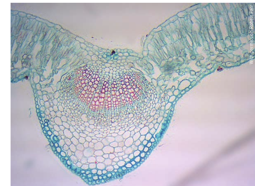
Tissue types in stems
Stems contain three main types of tissue:
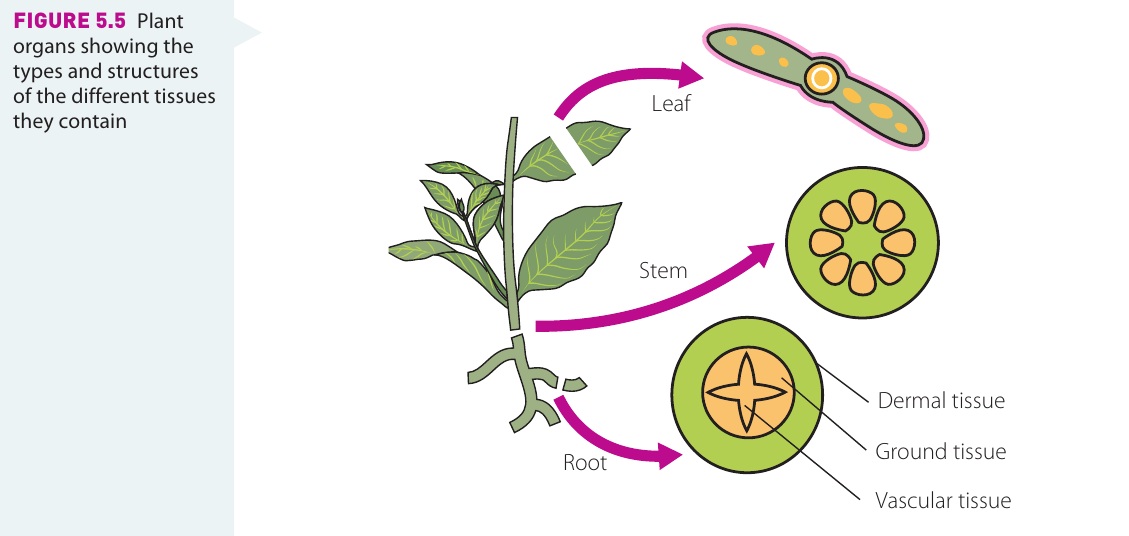
1. Dermal tissue
- Forms the outer layer of the stem
- Provides waterproofing
- Offers protection
- Controls gas exchange
2. Vascular tissue
- Composed of xylem and phloem tissues
- Arranged in structures called vascular bundles
- Extends from roots to leaves
- Provides structural support and enables transport of materials
- Water and mineral ions absorbed by roots travel through xylem in the stem to reach the leaves
- Products of photosynthesis move from leaves to all plant parts through phloem in the stem
3. Ground tissue
- Fills in around the vascular tissue
- Functions include storage, photosynthesis, and additional support
The arrangement of tissues in stems provides both strength and flexibility. Vascular bundles not only transport materials but also contribute to the stem's structural integrity, allowing plants to grow tall and support leaves in optimal positions for light capture.
The shoot system: leaves
Leaves are the primary organs for two critical processes:
- Photosynthesis: producing glucose using sunlight and carbon dioxide
- Transpiration: evaporation of water from the leaf, which aids water movement from roots and cools the plant
Absorbing sunlight
The structure of leaves is perfectly adapted for maximum light absorption:
Thin, flat structure:
- Large surface area maximizes absorption of light energy
- Thinness ensures no internal cell is too far from the surface to receive light
- The outermost epidermis layer is transparent, allowing sunlight to penetrate to photosynthetic cells beneath
Mesophyll cells:
The mesophyll (middle layers of the leaf) contains the cells responsible for most photosynthesis. Two types of mesophyll cells exist:
- Palisade cells
- Elongated, column-shaped cells
- Densely packed with chloroplasts
- Main photosynthetic cells in leaves
- Positioned vertically, immediately below the upper epidermis
- Exposed to maximum sunlight
- High chloroplast number ensures maximum photosynthesis rate
- Spongy mesophyll cells
- Second most important photosynthetic cells
- Located between palisade cells and lower epidermis
- Contain fewer chloroplasts than palisade cells
- Irregular in shape and distribution
- Spaces between cells allow easy gas movement
Palisade cells are the primary site of photosynthesis in most plants. Their vertical orientation, position directly beneath the upper epidermis, and dense concentration of chloroplasts make them perfectly designed for capturing light energy and converting it into chemical energy.
Adaptations to different environments:
Environmental Adaptations of Leaves:
Rainforest floor plants:
- Very large, thin, flat, dark-colored leaves
- Adapted to absorb maximum sunlight in low-light conditions
- Less concern about water loss due to high humidity
Hot, dry environment plants:
- Very small leaves with minimal surface area
- Can still absorb necessary sunlight while minimizing water loss through evaporation
- Many Australian plants show this adaptation
Gaseous exchange
Epidermis and cuticle:
- The epidermis (protective layer of cells) covers both upper and lower leaf surfaces
- Simple, flattened cells protect delicate inner tissues
- Epidermal cells are transparent to allow light to reach cells below
- Can secrete a waterproof cuticle to prevent water evaporation
Guard cells and stomata:
Within the epidermis are specialized cells that control gas exchange:
- Guard cells: Bean-shaped cells that occur in pairs
- Stoma (plural: stomata): A pore or opening surrounded by two guard cells
- Guard cells change shape to open and close stomata
- Stomata usually occur on the lower (under) surface of the leaf
- Control exchange of gases (carbon dioxide and oxygen)
- Control loss of water vapour
Guard cells act as gatekeepers for the leaf, regulating the opening and closing of stomata. This control is critical for balancing two competing needs: obtaining carbon dioxide for photosynthesis while minimizing water loss through transpiration.
In hot, dry habitats, waxy cuticles and other leaf characteristics reduce water loss through evaporation.
Transport in leaves
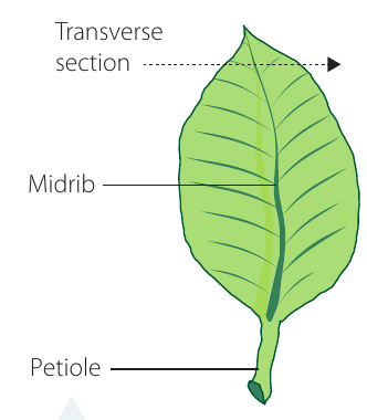
Vascular tissue in leaves is organized into veins:
- The main vein is called the midrib
- Many smaller veins branch out from the midrib
- Veins contain xylem and phloem tissue
- Distribution throughout the leaf ensures no cells are too far from transport pathways
- Vascular tissue also provides important structural support for the thin leaf blade
The extensive network of veins in a leaf serves dual purposes: ensuring efficient transport of materials to and from every cell, and providing the mechanical support necessary to keep the thin leaf blade spread out for maximum light exposure.
Cellular respiration in plants
Important concept: Plants carry out cellular respiration as well as photosynthesis.
Cellular respiration is a function of all living cells. In leaves, it occurs continuously but is "masked" or hidden by photosynthesis during the day.
A common misconception is that plants only photosynthesize. In reality, all plant cells carry out cellular respiration continuously, just like animal cells. The difference is that during daylight hours, photosynthesis occurs at a much higher rate, masking the effects of respiration.
Gas exchange patterns
During the day:
- Oxygen needed for cellular respiration comes from oxygen produced during photosynthesis
- Photosynthesis usually occurs at a greater rate than respiration
- Excess oxygen (not used in respiration) is released to the environment
- Carbon dioxide from cellular respiration is used as a reactant in photosynthesis
- When photosynthesis rate is high, plants absorb additional carbon dioxide from the air
- Net gas exchange observed: That associated with photosynthesis ( in, out)
At night:
- Photosynthesis stops (no sunlight)
- Cellular respiration continues
- Net gas exchange observed: That associated with respiration ( in, out)
Gas Exchange in Plants:
Daytime:
- Photosynthesis rate > Respiration rate
- Net exchange: absorbed, released
- Oxygen from photosynthesis supplies respiration needs
Nighttime:
- Only respiration occurs
- Net exchange: absorbed, released
- Pattern matches typical cellular respiration
Imaging technologies for studying plant structure
Advanced technologies have greatly enhanced our understanding of plant structure and function.
Modern imaging methods
Three-dimensional (3D) imaging:
- Digital cameras take photos from different angles
- Computer programs combine photos to produce 3D images
- Allows measurement and study of external plant structure
MRI (Magnetic Resonance Imaging):
- Uses radio waves and magnetic fields
- Takes series of images to produce computer-generated 3D images
- Can study root structure in plants grown in clear containers
- Provides detailed analysis beyond simple observation
PET (Positron Emission Tomography) and NT (Neutron Tomography):
- Detect radiation from radioisotopes
- Can be combined with MRI images
- Provide functional information about transport and processes
X-ray Computed Microtomography (Micro-CT):
- Similar to medical CT scans but smaller scale and higher resolution
- Non-destructive process
- Sample rotated in X-ray beam; hundreds of images recorded from different angles
- Images reconstructed into 3D computer-generated model
- Allows study of internal tissue spatial arrangement
Modern imaging technologies have revolutionized plant biology by allowing scientists to study living plants non-destructively. These tools reveal structures and processes that were previously invisible or required destroying the specimen to observe.
Radioisotopes as tracers
Radioisotopes are isotopes that emit radiation. They serve as useful tracers because technologies can detect and track their movement through biological systems.
Applications in plant research:
- Determining oxygen source in photosynthesis: Plants given water with radioactive oxygen showed all radioactive oxygen released as gas, proving water (not ) is the oxygen source
- Tracing glucose pathways: Carbon-14 added to carbon dioxide supply becomes incorporated into glucose molecules; radiation emitted by carbon-14 shows where glucose moves and accumulates
- Modern systems like RRIS (Real-time Radioactive Imaging System) visualize substance movement in phloem non-destructively
- PlanTIS (PET scanner for plants) traces carbon-11 incorporated into glucose molecules
Using Radioisotopes to Trace Photosynthesis:
Scientists provided plants with water containing radioactive oxygen atoms. After photosynthesis occurred, they measured where the radioactive oxygen appeared.
Result: All radioactive oxygen was found in the gas released by the plant, not in the glucose molecules.
Conclusion: This proved that the oxygen released during photosynthesis comes from water molecules, not from carbon dioxide, resolving a long-standing question about the photosynthetic process.
Key Points to Remember:
-
Autotrophs produce their own organic nutrients; heterotrophs must obtain all nutrients externally
-
Vascular plants have xylem and phloem for transport; non-vascular plants rely on diffusion and osmosis
-
Roots maximize surface area through branching, root hairs, and flattened epidermal cells to efficiently absorb water (by osmosis) and minerals (by diffusion or active transport)
-
Stems contain three tissue types: dermal (protection), vascular (transport), and ground (storage, support, photosynthesis)
-
Leaves are thin and flat to maximize light absorption; palisade cells (many chloroplasts, vertical position) are the main photosynthetic cells
-
Guard cells control stomata to regulate gas exchange ( in, out during day) and water loss
-
All plant cells respire continuously, but during the day, photosynthesis masks this process; net gas exchange during daylight reflects photosynthesis