Animal Tissues (Grade 10 NSC Matric Life Sciences): Revision Notes
Animal Tissues
Introduction to animal tissues
Animal bodies, including human bodies, are made up of four different types of tissue that work together to keep organisms functioning properly. These tissues are groups of similar cells that perform specific functions. Understanding these tissues is essential for comprehending how animal bodies are organised and how they carry out life processes.
Definition: Tissues are groups of similar cells that perform a particular function. This organisation allows for specialisation and efficiency in biological systems.
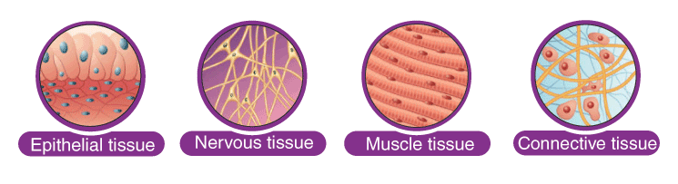
The four main types of animal tissue are:
- Epithelial tissue - forms protective barriers and covers surfaces
- Connective tissue - provides support, structure and connection between body parts
- Muscle tissue - enables movement throughout the body
- Nervous tissue - carries electrical and chemical signals for communication
Each tissue type has unique structural features that allow it to perform its specific functions effectively. You'll need to be able to recognise these tissues under a microscope and understand how their structure relates to their function.
Epithelial tissue
Epithelial tissue forms the outer layer of your body (skin) and lines many internal cavities and organs. This tissue acts as a protective barrier between your internal environment and the external world.
General functions of epithelial tissue
Epithelial tissue serves several important purposes:
- Provides a barrier between the external environment and internal organs
- Specialised for secretion and absorption of substances
- Protects organisms from microorganisms, injury, and fluid loss
- Helps remove waste products such as sweat from the skin
Classification of epithelial tissue
Essential Classification System
Epithelial tissues are classified based on two main characteristics that you must understand for identification:
- Number of cell layers (simple vs stratified)
- Shape of the cells (squamous vs cuboidal vs columnar)
Understanding this classification system helps you identify different types under a microscope.
Number of layers:
- Simple epithelium - consists of a single layer of cells
- Stratified epithelium - consists of two or more layers of cells
Cell shape:
- Squamous - flattened, thin cells
- Cuboidal - cube-shaped cells
- Columnar - tall, elongated cells
Types of epithelial tissue
Simple squamous epithelium
Location: Capillaries, alveoli (air sacs) in lungs; stratified form found in skin
Structure: Thin and flat cells that are elliptically shaped, lying on a basement membrane. Simple squamous epithelium is one cell thick. Stratified squamous epithelium consists of many layers.
Function: Responsible for diffusion processes. The thin structure allows for easy movement of substances across the cells.
The extremely thin nature of simple squamous epithelium makes it perfect for areas where substances need to pass through quickly, such as gas exchange in the lungs or nutrient exchange in capillaries.
Simple cuboidal epithelium
Location: Kidney tubules and glands responsible for excretion
Structure: Cube-like structure; may occasionally have structures called microvilli on the surface to aid absorption
Function: Serves a protective function against bacteria and prevents water loss by lining various structures
The cube shape of these cells provides more space for cellular organelles compared to squamous cells, making them well-suited for active processes like secretion and absorption.
Simple columnar epithelium
Location: Digestive tract, reproductive organs
Structure: Elongated cells with nuclei located at the base of the cell. Cells are connected by tight junctions and receive nutrients from the basement membrane
Function: Main function is protective, preventing bacterial infection. Can also secrete mucus to protect surfaces from damage
The tall, column-like shape provides even more internal space for organelles, making these cells excellent for secretion and absorption. The tight junctions between cells ensure that substances must pass through the cells rather than between them.
Muscle tissue
Muscle tissue enables movement in animals, from the beating of your heart to the voluntary movements of your limbs. There are three distinct types of muscle tissue, each with specific characteristics suited to their particular functions.
Types of muscle tissue
1. Skeletal muscle
Skeletal muscle is under voluntary control, meaning you can consciously decide when to contract these muscles. This tissue has a striated (striped) appearance due to regular patterns of proteins responsible for contraction.
Key characteristics:
- Voluntary control - you can consciously control when these muscles contract
- Striated appearance - shows regular striped patterns under a microscope
- Anchored by tendons - connects to bones to enable movement
- Functions include - locomotion (walking, running) and maintaining posture
- Rapid contractions - contracts and relaxes in short bursts
2. Cardiac muscle
Cardiac muscle makes up the major tissue of the heart and is involuntary, meaning it contracts automatically without conscious control.
Key characteristics:
- Involuntary control - contracts automatically without conscious thought
- Striated appearance - similar striped pattern to skeletal muscle
- Branched structure - unlike skeletal muscle, cardiac muscle fibres branch and connect at irregular angles
- Coordinated contractions - the branched connections help coordinate heart contractions
- Continuous function - must work constantly throughout your lifetime
3. Smooth muscle
Smooth muscle is involuntary and non-striated, with a different structure from the other muscle types.
Key characteristics:
- Involuntary control - contracts automatically
- Non-striated - lacks the striped appearance of other muscle types
- Tapered ends - cells are spindle-shaped with pointed ends
- Location - found in blood vessel walls, digestive system, urinary tract, and trachea
- Function - responsible for rhythmic contractions like peristalsis (moving food through digestive system) and controlling blood pressure
- Sustained contractions - contracts for longer periods than striated muscle
Remember the muscle types:
- Skeletal: Voluntary + Striated
- Cardiac: Involuntary + Striated + Branched
- Smooth: Involuntary + Non-striated
Nervous tissue
Nervous tissue forms the communication network of the body, transmitting electrical and chemical signals between different body parts. This tissue makes up both the central nervous system (brain and spinal cord) and the peripheral nervous system (nerves throughout the body).
Function of nervous tissue
The primary function of nervous tissue is to transmit nerve impulses throughout the body. These impulses allow for:
- Communication between different body parts
- Response to environmental stimuli
- Coordination of body functions
- Control of muscle contractions and gland secretions
Structure of nervous tissue
Essential Neuron Structure
Nervous tissue consists of specialised cells called neurons that have unique structures adapted for signal transmission:
- Cell body (soma) - contains the nucleus and most organelles
- Dendrites - receive incoming signals from other neurons
- Axon - sends signals away from the cell body
- Myelin sheath - fatty substance that surrounds many axons, increasing signal speed and providing insulation
Types of neurons
There are three main types of neurons, each with specific functions:
Sensory neurons
- Structure: Unipolar neurons with receptor cells
- Function: Transmit signals from environmental stimuli to the central nervous system
- Process: Convert external stimuli (light, sound, touch, etc.) into electrical signals that travel to the brain and spinal cord
Motor neurons
- Structure: Multipolar neurons that connect to effectors like muscles
- Function: Carry impulses from the central nervous system to effectors (muscles or glands)
- Process: Receive signals from the brain/spinal cord and stimulate muscle contractions or gland secretions
Interneurons
- Structure: Multipolar neurons with highly branched dendrites and shorter axons
- Function: Connect sensory and motor neurons within the central nervous system
- Process: Process information and coordinate responses between sensory input and motor output
Worked Example: Reflex Arc
When you touch a hot surface:
- Sensory neurons detect the heat stimulus in your finger
- Interneurons in your spinal cord process this information
- Motor neurons immediately signal your muscles to pull your hand away
This demonstrates how all three neuron types work together for rapid responses!
Connective tissue
Connective tissue is found throughout the body, providing structural support, connecting different tissues and organs, and sometimes separating them. All connective tissue contains cells, fibres (such as collagen), and an extracellular matrix that gives each type its unique properties.
Types of connective tissue
Areolar (loose connective) tissue
- Structure: Jelly-like matrix with a network of elastic fibres
- Function: Holds organs in place, cushions and protects organs, acts as packing material
- Location: Surrounds blood vessels and nerves, found in the mesentry around the intestine
White fibrous tissue
- Structure: Consists of non-elastic fibres
- Function: Acts as a shock absorber, transfers or absorbs forces
- Location: Tendons, ligaments, and tough membrane sheaths around organs
Cartilage
- Structure: Rubbery matrix that can be flexible or rigid
- Function: Gives structure, shape and strength; reduces friction; provides support
- Location: Joints, nose, sternum, trachea
Bone tissue
- Structure: Made up of collagen fibres mineralised with calcium and phosphates
- Function: Provides strength and support; creates red and white blood cells
- Location: Bones found throughout the body
Each type of connective tissue has a specific matrix composition that determines its properties. For example, bone tissue is hard because of calcium and phosphate deposits, while cartilage is flexible due to its rubbery matrix.
Blood
Blood is classified as a specialised form of connective tissue because it originates from the same embryonic tissue as other connective tissues and contains fibres. However, unlike other connective tissues, blood is liquid and flows throughout the body.
Components of blood
Blood consists of several distinct components suspended in a liquid called plasma:
Red blood cells (erythrocytes)
Red blood cells are the most numerous cells in blood and have several unique characteristics:
- Shape: Biconcave (doughnut-shaped without a hole) which makes them flexible
- No nucleus: Lack a nucleus and most organelles, providing more space for haemoglobin
- Haemoglobin: Contains the protein haemoglobin with iron at its centre
- Function: Transport oxygen from lungs to tissues and carbon dioxide from tissues to lungs
- Lifespan: Approximately 120 days
- Flexibility: Can squeeze through narrow capillaries due to their shape
White blood cells (leukocytes)
White blood cells are part of the body's immune system and defend against disease:
- Production: Made in bone marrow and lymph nodes
- Structure: Have one or more nuclei and are larger and more irregular than red blood cells
- Function: Protect the body from diseases and infections
- Types: Include neutrophils, basophils, eosinophils, lymphocytes, monocytes, and macrophages
- Antibodies: Some white blood cells (lymphocytes) produce chemicals called antibodies that destroy disease-causing microorganisms
Platelets (thrombocytes)
Platelets are cell fragments that play a crucial role in blood clotting:
- Origin: Produced in bone marrow as fragments of larger cells
- Structure: Have no nucleus and are much smaller than red or white blood cells
- Function: Assist in blood clotting and prevent excessive bleeding
- Process: Clump together at wound sites to form clots that stop bleeding
Plasma
Plasma is the liquid component of blood that makes up about 55% of total blood volume:
- Composition: Contains dissolved proteins, hormones, urea, and carbon dioxide
- Functions:
- Transports nutrients, cells, and metabolic waste products
- Maintains blood volume and pressure
- Helps regulate body temperature
- Contains clotting factors that work with platelets
Blood Component Functions
Remember that blood has four main components, each with specific roles:
- Red blood cells: Oxygen and carbon dioxide transport
- White blood cells: Immune defence
- Platelets: Blood clotting
- Plasma: Transport medium and maintains blood properties
Practical investigation: tissue identification
Understanding animal tissues requires both theoretical knowledge and practical experience. Microscopic examination of tissue samples helps you identify different tissue types and understand their structures.
Key identification features
When examining tissues under a microscope, look for these distinguishing features:
Epithelial tissue:
- Cells closely packed together
- Little extracellular matrix
- Cells arranged in layers (simple or stratified)
- Cell shapes (squamous, cuboidal, or columnar)
Muscle tissue:
- Elongated cells (muscle fibres)
- Presence or absence of striations
- Single or multiple nuclei per cell
Nervous tissue:
- Star-shaped or elongated cells
- Long projections (dendrites and axons)
- Supporting cells around neurons
Connective tissue:
- Cells scattered in matrix material
- Visible fibres in many types
- Variable matrix consistency
Worked Example: Identifying Epithelial Tissue
When looking at a microscope slide:
- Check cell arrangement: Are cells tightly packed with little space between them?
- Count the layers: One layer = simple, multiple layers = stratified
- Observe cell shape: Flat = squamous, cube-like = cuboidal, tall = columnar
- Combine observations: "Simple squamous" = single layer of flat cells
This systematic approach ensures accurate identification!
Exam tips for tissue identification
Microscopy Success Tips
- Practice identifying tissues from microscope images
- Focus on key distinguishing features rather than memorising every detail
- Understand the relationship between structure and function
- Learn the locations where each tissue type is commonly found
- Be able to explain why each tissue's structure makes it suitable for its function
Key Points to Remember:
-
Animal bodies contain four main tissue types: epithelial (protection/barriers), connective (support/structure), muscle (movement), and nervous (communication)
-
Structure determines function: Each tissue's specific cellular arrangement and composition enables it to perform its particular role effectively
-
Epithelial tissue classification: Remember the two-part system - number of layers (simple vs stratified) and cell shape (squamous vs cuboidal vs columnar)
-
Muscle tissue variety: Skeletal (voluntary, striated), cardiac (involuntary, striated, branched), and smooth (involuntary, non-striated) each serve different movement needs
-
Blood is specialised connective tissue: Contains red blood cells (oxygen transport), white blood cells (immune defence), platelets (clotting), and plasma (transport medium) working together to maintain life