Muscle Structure and Function (Grade 10 NSC Matric Life Sciences): Revision Notes
Muscle Structure and Function
Hierarchical organisation of skeletal muscle
Understanding muscle structure requires looking at how muscles are organised from the smallest components up to the complete muscle. Think of this like Russian nesting dolls - each level contains and organises the smaller components within it.
The basic functional units of any muscle are called myofibrils. These are tiny thread-like structures that contain the actual machinery responsible for muscle contraction. Multiple myofibrils bundle together to form a muscle fibre, which is essentially a single muscle cell.
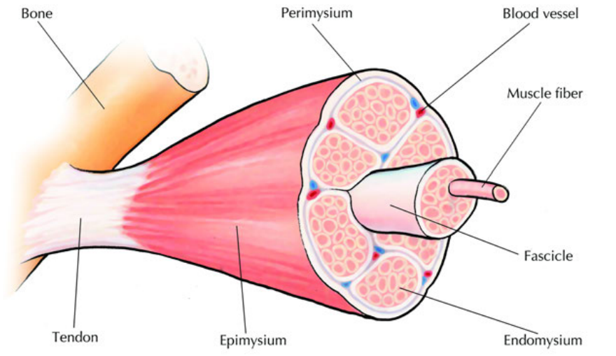
Several muscle fibres are then grouped together and surrounded by a thin layer of connective tissue called the perimysium. This creates structures called fascicles - you can think of fascicles as bundles of muscle fibres working as a team.
Finally, multiple fascicles are wrapped together by a tough outer layer called the epimysium to form the complete muscle that you can see and feel under your skin.
Understanding the Hierarchy is Essential:
The organisation follows this pattern: Myofibrils → Muscle fibres → Fascicles → Complete muscle
Each level is wrapped in protective connective tissue, and this hierarchical organisation allows for both strength and precise control of movement.
Anatomical components of muscle structure
Several important structures work together to make muscle function possible:
Connective tissues organise and protect the muscle:
- Epimysium - the tough outer wrapping that covers the entire muscle
- Perimysium - the thin layer that bundles muscle fibres into fascicles
- Endomysium - the delicate tissue that surrounds individual muscle fibres
Tendons are crucial for transferring the force created by muscle contractions. These strong, fibrous cords connect muscles directly to bones. When a muscle contracts, the tendon transfers this pulling force to move the bone, creating the movement you intended.
Blood vessels run throughout the muscle tissue, bringing essential oxygen and nutrients to the hardworking muscle cells and removing waste products like carbon dioxide.
Think of connective tissues as the "packaging system" of muscles - they keep everything organised, protected, and working together efficiently. Without these tissues, muscle fibres would be disorganised and unable to generate coordinated force.
Muscle contraction mechanism
The magic of muscle contraction happens at the molecular level within each myofibril. Every myofibril contains numerous sarcomeres, which are the actual contractile units that create the force for movement.
Each sarcomere consists of two types of protein filaments:
- Actin filaments - thin filaments that appear blue in diagrams
- Myosin filaments - thick filaments with special heads that appear red in diagrams
When your brain sends a signal for muscle contraction, something fascinating occurs: the actin and myosin filaments slide past each other, rather like pulling a rope hand-over-hand. The myosin heads grab onto the actin filaments and pull them towards the centre of the sarcomere.
The Sliding Filament Mechanism is Fundamental:
This is the core principle of how ALL skeletal muscle contraction works. The filaments don't stretch or change length - they slide past each other like telescoping parts. Understanding this mechanism is crucial for understanding muscle function.
The sarcomere and sliding filament theory
The sarcomere is beautifully organised with distinct regions that you can identify:
- Z-disks - the boundaries that mark the ends of each sarcomere
- I-band - the region containing only thin actin filaments
- H-zone - the central region containing only thick myosin filaments
- M-line - the central line where myosin filaments are anchored
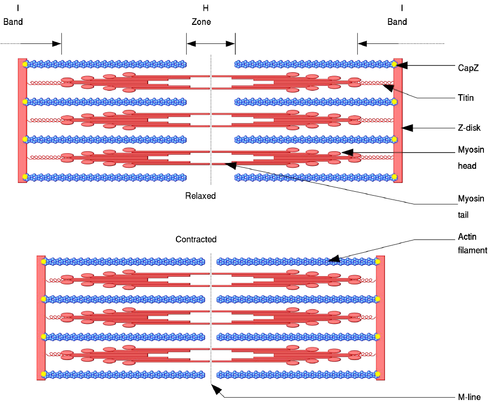
During muscle contraction, the sliding filament mechanism occurs:
- In the relaxed state: The actin and myosin filaments overlap only slightly, and the H-zone is clearly visible
- During contraction: The myosin heads bind to actin filaments and pull them towards the centre
- In the contracted state: The filaments slide past each other, the H-zone becomes much narrower, but importantly, neither the actin nor myosin filaments actually change length
This sliding action causes the entire sarcomere to shorten, and when millions of sarcomeres shorten simultaneously throughout a muscle, the whole muscle contracts and creates movement.
Why don't the filaments change length?
The actin and myosin filaments are made of proteins with fixed structures. They maintain their length because they're simply sliding past each other, like pulling two combs through each other. The shortening you see is the result of increased overlap, not stretching or compression of the filaments themselves.
Energy requirements for muscle function
Muscle contraction is an energy-demanding process that requires ATP (adenosine triphosphate) - the body's universal energy currency. Every time myosin heads bind to actin filaments and perform their pulling action, they consume ATP molecules.
This energy comes from cellular respiration, the process by which your muscle cells break down glucose and other nutrients in the presence of oxygen to produce ATP. This is why you breathe harder during exercise - your muscles need more oxygen to produce the ATP required for continued contraction.
When you exercise intensively and your muscles can't get enough oxygen, they may switch to less efficient energy production methods, which is why you might feel muscle fatigue or even muscle soreness after vigorous activity.
ATP is Absolutely Essential:
Without ATP, muscles cannot contract at all. This is why when an organism dies, the muscles become rigid (rigour mortis) - there's no ATP available to release the myosin heads from the actin filaments, so they remain locked in position.
Remember!
Key Points to Remember:
-
Muscle hierarchy: Myofibrils → Muscle fibres → Fascicles → Complete muscle, each level wrapped in protective connective tissue
-
Sliding filament mechanism: Muscle contraction occurs when actin and myosin filaments slide past each other within sarcomeres, without the filaments themselves changing length
-
Energy dependence: All muscle contraction requires ATP energy, which is produced through cellular respiration using oxygen and nutrients
-
Voluntary control: Skeletal muscle is under conscious control, allowing you to coordinate complex movements for activities like sports, writing, or dancing
-
Integrated system: Muscles work together with bones, joints, tendons, and ligaments to create smooth, coordinated locomotion