Musculoskeletal Tissues and Human Locomotion (Grade 10 NSC Matric Life Sciences): Revision Notes
Musculoskeletal Tissues and Human Locomotion
Introduction to the musculoskeletal system
The musculoskeletal system is a remarkable network of tissues that work together to provide your body with structure and enable movement. This system includes several key components: bones, cartilage, joints, tendons, ligaments, and muscles. Each component has a specific role, but they all work together like parts of a well-designed machine to help you walk, run, jump, and perform countless daily activities.

Think of the musculoskeletal system as an integrated machine where each part depends on the others. Just like removing one gear from a clock would stop it from working, problems with any single component can affect the entire system's ability to function properly.
Understanding how these tissues function individually and collectively is essential for appreciating how your body moves and maintains its shape. In the sections that follow, we'll explore each component in detail to see how they contribute to human locomotion.
Bones
Structure and composition
Bones serve as the fundamental framework of your body, providing structural support and serving as attachment points for muscles. Rather than being simple, static structures, bones are living, dynamic tissues that constantly undergo changes throughout your life.
Many people think of bones as dry, lifeless structures like those seen in museums. In reality, bones are very much alive - they contain living cells, blood vessels, and nerves, and they constantly remodel themselves throughout your life.
The composition of bone tissue is truly fascinating. Bones contain a dense arrangement of collagen fibres combined with mineral salts including calcium, magnesium, and phosphates. This unique combination gives bones their remarkable properties - the calcium salts provide hardness and rigidity, while the collagen fibres contribute flexibility and strength. This dual nature allows bones to be both strong enough to support your body weight and flexible enough to withstand the stresses of daily movement.
Microscopic structure of bones
When we examine bone tissue under a microscope, we discover an intricate internal architecture that maximises strength while minimising weight. The microscopic structure reveals a sophisticated system of tunnels and chambers that house living cells and blood vessels.
The key features of bone microstructure include Haversian canals, which are hollow tunnels running parallel to the length of the bone. Under microscopic examination, these canals appear as dark circles against a lighter background. Each Haversian canal is surrounded by concentric rings of compact bone called lamellae, creating a structure that resembles tree rings.
The organisation of bone tissue is remarkably efficient. The circular arrangement of lamellae around Haversian canals creates maximum strength with minimal material - similar to how corrugated cardboard gains strength from its internal structure.
Within the bone matrix, you'll find small fluid-filled cavities called lacunae. Each lacuna contains bone cells known as osteocytes, which are responsible for maintaining the bone tissue. These lacunae connect to each other and to the Haversian canal through a network of tiny interconnecting canals called canaliculi. Cytoplasm extends through these canals, creating a communication network that supplies osteocytes with oxygen and nutrients while removing waste products.
The Haversian canals, lacunae, osteocytes, and canaliculi work together to form functional units called Haversian Systems. Multiple Haversian Systems combine to create the structure of compact bones, demonstrating how microscopic organisation contributes to macroscopic function.
Bone formation and development
The process of bone formation, called ossification, is particularly important during growth and development. Interestingly, infants and young children don't have bones identical to those of adults. Instead, their skeletal system is primarily composed of cartilage - a firm, elastic, fibrous material.
As individuals grow and mature, this cartilage is gradually replaced by bone cells, which deposit crystals of calcium carbonate and calcium phosphate. This ossification process significantly increases the strength and rigidity of the skeletal system, transforming the flexible cartilaginous framework of childhood into the robust bony skeleton of adulthood.
Cartilage
Main features
Cartilage is a tough, semi-transparent, flexible tissue that plays crucial roles throughout your body. The basic structure consists of a tough matrix or jelly-like substance composed primarily of collagen (a protein) and proteins with special carbohydrate chains called proteoglycans. This matrix is enclosed by a fibrous covering called the perichondrium.
Living within this matrix are specialised cells called chondrocytes, which are responsible for producing and maintaining the cartilage matrix. These cells secrete a rubbery protein matrix called chondrin and occupy small fluid-filled spaces called lacunae scattered throughout the matrix.
Unlike other tissues, cartilage contains no blood vessels or nerves within the matrix itself, which affects how it heals and maintains itself. This is why cartilage injuries often take longer to heal compared to other tissues.
Types of cartilage
Your body contains three distinct types of cartilage, each with unique characteristics that suit them for specific functions:
| Cartilage | Appearance | Location | Function |
|---|---|---|---|
| Hyaline cartilage | Glass-like, bluish-white, few fibres | At ends of bones, forms C-shaped structures in trachea, joins ribs to sternum, larynx and tip of nose, temporary cartilage in bones | Reduces friction at joints, allows movement of ribs during breathing, forms permanent structures, allows bones to increase in length |
| Fibrocartilage | Many white collagen fibres | Discs between the vertebrae, in the rim of ball and socket joints, between pubic bones | Acts as shock absorbers, makes the socket deeper while still allowing movement |
| Elastic cartilage | Many yellow fibres in matrix | In the pinna of the ear, in the epiglottis | Maintains the shape of the ear, strengthens the epiglottis |
Each type of cartilage has evolved specific properties for its location. Hyaline cartilage's smooth surface reduces friction, fibrocartilage's strength handles compression, and elastic cartilage's flexibility maintains shape while allowing bending.
Relationship between cartilage and bone
The relationship between cartilage and bone is particularly evident during human development. In infants and young children, much of what will eventually become bone starts as cartilage. This cartilaginous template provides a flexible framework that can grow and change shape as the child develops.
As growth progresses, the cartilage is gradually replaced through the ossification process. This transformation greatly increases the strength of the skeletal system while maintaining the overall shape established by the original cartilaginous framework.
Joints
Understanding joints
A joint represents the meeting point where two bones come together. These structures are essential for movement, as they allow bones to move against each other in various directions and planes. Without joints, your skeleton would be a rigid, immovable framework.
Joints can be classified in several ways, but one useful classification system divides them into three main categories based on the amount of movement they allow:
- Fibrous joints connect bones where no movement is permitted. The bones of your skull (cranium) provide an excellent example of fibrous joints, where the bones are tightly connected to protect your brain.
- Cartilaginous joints allow slight, restricted movement. The discs between the vertebrae in your spine exemplify this type of joint, providing some flexibility while maintaining structural integrity.
- Synovial joints permit free movement in one or more directions. These are the most complex joints and include the joints of your pelvis, arms, and legs. They facilitate movements essential for daily activities like standing, sitting, walking, and running.
Synovial joints - structure and function
Synovial joints deserve special attention because they're the most complex and functionally important joints in your body. Most joints in your skeleton are synovial joints, also known as movable joints.
The structure of synovial joints is specially designed to enable smooth, friction-free movement. These joints are characterised by the presence of joint capsules containing synovial fluid. This fluid acts as a lubricant, preventing friction during movement and ensuring smooth operation of the joint.
Synovial fluid has the perfect consistency for joint lubrication - it's thick enough to stay in place but thin enough to allow smooth movement. It also helps nourish the cartilage since cartilage has no blood supply of its own.
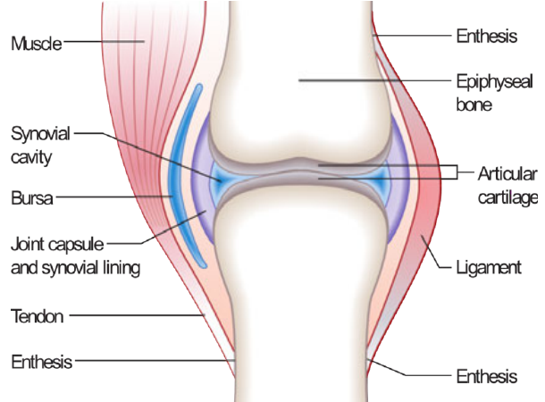
Key structural components of synovial joints include:
- Articular cartilage covering the bone ends to reduce friction
- Synovial cavity filled with lubricating synovial fluid
- Joint capsule and synovial lining that encloses and protects the joint
- Ligaments that provide stability and prevent excessive movement
- Tendons that connect muscles to bones
- Bursa - small fluid-filled sacs that provide additional cushioning
Types of synovial joints
Synovial joints come in four main varieties, each designed for specific types of movement:
- Ball and socket joints are found in structures like the shoulder, allowing comprehensive movement including forwards/backwards, up/down, and rotational movements.
- Hinge joints operate like door hinges and are found in structures such as the elbow. They allow the forearm to move up and down in a simple flexing motion.
- Pivot joints enable turning movements of the head in rotational motion from side to side.
- Gliding joints are found in areas like the foot and allow bones to slide over one another, enabling toes to flex.
Tendons and ligaments
Understanding connective tissues
Tendons and ligaments are both composed of dense bands of connective tissue, but they serve distinctly different functions in your musculoskeletal system. Understanding the differences between these structures is crucial for appreciating how movement occurs.
Many students confuse tendons and ligaments. Remember: Tendons connect muscles To bones (both start with 'T'), while Ligaments Link bones to other bones (both start with 'L').
Ligaments are connective tissues that join bone to bone. An excellent example is the anterior cruciate ligament (ACL) of the knee, which connects the thigh bone to the shin bone. Tendons are connective tissues that join muscles to bones. A familiar example is the Achilles tendon, which connects your calf muscle to your heel bone.
Comparing ligaments and tendons
| Ligaments | Tendons |
|---|---|
| Join bone to bone | Attach muscles to bones |
| Consist of white collagen fibres and a network of yellow elastic fibres | Consist of non-elastic collagen fibres which give tendons a white shiny appearance |
| Strong collagen fibres prevent dislocation at joints, and yellow elastic fibres allow flexibility at the joint | Parallel arrangement of strong collagen fibres in order to efficiently convert muscle contraction into movement of the skeleton |
The structural differences between ligaments and tendons reflect their different functions. Ligaments need both strength and flexibility to stabilise joints while allowing movement, which is why they contain both strong collagen fibres and elastic fibres. Tendons, however, need to efficiently transfer the force of muscle contractions to move bones, so they're composed of parallel arrangements of strong, non-elastic collagen fibres.
Antagonistic muscles
How muscles work in pairs
Voluntary muscles are typically connected to at least two bones, and understanding how they attach is key to understanding movement. The attachment point to the immovable bone is called the origin, while the attachment point to the movable bone is called the insertion.
Most muscles work in pairs, and when one muscle contracts, it needs both an agonist and an antagonist to function properly. This pairing system ensures smooth, controlled movement and allows you to return your limbs to their starting positions.
An agonist is a muscle that contracts to move a limb away from its starting position. An antagonist is a muscle that works in opposition to the agonist, responsible for returning the limb to its original position when the agonist relaxes.
This antagonistic relationship is essential because muscles can only exert pulling forces - they cannot push. Therefore, when one muscle contracts and pulls a bone in one direction, another muscle is required to pull it back to its starting position.
Example: biceps and triceps
Worked Example: Biceps and Triceps Action
The biceps and triceps muscles of your arm provide an excellent example of antagonistic muscle action.
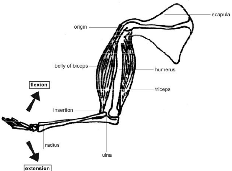
Step 1: Understanding the biceps
- Origin: scapula (shoulder blade)
- Insertion: radius and ulna (forearm bones)
- Action: When contracted, decreases angle between forearm and upper arm
- Classification: Flexor muscle
Step 2: Understanding the triceps
- Origin: humerus and scapula (three attachment points)
- Insertion: ulna
- Action: When contracted, increases angle between forearm and upper arm
- Classification: Extensor muscle
Step 3: Coordinated movement
- To bend arm: biceps contracts (agonist), triceps relaxes (antagonist)
- To straighten arm: triceps contracts (agonist), biceps relaxes (antagonist)
The biceps muscle has two attachment points: its origin is at the scapula (shoulder blade), and its insertion is at the humerus (upper arm bone). The biceps gets its name from having two distinct sections that join to form a single muscle body, then split again into two tendons - one inserting at the radius and the other at the ulna (the two bones of your forearm).
When your biceps muscle contracts, your forearm is lifted or bent, decreasing the angle between your forearm and upper arm. This ability to decrease the angle between joints makes the biceps a flexor muscle.
When your arm is already bent, the biceps cannot contract further since it's already in a contracted state. To straighten the arm, the triceps muscle (an extensor muscle) must contract. As the triceps contracts, it increases the angle between your forearm and upper arm, bringing about the straightening motion.
The triceps has three points of origin (two on the humerus and one on the scapula) and a single point of insertion on the ulna, which is why it's called the triceps (meaning three heads).
Human locomotion
Understanding locomotion
Locomotion refers to your ability to move from one place to another. This includes various types of movement such as running, swimming, jumping, and flying (though humans achieve flight through technology rather than biological adaptation). Human locomotion is accomplished through the coordinated use of our limbs and the musculoskeletal framework that supports them.

The photograph of marathon runners demonstrates human locomotion in action, showing how the skeletal framework described in this chapter facilitates complex, coordinated movement over extended periods. Notice how each runner's entire musculoskeletal system works together to maintain efficient movement patterns.
Structures involved in locomotion
Human movement requires the coordinated action of multiple musculoskeletal components:
Bones provide your body's supporting framework. They maintain your body's shape and provide surfaces for muscle attachment, creating the structural foundation necessary for all movement.
Joints serve as connection points between individual bones, allowing bones to move against each other and enabling the complex movements required for locomotion.
Ligaments connect bones together at the ends, forming joints. Most ligaments limit dislocation and prevent certain movements that could cause damage, while holding bones in position so they can work together in a coordinated manner.
Tendons connect muscles to bones, transferring the force generated by muscle contractions into movement of the skeletal system.
Muscles work in antagonistic pairs to move bones. When muscles contract, they pull on bones through their tendon connections, causing the bones to move at their joint connections.
Muscle structure and function
Types of muscle tissue
Your body contains three types of muscle tissue: skeletal, smooth, and cardiac muscle. In this chapter, we focus on skeletal muscle, which is the voluntary muscle you use for movement. Skeletal muscle can be controlled by your conscious will, making it the type you use for activities like running, skipping, and walking.
Basic muscle structure
The fundamental units of muscle are called myofibrils. These myofibrils combine to form muscle fibres (muscle cells), which are the basic contractile units of muscle tissue. Multiple muscle fibres are organised into bundles, and these bundles are surrounded by connective tissue to form complete muscles.
This hierarchical organisation - from myofibrils to muscle fibres to bundles to complete muscles - allows for the generation of significant force while maintaining the ability to produce precise, controlled movements. The arrangement ensures that when nerve signals trigger muscle contraction, the force is efficiently transmitted through the muscle structure to produce coordinated movement.
Summary
Key Points to Remember:
-
The musculoskeletal system includes bones, cartilage, joints, tendons, ligaments, and muscles working together to provide structure and enable movement.
-
Bones are living tissues with complex microscopic structures including Haversian systems, and they develop through ossification where cartilage is replaced by bone tissue.
-
Three types of cartilage (hyaline, fibrocartilage, and elastic) have different properties suited for their specific locations and functions in the body.
-
Muscles work in antagonistic pairs - when one muscle (agonist) contracts to produce movement, its partner (antagonist) must relax and then contract to return the limb to its original position.
-
Tendons connect muscles to bones while ligaments connect bones to bones, and both are essential for coordinated movement and joint stability.