Circulatory Systems in Animals (Grade 10 NSC Matric Life Sciences): Revision Notes
Circulatory Systems in Animals
Introduction to transport systems
All living organisms need oxygen and nutrients to survive, and they must remove carbon dioxide and other waste products. Transport systems are essential for moving these materials around the body. The circulatory system does much more than just transport nutrients and gases - it also carries hormones to target organs and helps the immune system by transporting white blood cells and antibodies throughout the body.
Simple, single-celled organisms can rely on diffusion to move materials in and out of their cells. However, larger, more complex organisms need specialised circulatory systems to transport materials efficiently to all parts of their bodies.
The need for specialised transport systems increases dramatically with organism size. While a single-celled amoeba can rely entirely on diffusion, a human with trillions of cells requires an intricate network of blood vessels stretching over 100,000 kilometres!
Types of circulatory systems
There are two main types of circulatory systems found in animals: open and closed systems.
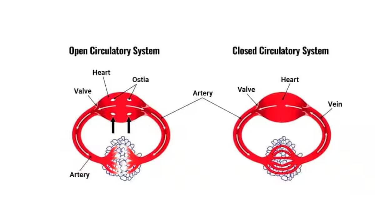
Open circulatory systems
In an open circulatory system, the blood vessels transport fluid into a body cavity where it flows freely around the organs. The blood directly bathes the organs, supplying them with oxygen and nutrients while removing waste products. This type of system moves blood quite slowly because it lacks the muscular walls found in closed systems.
Most invertebrates, including crabs, insects, and snails, have open circulatory systems. The blood flows at a much slower pace due to the absence of contained vessels throughout the body.
Closed circulatory systems
In a closed circulatory system, blood never leaves the blood vessels. Instead, materials are transferred from one blood vessel to another without the blood entering body cavities. Blood flows in one direction only, efficiently delivering oxygen and nutrients to cells while removing waste products.
Closed systems can be further divided into single and double circulation systems, depending on how many times blood passes through the heart in one complete circuit.
Key Difference: In open systems, blood directly contacts organs in body cavities. In closed systems, blood remains contained within vessels, making material exchange more controlled and efficient.
Single and double circulation systems
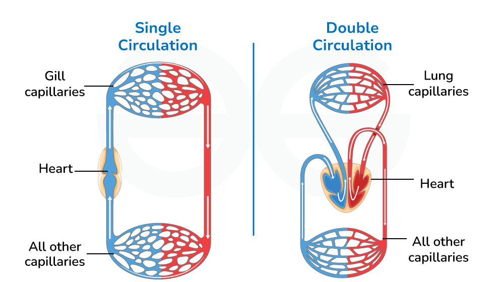
Single circulation systems
Fish have single circulation systems with a two-chambered heart consisting of one atrium and one ventricle. Here's how it works:
- The heart pumps deoxygenated blood to the gills
- At the gills, the blood picks up oxygen and releases carbon dioxide
- The now-oxygenated blood flows directly to the rest of the fish's body
- Deoxygenated blood returns to the heart to complete the circuit
Double circulation systems
Birds and mammals, including humans, have double circulation systems with four-chambered hearts. This system is called "double" because blood passes through the heart twice in one complete circuit.
The four chambers consist of:
- Left atrium and left ventricle (left side of heart)
- Right atrium and right ventricle (right side of heart)
The advantage of a four-chambered heart is that there's no mixing of oxygenated and deoxygenated blood, making the system much more efficient than single circulation systems.
Worked Example: Comparing Circulation Efficiency
Single Circulation (Fish):
- Blood passes through heart once per circuit
- Pressure drops significantly after passing through gills
- Lower overall efficiency for oxygen delivery
Double Circulation (Mammals):
- Blood passes through heart twice per circuit
- High pressure maintained for systemic circulation
- Much higher efficiency for oxygen delivery to body tissues
Human circulatory system
The human circulatory system consists of two separate circuits: the pulmonary circuit and the systemic circuit.
Pulmonary circulation
The pulmonary circulation carries deoxygenated blood from the heart to the lungs and returns oxygenated blood back to the heart.
Here's the pathway:
- Deoxygenated blood leaves the right ventricle through the pulmonary artery
- The pulmonary artery is the only artery that carries deoxygenated blood
- Blood travels to the lung capillaries where gas exchange occurs
- Carbon dioxide moves out of the blood into the air spaces (alveoli)
- Oxygen moves from the alveoli into the blood
- Oxygenated blood returns to the left atrium via the pulmonary veins
Remember the exception: The pulmonary artery carries deoxygenated blood (unlike all other arteries), and pulmonary veins carry oxygenated blood (unlike all other veins).
Systemic circulation
Systemic circulation transports oxygenated blood from the heart to all body organs and returns deoxygenated blood back to the heart.
The pathway works like this:
- Oxygenated blood leaves the left ventricle through the aorta (the body's largest artery)
- The aorta branches into smaller arteries that supply different organs
- These arteries branch further into tiny capillaries
- In the capillaries, oxygen moves into cells while carbon dioxide and waste products move into the blood
- Deoxygenated blood flows into venules, then larger veins
- Blood returns to the right atrium via two major veins: the superior vena cava (from head and arms) and inferior vena cava (from lower body)
Heart structure and function
External structure of the heart
The heart is a powerful muscle about the size of your clenched fist. It's located in your chest cavity, just behind your breastbone, in a space called the pericardial cavity. The heart is surrounded by a protective double membrane called the pericardium, with pericardial fluid between the layers to reduce friction during heartbeats.

The heart muscle itself is called the myocardium and consists of four chambers:
- Atria (singular: atrium) - the two upper chambers that receive blood
- Ventricles - the two lower chambers that pump blood out of the heart
The ventricles have thicker, more muscular walls than the atria because they need to pump blood with greater force. The left ventricle has the thickest walls since it must pump blood to the entire body, while the right ventricle only pumps blood to the lungs.
Internal structure of the heart
The heart's internal structure includes several important features that ensure blood flows in the correct direction:
Heart chambers:
- Right atrium: receives deoxygenated blood from the body
- Right ventricle: pumps deoxygenated blood to the lungs
- Left atrium: receives oxygenated blood from the lungs
- Left ventricle: pumps oxygenated blood to the body
Heart valves prevent blood from flowing backwards:
- Tricuspid valve: between right atrium and right ventricle
- Bicuspid valve (also called mitral valve): between left atrium and left ventricle
- Semi-lunar valves: at the base of the aorta and pulmonary artery
The valves are attached to strong tendinous cords (chordae tendineae) that connect to papillary muscles. These structures prevent the valves from opening the wrong way when the ventricles contract.
The valve names indicate their structure: "tricuspid" has three flaps, "bicuspid" has two flaps, and "semi-lunar" valves are shaped like half-moons.
Circulation of blood through the heart
Blood follows a specific pathway through the heart in one complete circuit:
- Deoxygenated blood enters the right atrium from the superior and inferior vena cava
- Blood flows through the tricuspid valve into the right ventricle
- The right ventricle pumps blood through the pulmonary valve into the pulmonary artery
- Blood travels to the lungs for gas exchange
- Oxygenated blood returns to the left atrium via pulmonary veins
- Blood flows through the bicuspid valve into the left ventricle
- The left ventricle pumps blood through the aortic valve into the aorta
- Blood travels to the rest of the body through the systemic circulation
Worked Example: Following Blood Through One Complete Circuit
Step 1: Deoxygenated blood returns from body tissues
- Superior vena cava (from head, neck, arms)
- Inferior vena cava (from lower body)
- Blood enters right atrium
Step 2: Right side circulation
- Right atrium → tricuspid valve → right ventricle
- Right ventricle → pulmonary valve → pulmonary artery → lungs
Step 3: Gas exchange at lungs
- CO₂ removed, O₂ added to blood
Step 4: Left side circulation
- Lungs → pulmonary veins → left atrium
- Left atrium → bicuspid valve → left ventricle
- Left ventricle → aortic valve → aorta → body tissues
Major organs and blood supply
Every organ in the body receives its own blood supply through specific arteries and returns blood through corresponding veins. This ensures all cells receive the oxygen and nutrients they need for proper function.
Key organ blood supplies:
- Brain: supplied by carotid and vertebral arteries; drained by jugular veins (receives 15% of total heart output)
- Liver: receives blood from hepatic artery and hepatic portal vein; drained by hepatic vein
- Kidneys: supplied by renal arteries; drained by renal veins (philtre waste from blood)
- Heart muscle: supplied by coronary arteries; drained by cardiac veins
The hepatic portal system is special because nutrient-rich blood from the digestive system must pass through the liver first before returning to the heart. This allows the liver to process and regulate nutrients entering the bloodstream.
The cardiac cycle
The cardiac cycle refers to the sequence of events that occur during one complete heartbeat. At rest, each heartbeat takes approximately 0.8 seconds, and the normal heart rate is about 72 beats per minute.
Phases of the cardiac cycle
Phase 1: Atrial systole (0.1 seconds)
- Both atria contract simultaneously
- Blood flows from the superior and inferior vena cava into the right atrium
- Blood flows from the pulmonary veins into the left atrium
- Blood is forced through the tricuspid and bicuspid valves into the ventricles
Phase 2: Ventricular systole (0.3 seconds)
- Both ventricles contract while atria relax
- Blood is forced upward, closing the tricuspid and bicuspid valves (creates "lubb" sound)
- Blood flows through semi-lunar valves into the pulmonary artery and aorta
- Atria begin filling with blood during this time
Phase 3: General diastole (0.4 seconds)
- All four chambers relax
- Ventricular pressure drops, closing the semi-lunar valves (creates "dubb" sound)
- Blood continues flowing into the atria from veins
- The cycle begins again
Heart sounds and pulse
The heart produces two distinct sounds during each beat:
- "Lubb" sound: caused by tricuspid and bicuspid valves closing when ventricles contract
- "Dubb" sound: caused by semi-lunar valves closing when ventricles relax
Your pulse is the wave of pressure created by the left ventricle pumping blood into the aorta. It can be felt at various points where arteries are close to the skin surface, such as the wrist or neck.
The timing of the cardiac cycle phases: 0.1s (atrial systole) + 0.3s (ventricular systole) + 0.4s (general diastole) = 0.8s total cycle time at rest.
Heart rate control and electrical activity
Control mechanisms
The heart has its own built-in pacemaker system that controls the cardiac cycle:
- Sinoatrial node (SA node): located in the right atrium wall, acts as the heart's natural pacemaker
- Atrioventricular node (AV node): located between atria and ventricles, delays electrical signals briefly
The electrical impulse starts at the SA node, spreads through both atria (causing them to contract), then reaches the AV node. After a brief pause, the signal spreads through specialised conducting fibres (bundles of His and Purkinje fibres) to both ventricles, causing them to contract.
This system works automatically (automaticity) without conscious control, though heart rate can change due to:
- Exercise and physical activity
- Emotional stress or excitement
- Hormones like adrenaline
- Nervous system signals
Electrocardiogram (ECG)
The heart's electrical activity can be measured using an electrocardiogram (ECG). A normal heart shows a very regular rhythm, while abnormal patterns indicate heart problems:
- Bradycardia: heart rate too slow (less than 60 beats per minute)
- Tachycardia: heart rate too fast (more than 100 beats per minute)
- Arrhythmia: irregular heart rhythm
Stroke volume and blood pressure
Stroke volume is the amount of blood pumped by the heart during each beat. During exercise, the heart increases both its stroke volume and heart rate to meet the body's increased demand for oxygen and nutrients.
Blood pressure is the force that blood exerts on blood vessel walls. Normal blood pressure is 120/80 mmHg, where:
- 120 is the systolic pressure (when ventricles contract)
- 80 is the diastolic pressure (when ventricles relax)
High blood pressure (hypertension) can be dangerous and increases the risk of heart attack, stroke, or aneurysm. Low blood pressure (hypotension) can cause dizziness and fainting due to insufficient blood supply to the brain.
Cardiac Output Formula:
This formula shows how the heart can increase blood flow by increasing either the volume pumped per beat or the number of beats per minute.
Blood vessels
Blood vessels form a network throughout the body, with each type specialised for its specific function.

Arteries
Arteries carry blood away from the heart and generally transport oxygenated blood (except for the pulmonary artery). They have thick walls with three layers:
- Outer layer: connective tissue for protection
- Middle layer: smooth muscle that can contract to control blood flow and pressure
- Inner layer: smooth endothelium that reduces friction
Arteries branch into smaller arterioles, which then branch into microscopic capillaries.
Capillaries
Capillaries are the smallest blood vessels, consisting of just a single layer of endothelial cells. This thin wall allows efficient exchange of materials between blood and tissues:
- Oxygen and nutrients move from blood into tissue fluid
- Carbon dioxide and waste products move from tissue fluid into blood
- Blood cells never directly contact body cells - exchange occurs through tissue fluid
Capillaries form intricate networks throughout tissues, ensuring every cell is close to a blood supply.
Capillaries are so narrow that red blood cells must pass through them in single file. This slow movement maximises the time available for gas and nutrient exchange.
Veins
Veins carry blood back to the heart and generally transport deoxygenated blood (except for pulmonary veins). They have thinner walls than arteries because blood pressure is much lower by the time it reaches veins.
Key features of veins:
- Two-layer walls: outer connective tissue and inner endothelium
- Large internal diameter to accommodate slower-moving blood
- Semi-lunar valves at intervals to prevent blood flowing backwards
- Located closer to the skin surface to release excess heat
Small capillaries join to form venules, which merge into larger veins that eventually return blood to the heart.
Comparison of blood vessel types
| Feature | Arteries | Capillaries | Veins |
|---|---|---|---|
| Blood flow direction | Away from heart | Through tissue beds | To the heart |
| Wall structure | Thick, three layers with muscle | Single layer of endothelium | Thin, two layers |
| Blood pressure | High pressure | Pressure drops significantly | Low pressure |
| Valves | Only in aorta and pulmonary artery | None | Semi-lunar valves present |
| Pulse detection | Strong pulse felt | No pulse | No pulse detected |
| Location | Deeper in tissues | At tissue level | Near skin surface |
Key Memory Aid: "Arteries Away, Veins to" - arteries carry blood away from the heart, veins carry blood back to the heart. The structure of each vessel type perfectly matches its function.
Lymphatic system
The lymphatic system is part of the circulatory system, consisting of a network of vessels that carry a clear fluid called lymph back to the heart. This system serves important functions:
- Fluid balance: returns excess tissue fluid to the bloodstream
- Immune function: transports white blood cells and philtres out pathogens
- Fat absorption: absorbs fats from the digestive system
The lymphatic system works alongside the cardiovascular system to maintain proper fluid levels in tissues and support immune responses against disease-causing organisms. Without it, tissues would become waterlogged and the immune system would be severely compromised.
Practical investigations
Understanding how the circulatory system works involves hands-on investigations that demonstrate key concepts.
Practical Investigation: Heart Rate and Exercise
Aim: To investigate how heart rate changes before, during, and after exercise
Method:
- Measure resting pulse rate for 1 minute
- Perform strenuous exercise (running in place for 2 minutes)
- Record pulse immediately after exercise
- Continue measuring pulse every minute for 5 minutes
- Plot results on a graph showing time vs. heart rate
Expected Results:
- Resting heart rate: ~72 beats per minute
- Heart rate increases to 120-150 beats per minute during exercise
- Heart rate gradually returns to normal during recovery
- Fitter individuals typically have lower resting heart rates and recover more quickly
Key Variables:
- Independent variable: time after exercise
- Dependent variable: heart rate (beats per minute)
- Constants: same person, same exercise intensity, same measurement method
Conclusion: This investigation demonstrates how the cardiovascular system adapts to meet the body's changing demands for oxygen and nutrients during physical activity.
Key Points to Remember:
-
Open circulatory systems allow blood to flow freely in body cavities (invertebrates), while closed systems keep blood contained in vessels (vertebrates)
-
Single circulation (fish) involves blood passing through the heart once per circuit, while double circulation (mammals and birds) involves blood passing through the heart twice per circuit
-
The four-chambered heart has two atria (receiving chambers) and two ventricles (pumping chambers), with valves ensuring one-way blood flow
-
Pulmonary circulation carries deoxygenated blood to the lungs and returns oxygenated blood to the heart, while systemic circulation delivers oxygenated blood to body tissues and returns deoxygenated blood to the heart
-
Blood vessels have specialised structures: arteries have thick muscular walls for high-pressure blood flow, capillaries have thin walls for material exchange, and veins have valves to prevent backflow of low-pressure blood
-
The cardiac cycle consists of three phases taking 0.8 seconds total: atrial systole (0.1s), ventricular systole (0.3s), and general diastole (0.4s)
-
Heart rate control is automatic via the SA node (pacemaker), but can be modified by exercise, stress, and hormones