Human Gaseous Exchange (Grade 11 NSC Matric Life Sciences): Revision Notes
Human Gaseous Exchange
The human respiratory system is a remarkable network designed specifically for the vital process of gas exchange. This system works continuously to supply your body with life-sustaining oxygen whilst removing the waste product carbon dioxide. Understanding how this intricate system functions will help you appreciate one of the most fundamental processes keeping you alive.
Overview of the human respiratory system
The human respiratory system consists of three main components that work together seamlessly:
- Air passages - the pathway that air travels through to reach the lungs
- Lungs - the main organs where gas exchange occurs
- Breathing muscles - the muscles that create the pressure changes needed for ventilation
This system is perfectly designed to carry out gaseous exchange efficiently. The journey of air from the atmosphere to the tiny air sacs in your lungs involves multiple specialised structures, each with specific adaptations for their role.
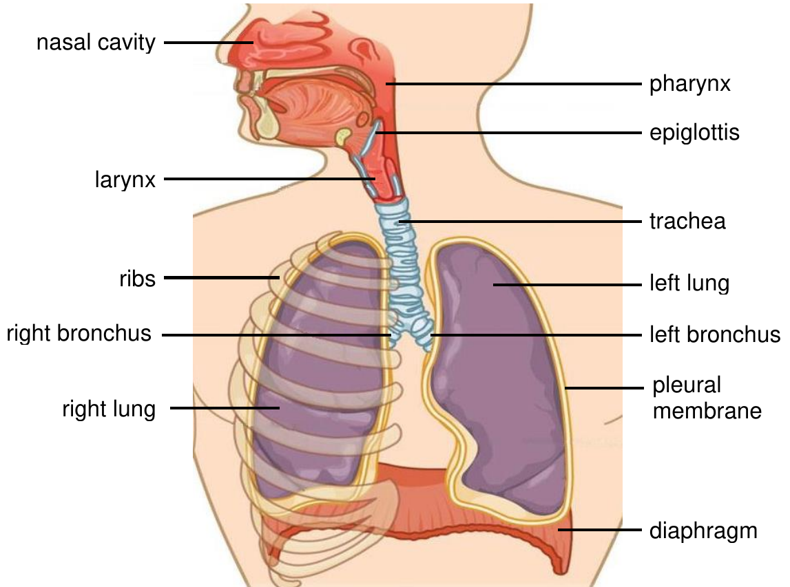
Structure and function of respiratory organs
Air passages
The air you breathe follows a specific pathway through your respiratory system. Each structure along this route has special features that prepare the air for gas exchange.
| Structure | Key Functions |
|---|---|
| Nostrils and nasal cavities | - Philtre incoming air using epithelial and goblet cells - Warm air using blood capillaries - Add moisture to prevent drying of respiratory surfaces - Trap dirt and particles using mucus and cilia |
| Pharynx | - Connects nasal cavity to larynx - Shared pathway for respiratory and digestive systems - Lined with protective mucous membranes |
| Larynx | - Contains vocal cords for sound production - Protected by cartilage structure (voice box) - Air passes over vocal cords during breathing |
| Epiglottis | - Cartilage flap above the larynx - Closes during swallowing to prevent food entering trachea - Essential safety mechanism for breathing |
| Trachea | - Main airway supported by C-shaped cartilage rings - Lined with ciliated epithelium and mucus-producing cells - Keeps airway open while allowing flexibility |
Bronchi and bronchioli
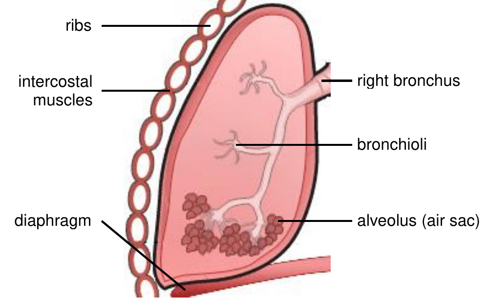
The trachea branches into two main bronchi that enter each lung. These structures become progressively smaller and more numerous:
- Bronchi have O-shaped cartilage rings for support and are lined with mucous membranes
- Bronchioli are smaller, narrower passages without cartilage reinforcement
- Each bronchus divides into many bronchioli, creating an extensive branching network
- This branching pattern maximises the surface area for air distribution
Alveoli - the site of gas exchange
At the end of each bronchiole are clusters of tiny air sacs called alveoli. These remarkable structures are perfectly adapted for their crucial role:
- Thin walls made of squamous epithelium (only one cell thick) allow rapid gas diffusion
- Moist surface provided by tissue fluid maintains optimal conditions for gas exchange
- Extensive capillary network surrounds each alveolus for efficient gas transport
- Large combined surface area from millions of alveoli maximises gas exchange
Breathing muscles
The mechanical process of breathing requires coordinated muscle action:
- Ribs protect the lungs and provide attachment points for breathing muscles
- Intercostal muscles between the ribs contract and relax to change chest volume
- Diaphragm is a dome-shaped muscle sheet below the lungs that creates major pressure changes
The mechanism of breathing
Breathing is a mechanical process driven by pressure differences between the atmosphere and your lungs. This process involves volume and pressure changes in the thoracic cavity through coordinated muscle contractions.
Key terminology
| Term | Definition |
|---|---|
| Diaphragm | Dome-shaped muscle separating chest and abdominal cavities; essential for breathing |
| Inhalation | Active process of breathing air into the lungs |
| Exhalation | Passive process of breathing air out of the lungs |
| Spirometer | Instrument measuring lung air volume during breathing |
Inhalation - breathing in
During inhalation, your body actively works to draw air into the lungs:
- Diaphragm contracts and flattens, moving downward
- External intercostal muscles contract, lifting the ribcage upward and outward
- Chest cavity volume increases, causing air pressure inside to decrease
- Atmospheric pressure becomes greater than lung pressure
- Air flows into the lungs to equalise the pressure difference
This is an active process requiring energy from muscle contractions.
Exhalation - breathing out
Exhalation occurs when breathing muscles relax:
- Diaphragm relaxes and moves upward to its dome shape
- External intercostal muscles relax, allowing ribcage to move down and inward
- Thoracic cavity volume decreases, increasing air pressure in the lungs
- Lung pressure exceeds atmospheric pressure
- Air is forced out to equalise pressure
This is typically a passive process when muscles simply relax.
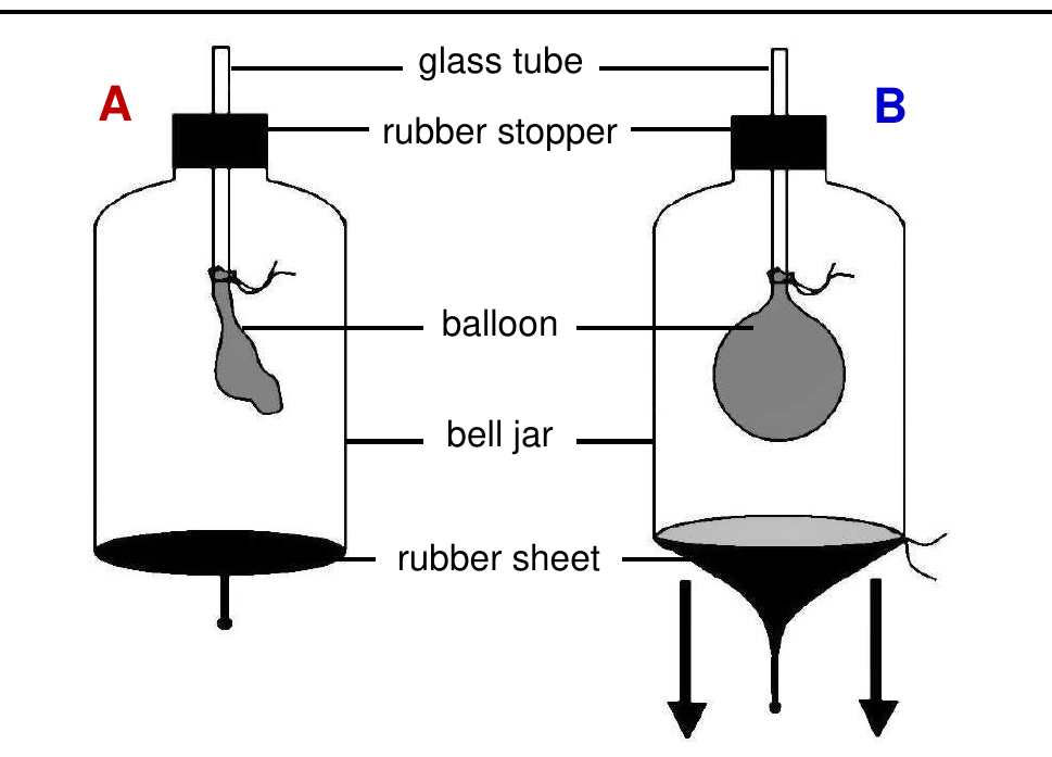
Demonstration: The Bell Jar Model
The bell jar model demonstrates these pressure relationships effectively. When the rubber sheet (representing the diaphragm) is pulled down, the balloon (representing lungs) inflates due to decreased pressure in the jar.
This shows how pressure changes drive the breathing process.
Composition of inhaled and exhaled air
The air composition changes significantly as it passes through your respiratory system, reflecting the gas exchange occurring in your lungs.
| Gas | Inhaled Air (%) | Exhaled Air - Sleeping (%) | Exhaled Air - Exercising (%) |
|---|---|---|---|
| Nitrogen (N₂) | 78 | 78 | 78 |
| Oxygen (O₂) | 21 | 16 | 12 |
| Carbon dioxide (CO₂) | 0.04 | 4 | 9 |
Key observations:
- Nitrogen levels remain constant because this gas is not used in cellular respiration
- Oxygen decreases from 21% to 16% (sleeping) or 12% (exercising) as it's absorbed by the blood
- Carbon dioxide increases dramatically from 0.04% to 4% (sleeping) or 9% (exercising) as it's released from the blood
- Exercise intensifies these changes due to increased cellular respiration demands
Exam tip: Remember that exhaled air is warmer (around 36.8°C) than inhaled air. You can demonstrate CO₂ presence in exhaled air using lime water, which turns milky when CO₂ bubbles through it.
Effect of exercise on breathing and pulse rate
Your respiratory and cardiovascular systems work together to meet your body's changing oxygen demands. When you exercise, both systems respond dramatically.
During exercise, the respiratory control centre in your medulla oblongata detects increased CO₂ levels and responds by:
- Increasing breathing rate - more breaths per minute
- Increasing breathing depth - larger volume of air per breath
- Coordinating with heart rate increases for efficient gas transport
The following data shows typical responses to different activity levels:
| Activity | Heart Rate (beats/min) | Breathing Rate (breaths/min) |
|---|---|---|
| At rest | 68-72 | 12 |
| Walking | 63-86 | 12 |
| Running | 112-120 | 14-16 |
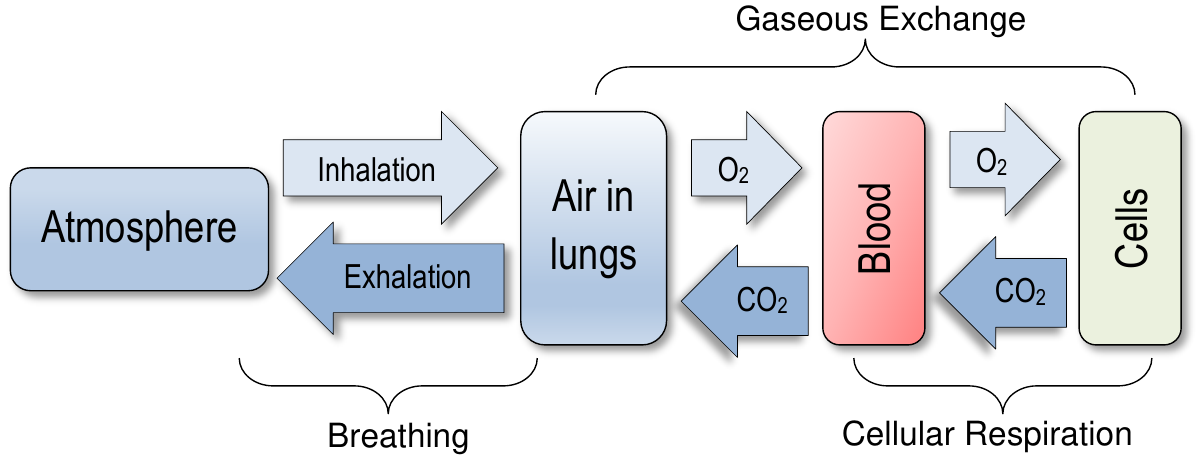
Internal and external gaseous exchange
Gas exchange occurs at two critical locations in your body, each serving different but complementary functions.
External gaseous exchange at the alveoli
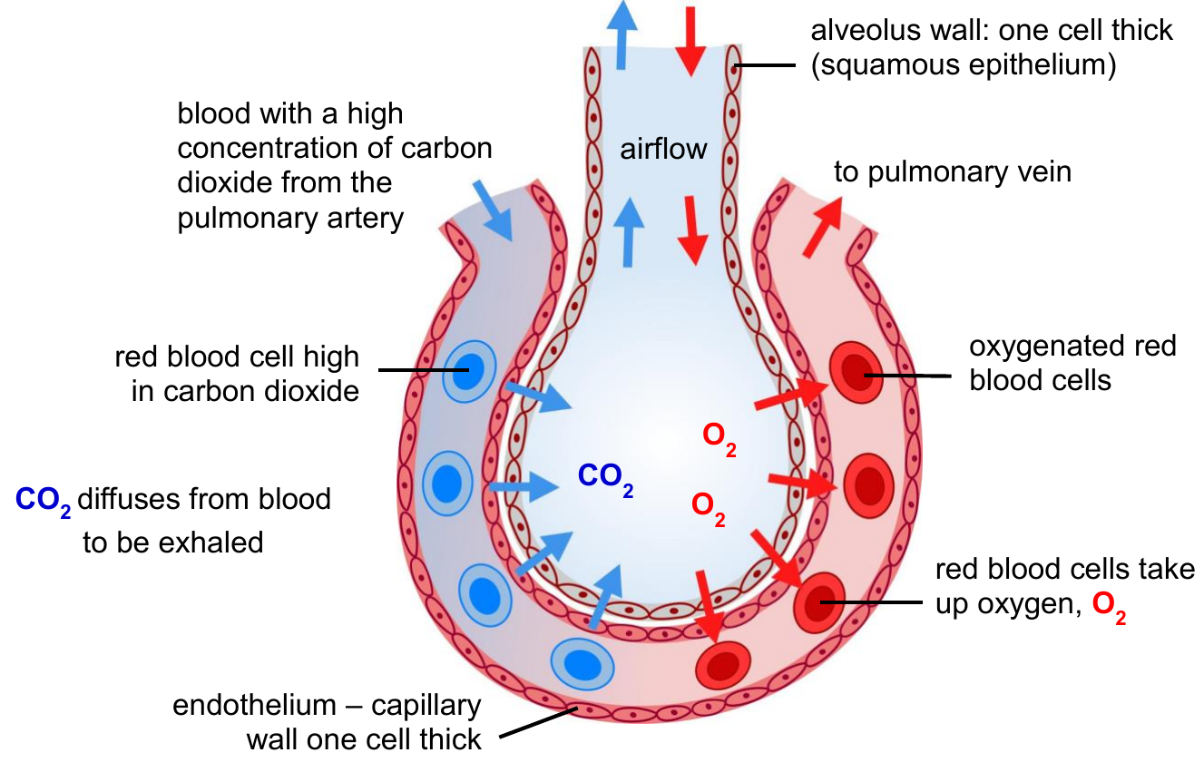
External gaseous exchange happens between the air in alveoli and the blood in surrounding capillaries. This process depends on concentration gradients:
- High oxygen concentration in inhaled alveolar air compared to deoxygenated blood
- Low carbon dioxide concentration in inhaled air compared to blood from tissues
- Oxygen diffuses from alveolar air into blood capillaries
- Carbon dioxide diffuses from blood into alveolar air for exhalation
The alveolar structure is perfectly adapted for this exchange:
- One-cell-thick walls minimise diffusion distance
- Extensive capillary network maximises contact with blood
- Moist surface maintains optimal diffusion conditions
- Large combined surface area increases exchange efficiency
Transport of respiratory gases
Your circulatory system efficiently transports gases throughout your body:
Oxygen transport:
- Most oxygen combines with haemoglobin in red blood cells (erythrocytes)
- Forms oxyhaemoglobin for transport to body tissues
- Iron atoms in haemoglobin molecules bind oxygen
Carbon dioxide transport:
- Most CO₂ is transported as bicarbonate ions in blood plasma
- Some CO₂ dissolves directly in plasma
- Blood carries CO₂ from tissues back to lungs for removal
Internal gaseous exchange at tissues
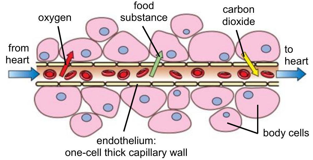
Internal gaseous exchange occurs between blood and body cells through capillary walls:
- Oxygenated blood arrives at tissue capillaries from the heart
- Oxygen diffuses from blood into cells (high to low concentration)
- Carbon dioxide diffuses from cells into blood (high to low concentration)
- Deoxygenated blood returns to heart and then to lungs
- Tissue fluid provides the moist medium necessary for gas exchange
This complete system ensures continuous oxygen delivery to cells and carbon dioxide removal, supporting cellular respiration throughout your body.
Homeostatic control of breathing
Your body maintains precise control over breathing through a sophisticated negative feedback mechanism. This system ensures that blood gas levels remain within safe limits automatically.
The Control Mechanism Process:
- Carbon dioxide levels increase in blood during cellular respiration
- Receptor cells in carotid arteries detect elevated CO₂ concentrations
- Nerve impulses travel to the medulla oblongata in the brain
- Medulla oblongata stimulates breathing muscles and heart
- Breathing rate and depth increase, heart beats faster
- More CO₂ is exhaled from the lungs, more O₂ is inhaled
- CO₂ levels return to normal, completing the feedback loop
This homeostatic control involves:
- Increased heart rate for faster blood circulation and gas transport
- Deeper, more frequent breathing for enhanced oxygen intake and CO₂ removal
- Automatic adjustment to meet changing metabolic demands
Key concept: This system responds primarily to CO₂ levels rather than oxygen levels, making it extremely sensitive to metabolic changes.
Effect of altitude on gaseous exchange
Altitude significantly affects your respiratory system's efficiency. Understanding these effects helps explain why athletes sometimes train at high altitudes.
Altitude refers to height above sea level, measured in metres. At higher altitudes:
- Air becomes 'thinner' with lower oxygen concentration
- Less oxygen available for gas exchange in the lungs
- Concentration gradient between alveolar air and blood decreases
- Oxygen absorption by red blood cells becomes less efficient
Physiological adaptations to altitude
Your body compensates for reduced oxygen availability through several adaptations:
| Measurement | Before Training Camp | After Training Camp |
|---|---|---|
| Red blood cell count (millions/mm³) | 4.69 | 5.37 |
| Haemoglobin levels (g/dL) | 14.8 | 16.8 |
These changes represent your body's remarkable ability to adapt:
- Increased red blood cell production provides more oxygen-carrying capacity
- Higher haemoglobin concentrations enhance oxygen transport efficiency
- Enhanced oxygen delivery to tissues despite lower atmospheric oxygen
Practical application: Athletes training at altitude develop these adaptations, giving them advantages when competing at sea level where oxygen is more abundant.
Diseases of the respiratory system
Various factors can negatively affect your respiratory system's function:
- Pathogens (viruses and bacteria) can cause infections
- Environmental pollutants (pollen, smoke) can trigger allergic responses
- Carcinogens (cancer-causing substances) can damage respiratory tissues
Understanding these threats emphasises the importance of protecting your respiratory health through good hygiene, avoiding pollutants, and maintaining a healthy lifestyle.
Key Points to Remember:
-
The respiratory system has three main parts: air passages, lungs, and breathing muscles, each with specialised functions for efficient gas exchange.
-
Breathing is a mechanical process: Inhalation is active (requires muscle contractions), while exhalation is typically passive (muscles relax).
-
Gas exchange occurs in two locations: External exchange at alveoli between air and blood, and internal exchange at tissues between blood and cells.
-
Homeostatic control maintains balance: Your medulla oblongata automatically adjusts breathing based on CO₂ levels through negative feedback mechanisms.
-
Exercise and altitude affect gas exchange: Both increase oxygen demands, leading to physiological adaptations like increased breathing rate, heart rate, and red blood cell production.