Technologies to Study Cell Structure (HSC SSCE Biology): Revision Notes
Technologies to Study Cell Structure
Introduction to microscopy
Scientists have been curious about viewing tiny structures for centuries. In the 1500s, they began using handheld magnifying glasses to examine objects more closely. However, to see even finer details, they needed more powerful tools.
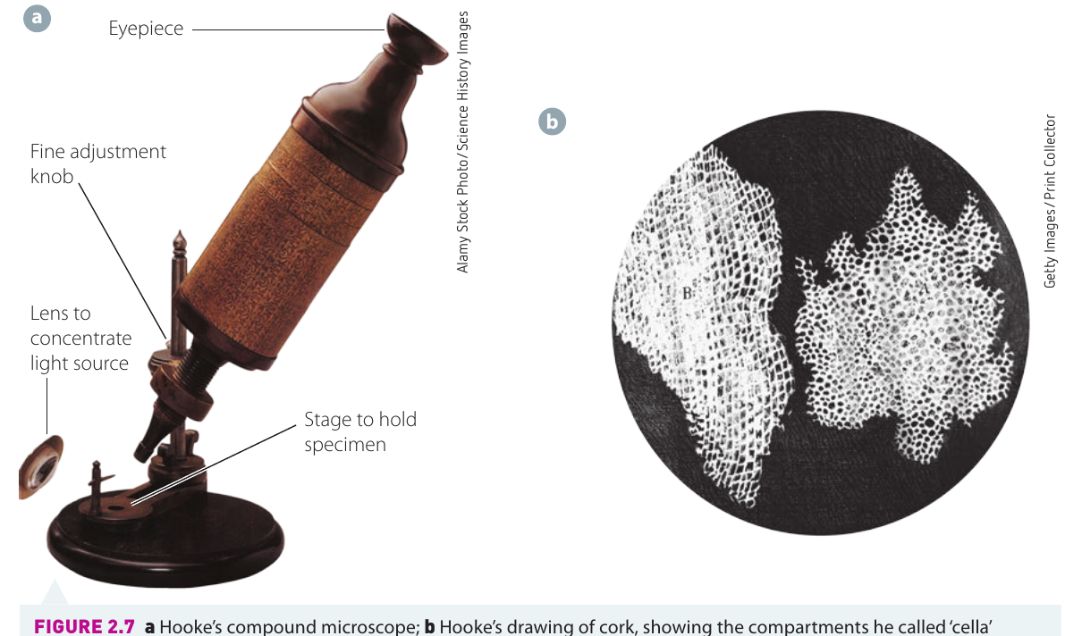
The breakthrough came with the invention of the compound microscope. This early device consisted of two convex lenses positioned at opposite ends of a barrel. It was the ancestor of the modern light microscope we use in laboratories today.
Over time, scientists made improvements to these compound microscopes. They added stands to hold them steady, focus knobs for adjusting the image, and light sources for better illumination. These enhancements allowed scientists to study the structure of organisms in much greater detail.
Robert Hooke's discovery
In the 1660s, a scientist named Robert Hooke used an improved compound microscope to examine a thin slice of cork. When he looked at the cork under magnification, he saw tiny compartments that reminded him of small rooms. He called these compartments "cella" (the Latin word for small room), which eventually led to our modern term "cell."
The word "cell" that we use today in biology comes directly from Hooke's observation. He chose the Latin word "cella" because the tiny compartments in cork reminded him of the small rooms (cells) that monks lived in at monasteries. This makes "cell" one of the oldest terms in biology!
Modern microscopes
As technology advanced, microscopes became increasingly sophisticated. Today, we have several types of microscopes, each with unique capabilities for studying cells.
Light microscopes
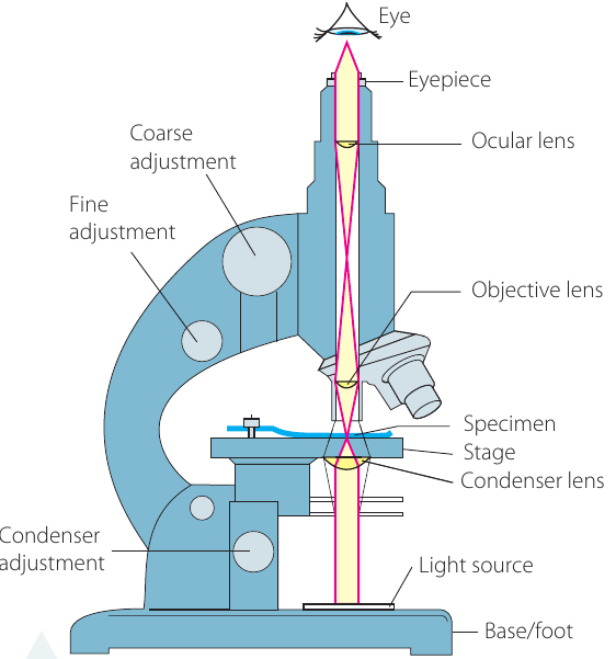
The compound light microscope is the type you typically use in school science classes. Understanding how it works helps us appreciate what we can see with it.
How light microscopes work
In a compound light microscope, light from a source passes upward through a condenser lens. This light then travels through the thin specimen you're examining. After passing through the specimen, the light enters a convex objective lens, which magnifies the image. Finally, you view the magnified image through the ocular lens (eyepiece).
Key specifications
Magnification refers to how much larger the image appears compared to the actual object. Light microscopes can magnify objects up to , depending on the lenses used.
However, magnification alone isn't enough to see cellular details clearly. Resolution (also called resolving power) is equally important. Resolution is the ability to distinguish between two separate objects. It represents the smallest distance between two objects where each can still be observed as separate entities.
Understanding the Difference: Magnification vs Resolution
Many students confuse magnification with resolution, but they are different:
- Magnification = How much bigger something appears
- Resolution = How much detail you can see (the ability to distinguish two separate points)
You could magnify an image , but if the resolution is poor, the image will just be a blurry, enlarged version with no additional detail!
For compound light microscopes, the maximum resolution is (nanometres). This means the best light microscopes can only distinguish two separate structures if the distance between them is or more. If two objects are closer than apart, they will appear as a single object.
Advantages of light microscopes
One significant advantage of light microscopes is their versatility. Both living and non-living specimens can be viewed using a compound light microscope. This makes them invaluable for observing living cells and their behaviours.
Fluorescence microscopes
Fluorescence microscopes are similar to light microscopes but have special features that allow scientists to target and observe specific parts of cells.
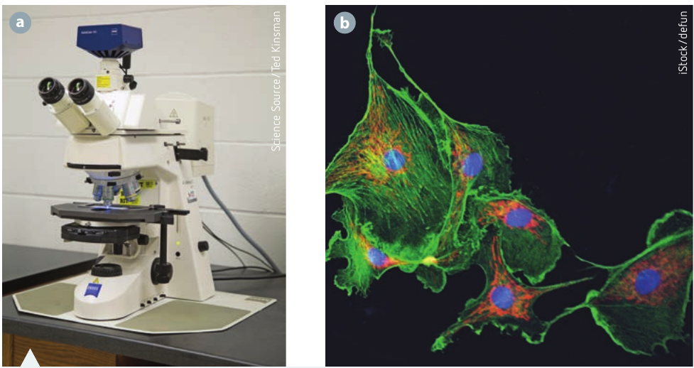
How fluorescence microscopy works
To use a fluorescence microscope, scientists first label the sample with a fluorescent substance. This substance is carefully chosen to attach to the specific structures the scientist wants to observe.
When the sample is illuminated with high-intensity light, the fluorescent substance absorbs this light and then emits its own light (fluorescence). The emitted fluorescent light passes through special filters that separate it from surrounding light. This allows the viewer to see only the areas of the sample that are fluorescing.
Applications
This technique is particularly useful for:
- Identifying specific proteins within cells
- Observing cellular structures beyond the normal resolution limit of light microscopes
- Tracking the location of particular molecules
- Studying cellular processes in real-time
Fluorescence microscopy is widely used in modern biological research because it allows scientists to "tag" specific molecules with fluorescent markers. This means you can watch exactly where particular proteins go inside a living cell, making it an incredibly powerful tool for understanding cellular processes!
Electron microscopes
Since the 1950s, electron microscopes have revolutionised our understanding of microscopic structures. These powerful instruments use completely different technology compared to light microscopes.
Basic principles
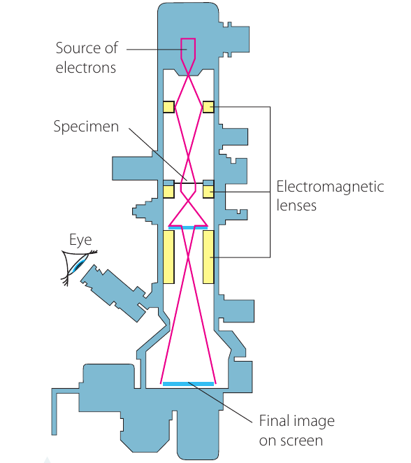
Instead of using light rays and glass lenses, electron microscopes use:
- An electron beam instead of light
- Electromagnets instead of glass lenses
The interaction between electrons and the specimen creates a viewable image on a screen.
Why electron microscopes are superior
Electron microscopes achieve much greater magnification than light microscopes. They also have substantially higher resolving power because electrons have much shorter wavelengths than visible light.
These capabilities have revealed structures at both the cellular and subcellular levels. Many cell organelles were seen for the first time using electron microscopes. Materials previously thought to have little internal structure were shown to have elaborate organisation. Features as small as one-tenth of a nanometre (one ten-billionth of a metre) can be observed, including individual atoms.
Worked Example: Comparing Wavelengths
The wavelength of visible light is approximately .
The wavelength of electrons used in electron microscopes is approximately .
This means electrons have a wavelength that is:
This dramatically shorter wavelength is why electron microscopes can achieve much higher resolution than light microscopes!
Transmission electron microscope (TEM)
The transmission electron microscope (TEM) is the most common type of electron microscope. In a TEM, electrons are transmitted through (pass through) the specimen.
Key specifications:
- Produces two-dimensional images
- Can magnify up to
- Has a resolution of approximately
The high resolution allows scientists to see internal structures of organelles in remarkable detail.
Scanning electron microscope (SEM)
The scanning electron microscope (SEM) works differently. It bombards solid specimens with a beam of electrons. These electrons cause secondary electrons to be emitted from the surface layers of the specimen.
Key specifications:
- Creates three-dimensional images of surfaces
- Has slightly lower resolution (approximately ) than TEM
- Provides excellent surface detail

The three-dimensional images from SEMs are particularly useful for studying surface structures and textures.
Disadvantages of electron microscopes
Despite their powerful capabilities, electron microscopes have some limitations:
Critical Limitations of Electron Microscopes
1. Living specimens cannot be viewed: The specimen must be placed in a vacuum for viewing because air would interfere with the flow of electrons. This means no living tissue can be examined.
2. Risk of artefacts: The specimen preparation is complex and may introduce artefacts into the image. An artefact is something introduced during preparation that wouldn't normally be present in the specimen. Scientists can identify artefacts by comparing samples prepared using different methods.
3. Size and cost: Electron microscopes are much larger, more expensive, and require higher maintenance costs compared to light microscopes.
Computer-enhanced technology
Modern technology has added another dimension to microscopy. Digital processing of microscope images allows scientists to view cells in innovative ways.
Digital image processing
Computer software can create three-dimensional representations of cell structures, providing a much deeper understanding of how cells are organised and function. Advanced computer technologies also help construct models showing how molecules interact during cellular reactions.
Confocal laser scanning microscopy
Confocal laser scanning microscopy is a sophisticated technique that creates three-dimensional images of specimens.
How it works
A laser produces a narrow, intense beam of light focused to a pinpoint on the sample. The microscope captures only the light from this precise focal point, while excluding all out-of-focus areas.
This focusing process occurs many times at different levels throughout the specimen. An image reconstruction program then combines data from all these different levels to construct a three-dimensional image.
Advantages
Confocal laser scanning microscopes are particularly useful for:
- Imaging structural components of cells
- Creating detailed 3D models
- Studying intact specimens without destroying them
Unlike electron microscopes, confocal microscopy can be used on living specimens, making it ideal for studying dynamic cellular processes as they happen. The computer combines multiple "optical slices" taken at different depths to create a complete 3D image!
Comparison of microscope types
| Microscope Type | Magnification | Resolution | Living Specimens? | Image Type |
|---|---|---|---|---|
| Light | Up to | Yes | 2D | |
| Fluorescence | Up to | Yes | 2D (specific structures) | |
| TEM | Up to | No | 2D | |
| SEM | Up to | No | 3D | |
| Confocal | Variable | Better than standard light | Yes | 3D |
Key Points to Remember:
-
Early compound microscopes with two convex lenses allowed Robert Hooke to discover and name cells in the 1660s.
-
Magnification shows how much bigger an image appears, while resolution determines how much detail you can see (the minimum distance between two objects that can be distinguished as separate).
-
Light microscopes use light rays and glass lenses, can magnify up to with resolution, and can view both living and non-living specimens.
-
Electron microscopes use electron beams and electromagnets instead of light and lenses. They achieve much higher magnification and resolution but cannot view living specimens due to the vacuum requirement. TEM produces 2D images by passing electrons through specimens, while SEM creates 3D images by bouncing electrons off surfaces.
-
Computer-enhanced technologies like confocal laser scanning microscopy combine multiple images taken at different levels to create detailed three-dimensional models of cell structures, allowing scientists to study intact specimens in unprecedented detail.