Types of Cells (HSC SSCE Biology): Revision Notes
Types of Cells
Introduction to cells
Living organisms are remarkable biological systems whose secrets have fascinated scientists for centuries. The development of microscopy technology revolutionised our understanding of life by revealing the cell as the fundamental unit of all living organisms.
Whilst all cells share certain basic similarities, they differ significantly in their structure, function, and organisation. Some organisms consist of a single cell that must perform all life functions independently. Other organisms are composed of many specialised cells, each with specific structures and roles, working together to maintain the organism's overall function.
The invention of the microscope in the 17th century enabled scientists to observe cells for the first time, fundamentally changing our understanding of biology. Today, advanced microscopy techniques continue to reveal new details about cellular structure and function.
Classification of cells
Despite the enormous diversity of cells in nature, all cells can be classified into two fundamental types: prokaryotic cells and eukaryotic cells. Prokaryotic cells are considered 'primitive' because they have a much simpler internal organisation compared to eukaryotic cells. Interestingly, prokaryotic cells vastly outnumber eukaryotic cells on Earth.
Three Essential Features Shared by All Cells
Although prokaryotic and eukaryotic cells have many differences, they share three essential features:
- A cell membrane that controls the movement of substances into and out of the cell
- Cytoplasm, a fluid-like substance that fills the cell
- Ribosomes, tiny structures that manufacture proteins
These common features reflect the shared ancestry of all living organisms.
Prokaryotic cells
Definition and characteristics
The term 'prokaryotic' comes from the Greek words pro (meaning 'before') and karyon (meaning 'nucleus'). This name reflects the fact that these cells existed before the evolution of cells with a true nucleus.
Prokaryotic cells are remarkably small, typically measuring between and micrometres (m) in diameter. This is approximately 10 to 100 times smaller than most eukaryotic cells.
Essential components
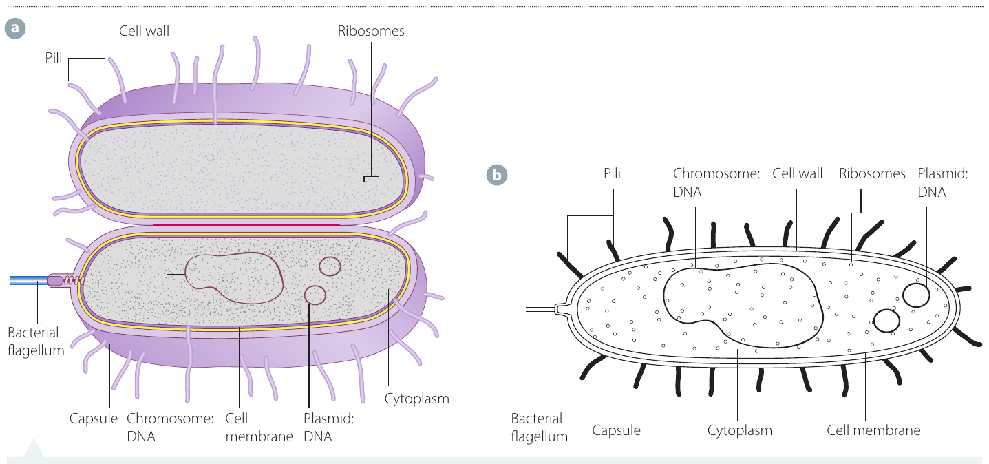
All prokaryotic cells contain four fundamental structures:
- Cell membrane - Controls the passage of materials entering and leaving the cell
- Cytoplasm - A gel-like fluid where chemical reactions occur
- Ribosomes - Structures that synthesise proteins
- Genetic material - DNA that carries hereditary information
Key Distinguishing Feature of Prokaryotic Cells
A distinctive feature of prokaryotic cells is the absence of a membrane surrounding their genetic material, meaning they lack a true nucleus. Most of the DNA is organised into a large loop called the bacterial chromosome, whilst smaller circular pieces of DNA called plasmids exist separately.
Unlike eukaryotic cells, the internal structures in prokaryotic cells are not enclosed by membranes—they simply float freely within the cytoplasm.
Additional structures
Some prokaryotic cells possess additional features that provide advantages in certain environments:
- Cell wall - A protective outer layer that provides structural support and shape. The composition varies depending on the type of prokaryotic cell.
- Capsule - An outer layer made of complex carbohydrates that offers extra protection.
- Pili (singular: pilus) - Hair-like projections on the cell surface that enable the cell to attach to surfaces or other cells.
- Flagella (singular: flagellum) - Whip-like tails that propel the cell through liquid environments.
Organisation and examples
Most prokaryotic organisms are unicellular, meaning they consist of just one cell that performs all necessary life functions. However, some bacterial species form colonies where multiple prokaryotic cells cluster together and cooperate.
Prokaryotic organisms are divided into two major groups:
Bacteria - Found in diverse environments worldwide, bacteria can be either beneficial (such as those in our digestive system) or harmful (disease-causing pathogens).
Archaea - These unicellular organisms thrive in extreme environments, including hydrothermal vents on the ocean floor and hot springs. They were once thought to be bacteria but are now recognised as a distinct group.

Eukaryotic cells
Definition and characteristics
The term 'eukaryotic' derives from the Greek words eu (meaning 'true' or 'proper') and karyon (meaning 'nucleus'), indicating these cells possess a true nucleus.
Eukaryotic cells are substantially larger and more complex than prokaryotic cells. They typically range from to micrometres (m) in size—approximately 10 to 100 times larger than prokaryotic cells.
Key features
The defining characteristic of eukaryotic cells is the presence of a membrane-bound nucleus containing the cell's genetic material. This nucleus is surrounded by a double membrane that separates the DNA from the rest of the cell's contents.
Organelles: The Key to Eukaryotic Complexity
All internal structures within eukaryotic cells are enclosed by membranes and are called organelles. Each organelle performs a specific function, contributing to the cell's overall operation.
These specialised compartments allow different biochemical processes—such as cellular respiration and photosynthesis (in plant cells)—to occur simultaneously in different locations within the cell. This compartmentalisation is what makes eukaryotic cells so much more complex and efficient than prokaryotic cells.
Organisation and examples
Organisms containing eukaryotic cells, known as eukaryotes, can be either unicellular or multicellular.
Unicellular eukaryotes include:
- Paramecium - A ciliated protozoan found in freshwater
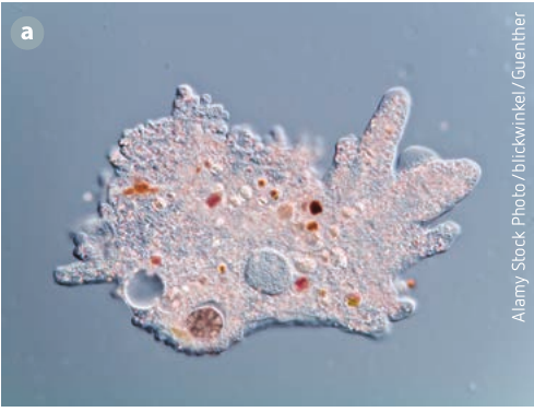
- Amoeba - A shape-changing organism that moves using pseudopodia
- Euglena - A photosynthetic organism with a flagellum for movement

Multicellular eukaryotes include all plants and animals. These organisms are composed of many different types of specialised eukaryotic cells, each designed for particular functions within the organism.
Comparing prokaryotic and eukaryotic cells
Understanding the differences and similarities between these two cell types is essential for grasping cellular biology. The table below summarises the key distinctions:
| Characteristic | Prokaryotic Cells | Eukaryotic Cells |
|---|---|---|
| Membrane-bound nucleus | Absent | Present |
| Membrane-bound organelles | Absent | Present |
| Cellular organisation | Primarily unicellular | Unicellular or multicellular |
| Size | - m | - m |
| Genetic material organisation | DNA in cytoplasm (chromosome and plasmids) | DNA enclosed within nucleus |
| Examples | Bacteria, Archaea | Plants, animals, fungi, protists |
Both cell types share three fundamental components: cell membrane, cytoplasm, and ribosomes. These common features reflect the shared ancestry of all living organisms and demonstrate that despite their differences, all cells have certain basic requirements for life.
Observing cells with microscopy
Light microscopes are valuable tools for examining both prokaryotic and eukaryotic cells. However, they have limitations in the level of detail they can reveal.
Using a light microscope, students can typically observe:
- Cell membranes
- Nuclei (in eukaryotic cells)
- Cytoplasm
- Chloroplasts (in plant cells)
- Cell walls (in plant cells and bacteria)
Smaller structures require more powerful electron microscopes to visualise clearly. When preparing specimens for microscopy, stains such as methylene blue are often used to make cellular structures more visible against the background.
Calculating total magnification
When using a compound light microscope, the total magnification is determined by multiplying the magnification of the eyepiece (ocular lens) by the magnification of the objective lens currently in use:
Worked Example: Calculating Total Magnification
If the ocular lens magnification is and the objective lens magnification is :
Therefore, the specimen would appear 400 times larger than its actual size.
The following table shows common magnification combinations:
| Ocular Lens Magnification | Objective Lens Magnification | Total Magnification |
|---|---|---|
Exam Tip: Showing Your Working
Always show your working when calculating magnification in examinations. Remember to include the multiplication symbol () in your final answer. Examiners award marks for clear working, even if your final answer contains a minor calculation error.
Remember!
Key Points to Remember:
- All cells are classified as either prokaryotic (simple, no nucleus) or eukaryotic (complex, with nucleus).
- Prokaryotic cells ( - m) are much smaller than eukaryotic cells ( - m).
- Both cell types possess a cell membrane, cytoplasm, and ribosomes as fundamental components.
- Prokaryotic cells lack membrane-bound organelles, whilst eukaryotic cells contain many specialised membrane-bound organelles.
- Total microscope magnification is calculated by multiplying the ocular lens magnification by the objective lens magnification.