Gas Exchange Structures (HSC SSCE Biology): Revision Notes
Gas Exchange Structures
Introduction to gas exchange
Gas exchange is a vital process in all living organisms. Every living cell requires a continuous supply of oxygen () and must eliminate carbon dioxide (). Both plants and animals have evolved specialised structures that facilitate the efficient movement of these gases between the organism and its environment.
The surface across which gases move is called the respiratory surface. Gas movement occurs through diffusion, where molecules move from areas of high concentration to areas of low concentration.
In small organisms like unicellular organisms or tiny multicellular organisms, substances can move directly across the cell membrane or thin body wall. However, larger multicellular organisms need specialised gas exchange structures and transport systems to ensure all cells receive oxygen and can remove carbon dioxide efficiently.
Gas exchange in plants
Leaf adaptations for gas exchange
Plant leaves are excellently adapted for gaseous exchange. Their structure includes several features that maximise efficiency:
- Large, flat shape: This maximises the surface area compared to the volume of each leaf, allowing more gas exchange to occur
- Open air spaces: Found within the leaf, these are formed by the irregular arrangement of spongy mesophyll tissue. These spaces increase surface area further and allow gases to move freely through much of the leaf without having to pass through cells
- Moist cell surfaces: The surfaces of cells inside the leaf are moist, allowing gases to dissolve. This dissolved state makes diffusion more effective
Most gas exchange in plants occurs through two types of structures: stomata and lenticels.
Stomata
The waxy cuticle covering leaves is impermeable to both water and gases. To allow gas exchange, leaves have evolved pores called stomata (singular: stoma or stomate). These are openings in the epidermis through which oxygen and carbon dioxide can pass.
Distribution of stomata:
- Mostly found on the undersurface (lower epidermis) of leaves
- Upper epidermis may have some stomata, but usually far fewer
- Distribution varies with plant habitat:
- Australian eucalypts have vertically hanging leaves with stomata on both surfaces
- Floating water plants typically have stomata only on their upper surfaces
- Underwater plants often lack stomata entirely
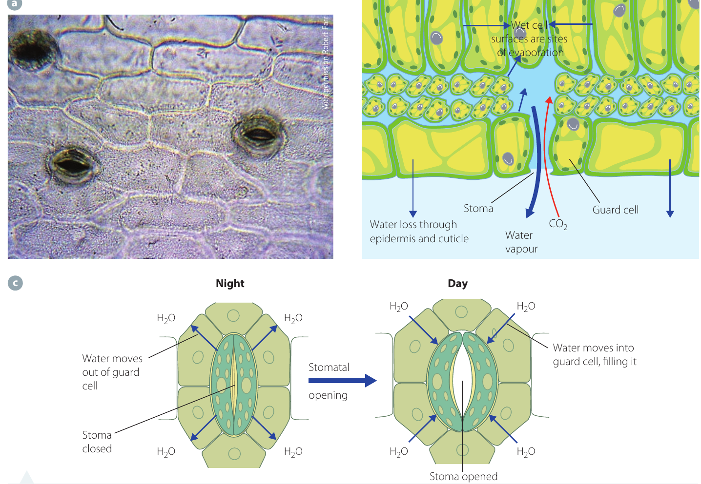
Structure of stomata
Each stoma is bordered by two specialised guard cells. These cells differ from other epidermal cells in important ways:
- Bean-shaped appearance
- Contain chloroplasts (unlike other epidermal cells)
- Inner wall is thicker than the outer wall

The presence of chloroplasts in guard cells is unique among epidermal cells. This allows guard cells to produce their own glucose through photosynthesis, which plays a role in the mechanism that controls stomatal opening and closing.
Opening and closing mechanism
Plants must balance their need for gas exchange against the risk of losing too much water through transpiration. The stomata can open and close to help manage this balance.
When stomata open:
- Guard cells fill with water and become turgid (swollen and firm)
- The thin, elastic outer walls stretch outwards
- The thick, inelastic inner walls cannot bulge, so they are pulled apart
- The pore between them widens
- Gases can now diffuse through the opening
When stomata close:
- Guard cells lose water and become flaccid (limp)
- The outer walls no longer bulge outwards
- The inner walls move back together
- The pore closes
- No gas exchange or water loss can occur
The exact mechanism that controls water movement into and out of guard cells is still being researched. Current scientific theories suggest it involves the movement of potassium ions.
Environmental factors affecting stomata
Several environmental factors influence whether stomata are open or closed:
Light
- This is the primary factor controlling stomata
- Generally, stomata open at daybreak and close at night
- This pattern aligns with photosynthesis, which requires during daylight
Temperature
- When temperature increases, more water vapour is lost through open stomata
- If water loss exceeds water uptake by roots, the plant's water content falls
- Guard cells lose water and stomata close to prevent further water loss
Water availability
- When water is scarce, photosynthesis becomes limited
- concentration inside the leaf rises
- High internal causes stomata to close, restricting further entry
- Conversely, if internal decreases, stomata open to allow more to enter
Humidity
- When air is saturated with water vapour (high humidity), the rate of water evaporation from leaves is reduced
- This allows stomata to remain open longer without excessive water loss
These environmental factors work together in an interconnected system. For example, high light intensity might normally cause stomata to open, but if temperature is also high and water availability is low, the stomata may close to prevent excessive water loss, even during the day.
Lenticels
While stomata handle gas exchange in leaves, woody parts of plants need a different solution. Lenticels are pores that enable gaseous exchange in the woody stems, trunks, and branches of trees and shrubs.
Characteristics of lenticels:
- Appear as small dots to the naked eye
- Under microscopic examination, they are revealed as clusters of loosely packed cells in the cork layer of bark
- Gas diffusion through lenticels is relatively slow compared to stomata
Lenticels allow living cells within woody stems to exchange and with the atmosphere, supporting cellular respiration even in mature, bark-covered plant parts.
Gas exchange in animals
Overview and importance
Gaseous exchange in animals involves the movement of gases between the internal environment (inside cells and bloodstream) and the external environment (atmosphere or water). This process is essential because:
- Oxygen is required by all cells for cellular respiration, which releases energy from nutrients
- Carbon dioxide is produced during cellular respiration and must be removed, as high concentrations become toxic, changing cellular pH and interfering with enzyme function
Carbon dioxide toxicity: High concentrations of are particularly dangerous because they change cellular pH (making it more acidic) and interfere with enzyme function. This is why efficient removal of is just as important as oxygen uptake.
The respiratory system contains specialised organs and tissues that allow organisms to take in oxygen and remove carbon dioxide.
Common characteristics of gas exchange structures
Despite the diversity of respiratory structures across different animal groups, all effective gas exchange surfaces share four essential characteristics:
- Large surface area
- Enhanced through folding, branching, or flattening
- Allows faster rate of diffusion
- Supplies oxygen and removes carbon dioxide more efficiently
- Thin, moist surface
- Ensures oxygen and carbon dioxide dissolve for easier diffusion
- Thinness decreases the distance gases must travel
- Moisture is essential for gas molecules to dissolve
- Close proximity to transport system
- Located near blood vessels or other transport structures
- Enables rapid transport of gases to and from all body cells
- Maintains efficient distribution throughout the organism
- Maintained concentration gradient
- Greater concentration of required gas on one side of the membrane than the other
- Ensures continuous diffusion in the correct direction
- Supports ongoing gas exchange
In larger terrestrial animals, gas exchange systems are internal. This internal location prevents dehydration of the delicate, moist respiratory surfaces, which must remain wet to function properly.
Gaseous exchange in mammals
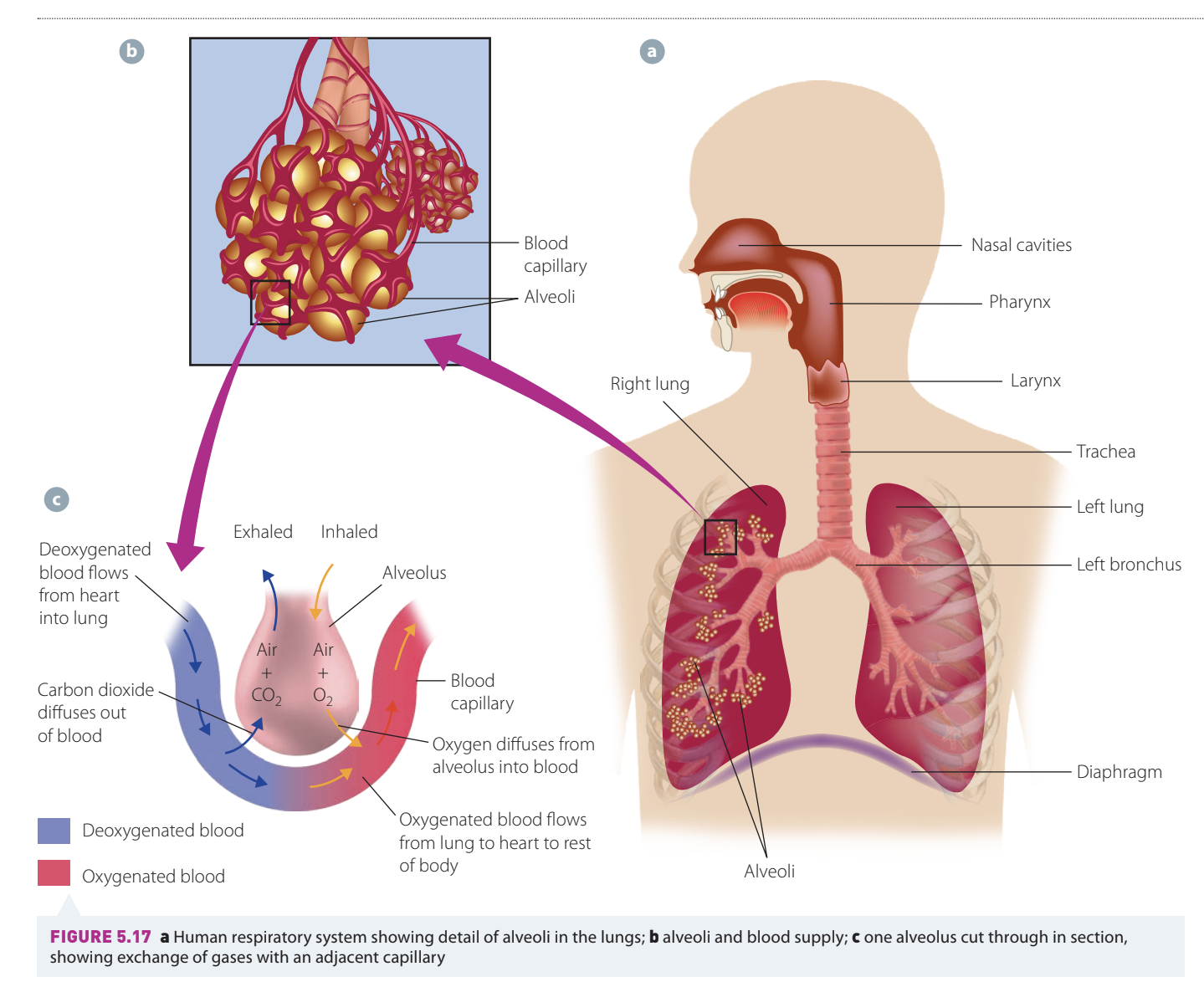
The alveoli
In mammals, the specialised gas exchange surfaces are located in the lungs. These structures are called alveoli (singular: alveolus).
Structure:
- Each alveolus is a thin-walled air sac
- Connected to the external environment through the respiratory pathway (nose/mouth → trachea → bronchi → bronchioles)
- Surrounded by tiny, thin-walled blood vessels called capillaries
Features that make alveoli efficient
The alveoli possess all four characteristics of effective gas exchange surfaces:
1. Massive surface area
- Human lungs contain approximately 300 million microscopic alveoli
- Supplied by around 280 million capillaries
- Each alveolus has folding of its thin interior lining, further increasing surface area
- This enormous surface area allows rapid gas exchange for the entire body
2. Extremely thin barrier
- Each alveolus has a lining made of flattened cells arranged in a single layer
- Facilitates efficient diffusion of gases across a very small distance (less than 1 μm)
- Both the alveolar wall and capillary wall are just one cell thick
3. Moist surface
- The entire respiratory system surface is kept moist
- Air inside alveoli is saturated with water vapour
- Mucus-lined epithelium reduces water evaporation
- Ensures oxygen and carbon dioxide are in dissolved form for efficient diffusion
4. Excellent blood supply
- Numerous blood capillaries closely surround the outside of each alveolus
- Ensures all alveoli are in close contact with blood
- Maintains concentration gradients for continuous gas exchange
The efficiency of alveoli is remarkable. The total surface area of all alveoli in human lungs is approximately 70 square metres—about the size of a tennis court! This massive surface area is packed into two organs that fit comfortably inside your chest.
Gas movement in alveoli
The movement of gases between alveolar air and blood occurs by diffusion down concentration gradients:
Oxygen movement:
- Inhaled air contains approximately 20% oxygen
- Oxygen concentration in alveolar air is higher than in blood arriving from the body
- Oxygen diffuses from alveoli → through alveolar wall → through capillary wall → into blood
- Blood becomes oxygenated and returns to the heart for distribution
Carbon dioxide movement:
- Carbon dioxide is a by-product of cellular metabolism
- Blood arriving at lungs has high concentration
- concentration in blood is higher than in alveolar air (which has only 0.04% )
- Carbon dioxide diffuses from blood → through capillary wall → through alveolar wall → into alveolar air
- is then exhaled
Comparing inhaled and exhaled air:
| Gas | Inhaled air | Exhaled air |
|---|---|---|
| Oxygen | ~20% | ~15% |
| Carbon dioxide | ~0.04% | ~4% |
This shows that we extract about 5% of the oxygen from inhaled air, and our exhaled air contains 100 times more carbon dioxide than atmospheric air. This demonstrates the efficiency of gas exchange in the alveoli.
Respiratory systems in other animals
Different animals have evolved respiratory structures suited to their specific environments.
Fish
Aquatic animals face a challenge: gases have low solubility in water, so their concentration in water is much lower than in air. Fish have evolved gills to extract the maximum possible oxygen from water.
Key features of fish gills:
- Located behind the head, protected by gill covers (opercula)
- Possess all characteristics of efficient respiratory surfaces
- Well supplied with blood capillaries
- Require water to flow constantly over them for maximum oxygen uptake
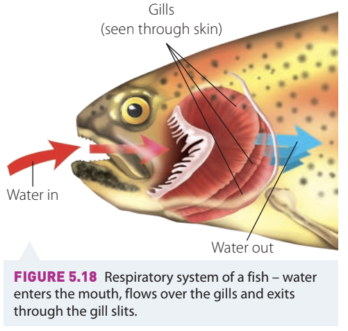
How gills work:
- Water enters through the fish's mouth as it swims or actively pumps water
- Water flows in one direction only over the gill filaments
- As water passes over gills, gaseous exchange occurs
- Oxygen diffuses from water into blood in gill capillaries
- Carbon dioxide diffuses from blood into water
- Water exits through the gill slits
The one-way flow of water over gills, combined with blood flowing in the opposite direction (counter-current system), maximises oxygen extraction from water. This counter-current mechanism allows fish to extract up to 80% of dissolved oxygen from water, far more efficient than if water and blood flowed in the same direction.
Insects
Terrestrial insects face the challenge of preventing water loss from their internal respiratory surfaces while still exchanging gases efficiently.
Key features of insect respiratory systems:
- Take in and expel air through spiracles (breathing pores)
- Spiracles have valves to regulate opening and closing, minimising water loss
- Body covering is largely impermeable to gases
- Do not have lungs or blood capillaries
- Small body size allows a simpler gas exchange system
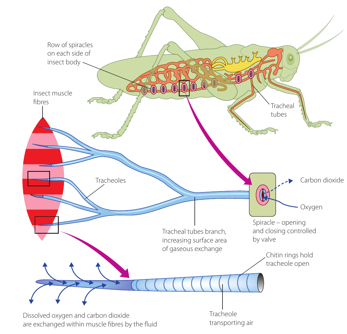
Structure and function:
- Spiracles
- Opening pores on the body surface
- Arranged in rows along the sides of the insect body
- Controlled by valves to prevent excessive water loss
- Tracheal tubes (tracheae)
- Branching air tubes that carry air directly into the body
- Air enters through spiracles and is drawn into these tubes
- Kept open by spiral rings of chitin (a tough, supportive substance)
- Branch extensively to increase surface area
- Tracheoles
- Smaller tubules that branch from the tracheae
- Create a very large surface area for gas exchange
- Bring air directly to and from individual body cells
- Ends are filled with watery fluid in which gases dissolve
Unique feature: Unlike all other internal respiratory systems, the insect tracheal system does not involve blood or blood capillaries in gas transport. Oxygen dissolved in the fluid at the ends of tracheoles diffuses directly into cells, and carbon dioxide diffuses directly from cells into the tracheoles. This direct delivery system is highly efficient for small organisms.
Ventilation:
- The rate of respiration is controlled by the number of open spiracles (more open during activity)
- Muscular movements of the thorax and abdomen during activity help ventilate the tracheal system
- Body movements during flying assist in moving air through the system
Key Points to Remember:
-
Gas exchange occurs across respiratory surfaces by diffusion, moving gases between an organism and its environment.
-
All efficient gas exchange surfaces share four characteristics: large surface area, thin and moist surfaces, close proximity to a transport system, and a maintained concentration gradient.
-
In plants, gas exchange occurs mainly through stomata (controlled by guard cells that open when turgid and close when flaccid) and lenticels in woody parts.
-
In mammals, approximately 300 million alveoli in the lungs provide an enormous surface area for gas exchange, with oxygen diffusing into blood and carbon dioxide diffusing out.
-
Different animals have specialised structures: fish use gills to extract oxygen from water with a counter-current system, while insects use a tracheal system with spiracles and branching tubes that bring air directly to cells without involving blood.