Transport Systems in Animals (HSC SSCE Biology): Revision Notes
Transport Systems in Animals
Introduction
Multicellular organisms require a constant supply of nutrients and oxygen, and they must continuously remove waste products. A circulatory system is the most efficient way to achieve this transport function throughout the body.
The evolution of transport systems was a crucial adaptation that allowed organisms to grow larger and more complex. Without efficient circulation, organisms would be limited in size because materials could only move by diffusion alone.
Open and Closed Circulatory Systems
Transport systems in multicellular animals can be divided into two main types: open and closed circulatory systems. Both types contain similar components:
- A heart (driving mechanism)
- A transport fluid
- A system of vessels
Key Difference Between Systems:
The fundamental distinction between open and closed circulatory systems lies in how the transport medium moves through the body:
- Closed circulatory system: The transport medium remains enclosed within vessels at all times
- Open circulatory system: The transport fluid leaves the vessels, enters body cavities, and comes into direct contact with organs
Open circulatory systems
Open circulatory systems are characteristic of invertebrate animals such as spiders, insects, crabs, and snails. These systems have the following features:
- Made up of one or more hearts and open-ended blood vessels
- Not sealed - the heart pumps blood into a cavity that surrounds organs
- Blood returns to the heart through special openings
- Exchange only nutrients and wastes with cells (gases are exchanged via a different system)
- Less efficient than closed systems because fluid pressure is low, causing slower circulation
The transport fluid in an open circulatory system is called haemolymph, which is a mixture of blood and tissue fluid.
How open circulatory systems work
When the long, pulsating vessel (the heart) contracts, it pumps haemolymph away from the heart into shorter vessels near the head end of the body. These vessels empty into large spaces in the body cavity called sinuses.
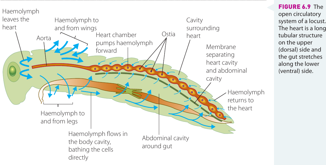
Haemolymph flows into the sinuses in the body cavity, bathing the cells directly. The movement of the organism aids the flow of haemolymph around the cells. Exchange of nutrients and wastes occurs by direct diffusion between the haemolymph and the cells.
When the heart expands, haemolymph returns to the heart by moving from the posterior (rear) sinuses back into the open end of the tubular heart, or it may enter the heart through tiny holes in the sides called ostia. This creates a continuous circulation pattern, albeit a slower one than in closed systems.
Closed circulatory systems
Closed circulatory systems are found in all vertebrate animals, including fish, frogs, reptiles, birds, and mammals (including humans). These systems consist of:
- Blood vessels
- A heart
- Blood (transport medium) that remains in vessels at all times
In mammals, the main transport system is known as the cardiovascular system. It transports nutrients and oxygen to all cells and carries wastes away from cells.
The heart pumps blood around the body, and the blood never flows through body cavities - it is always contained within vessels. The heart may have:
- Two chambers (as in fish)
- Three chambers (as in frogs and some reptiles)
- Four chambers (as in other reptiles, all birds, and mammals)

Blood vessels in closed systems
Blood flows through three types of blood vessels:
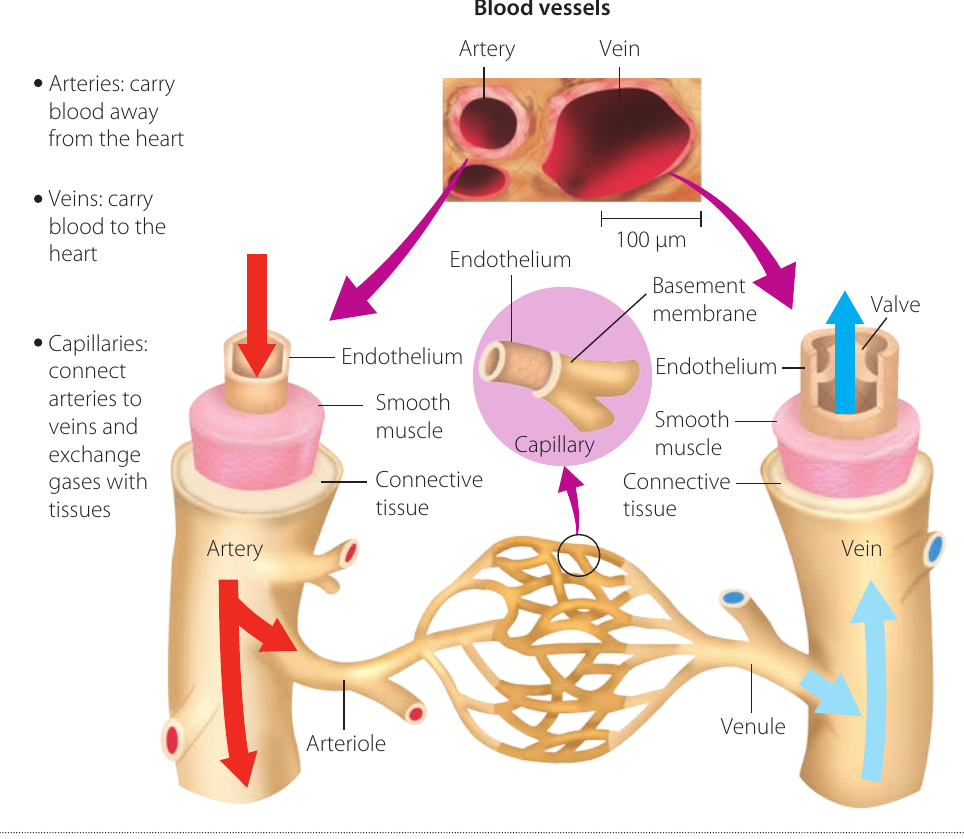
- Arteries: Carry blood away from the heart to the organs
- Veins: Carry blood from body organs towards the heart
- Capillaries: Form a link between arteries and veins
Arteries branch into smaller arterioles, which subdivide further into a network of capillaries. These capillaries branch extensively throughout the tissues, ensuring no cell is very far from a capillary.
The exchange of nutrients, wastes, and gases takes place between blood in the capillaries and the fluid surrounding the cells, called interstitial fluid or tissue fluid. This fluid is the crucial link between the capillaries and the cells, acting as an intermediary medium.
Capillaries join up to form venules, which in turn join up to form veins, returning blood to the heart.
The pathway of blood flow is: arteries → arterioles → capillaries → venules → veins → heart
Advantages of closed circulatory systems
In a closed circulatory system, the muscular heart pumps blood under high pressure, ensuring efficient transport. This suits large, active animals such as vertebrates. A four-chambered heart is the most efficient pumping mechanism because it keeps oxygenated blood and deoxygenated blood separate.
The separation of oxygenated and deoxygenated blood in a four-chambered heart is crucial for maintaining high metabolic rates. This design prevents the mixing of oxygen-rich and oxygen-poor blood, ensuring that tissues receive blood with maximum oxygen content.
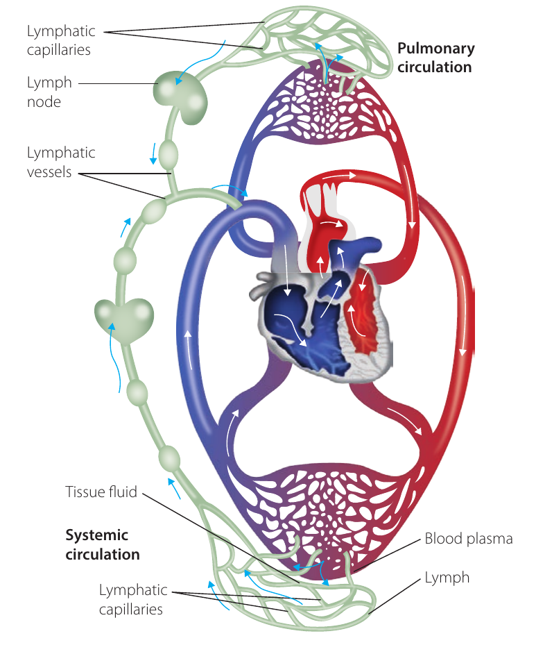
Lymphatic System
The lymphatic system forms part of the transport system in mammals and works alongside the cardiovascular system.
Function of the lymphatic system
As blood passes through tissues, fluid diffuses out of the capillaries. To prevent this interstitial fluid from building up in the tissues, lymph vessels in the tissues absorb it. This fluid, along with other substances such as white blood cells and the end products of lipid digestion, is known as lymph.
The lymph flows in the lymph vessels in one direction only, from the tissues to the heart. This movement is assisted by:
- The contraction of muscles in close proximity to the vessels
- Valves present in the lymph vessels to prevent the lymph flowing backwards
The lymphatic vessels from all regions of the body eventually join up to form two main lymphatic channels. In the region of the shoulders, these lymphatic channels drain into the veins, allowing the lymph fluid to rejoin the blood. This completes the circulation cycle and ensures that fluid lost from capillaries is eventually returned to the bloodstream.
Benefits of the lymphatic system
Key Functions of the Lymphatic System:
- Prevents the build-up of excess fluid in the tissues
- Helps to maintain the volume of the blood and therefore blood pressure
- Plays an important role in the defence of the body
Blood as a Medium of Transport
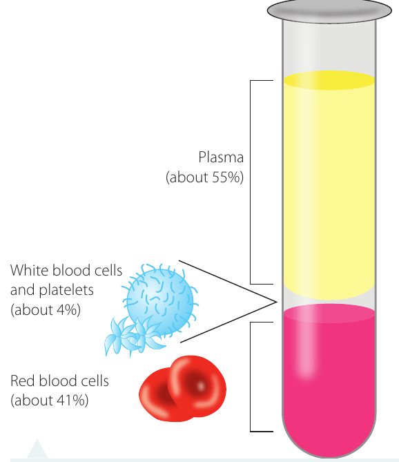
Blood is the fluid transport medium that flows through the heart and blood vessels of the cardiovascular system in vertebrates. It consists of two main components:
- Blood cells (45%)
- Plasma (55%)
When blood is centrifuged, 55% is found to be the watery plasma that collects on top of the cells, with many substances dissolved in it. The remaining 45% is the heavier red and white blood cells that collect below the plasma.
Properties of human blood
Blood Properties:
- Temperature: (carries heat and is higher than overall body temperature)
- pH: (slightly alkaline)
- Volume: Approximately litres in an adult human
For the normal functioning of the body and its enzymes, these levels of temperature, pH, and blood volume must be carefully maintained.
Functions of blood
Blood performs several important functions:
- Distributes heat around the body
- Transports nutrients and gases required by the body
- Removes wastes to be excreted from the body
- Carries other chemicals such as hormones
- Carries antibodies to fight infections
- Carries clotting factors
- Transports many other substances required by the body to function efficiently
Red blood cells
Red blood cells (erythrocytes) are the most abundant type of blood cell, with approximately million red blood cells per millilitre (mL) of blood.
Structure of red blood cells
Red blood cells form in bone marrow. During development:
- At first, each cell has a nucleus
- As the cell matures, the nucleus disintegrates
- A red pigment called haemoglobin develops inside the cell
The absence of a nucleus allows the red blood cell to carry more haemoglobin (an oxygen carrier). This is a crucial adaptation that maximizes the oxygen-carrying capacity of each cell.
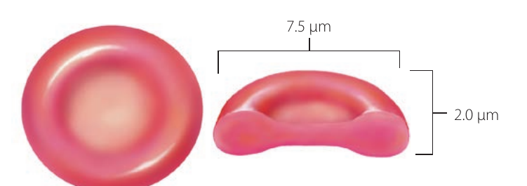
Mature red blood cells have the following characteristics:
- Diameter: Approximately µm (micrometres)
- Shape: Round, biconcave, and slightly flattened towards the centre (similar to a donut without the hole completely removed)
This biconcave shape makes red blood cells more pliable and elastic so that they can squeeze through capillaries that are sometimes narrower than their actual size.
Function and lifespan
- Main function: To transport oxygen
- Lifespan: Approximately months
- When they die, they are broken down and replaced by newly formed blood cells from the bone marrow
White blood cells
White blood cells (leucocytes) are also produced in bone marrow. There are approximately white blood cells per mL of human blood.
Characteristics of white blood cells
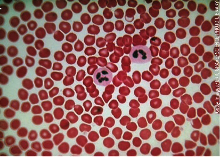
White blood cells differ significantly from red blood cells:
- Larger than red blood cells (about 50% bigger)
- Less abundant than red blood cells
- All contain a nucleus (in some white blood cells, it may be an unusual shape)
- In prepared microscope slides, the staining technique imparts a purple colour to the nucleus
- Several different types exist, each carrying out a specific function in defending the body
Function
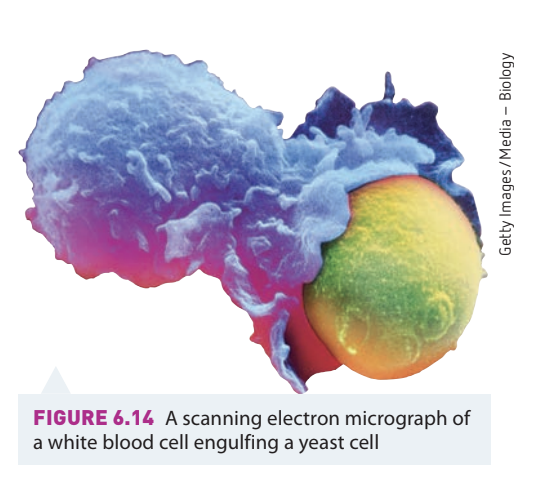
White blood cells function as part of the immune system. Their main role is in the defence of the body against invading foreign bodies. They are found in the tissues as well as in the blood.
Some white blood cells live for only a few minutes to days, while others can live for years. Leucocytes can pass through capillaries by squeezing between the cells that make up the wall of the capillary, allowing them to reach regions of damaged cells.
Platelets
Platelets (thrombocytes) are fragments of special cells, also produced in the bone marrow.
Characteristics of platelets
- Shape: Crescent-shaped
- Size: About half the size of red blood cells
- Count: Approximately per mL of blood
Function in blood clotting
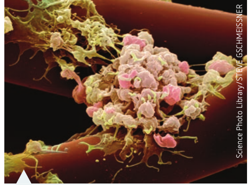
Platelets have a crucial function in the clotting of blood. The clotting process works as follows:
- Platelets stick to each other and to the fibres that develop at the site of a wound when blood is exposed to air
- The contact between fibres and platelets causes the platelets to break open and release an enzyme called thromboplastin
- This sets in progress a sequence of steps to seal the blood vessels and cause blood to clot, preventing excessive blood loss
Warning About Abnormal Clotting:
If blood clots form inside blood vessels without exposure to air, this causes blockages to circulation, as occurs in deep vein thrombosis (DVT). This is a serious medical condition that requires immediate attention, as clots can break free and travel to vital organs.
Plasma
Plasma is the yellow, watery fluid part of blood. It consists of:
- Approximately 90% water
- Approximately 10% proteins and other substances
Plasma makes up most of the volume of blood and it carries many substances in either dissolved or suspended form.
Substances carried by plasma
Besides carrying blood cells, plasma also transports:
- Plasma proteins:
- Clotting factors
- Immunoglobulins (antibodies to fight infections)
- Albumen
- Enzymes
- Nutrients (end products of digestion):
- Amino acids (from digested proteins)
- Glucose (from digested carbohydrates)
- Glycerol and fatty acids (from digested lipids)
- Cholesterol
- Gases:
- Oxygen
- Carbon dioxide
- Excretory waste products:
- Nitrogenous wastes such as urea, uric acid, and ammonia
- Ions:
- Mainly sodium chloride
- Calcium and magnesium phosphates
- Regulatory substances:
- Hormones (chemical messenger molecules involved in the coordination of body systems)
- Other substances:
- Vitamins
Blood Vessels
Blood vessels are an integral part of the transport system in mammals. Their function is to carry blood around the body, transporting nutrients, gases, and wastes.
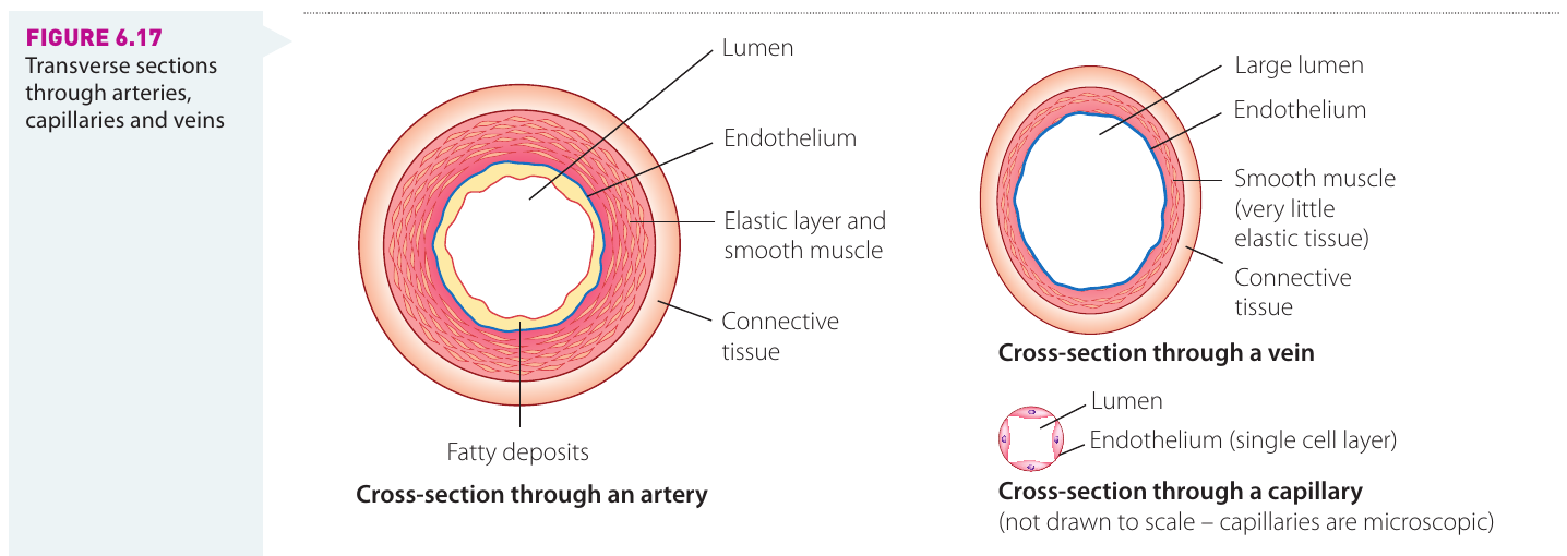
Each of the three types of blood vessels - arteries, capillaries, and veins - has a specific structure related to its function. These three vessel types share a similar basic structure, but they differ in terms of:
- The layers of tissue that make up the wall of each vessel
- The size of the lumen (central cavity)
Arteries
Arteries carry blood away from the heart to the organs.
Structure
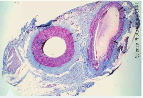
The blood that enters arteries is under very high pressure as it is pumped out of the heart in regular bursts. The walls of arteries have the following features:
- Much thicker than those of veins to withstand this pressure
- Highly elastic to allow expansion when a pulse of blood moves through
- Contain substantial amounts of smooth muscle and elastic tissue
- Contract back to the original diameter after expansion
Function
The Elasticity Advantage:
The elasticity of the artery wall serves two purposes:
- Allows the artery to expand when a pulse of blood moves through
- The contraction squeezes the blood forward and propels it along
This elastic recoil mechanism helps maintain continuous blood flow even between heartbeats, smoothing out the pulsatile flow from the heart.
Veins
Veins return blood to the heart.
Structure
Veins have the following structural features:
- Walls that are thinner than those of arteries (blood flows under lower pressure)
- Very few elastic fibres (they do not need to stretch and recoil)
- Internal diameter much wider than that of an artery
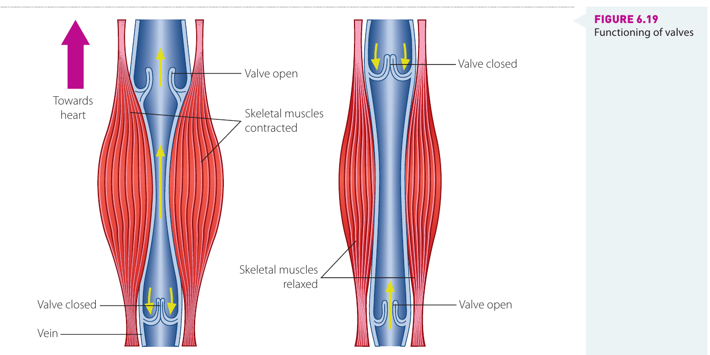
- Valves situated at regular intervals along their lengths
Function and blood flow mechanisms
Since blood seeps into veins and is not pumped, two mechanisms prevent the backflow of blood:
Mechanisms Preventing Backflow:
-
Muscle contraction: When the muscles in the tissue that surrounds the veins contract, the walls of the veins are compressed, propelling the blood towards the heart
-
Valves: Work like one-way swing doors
- They open to allow blood to flow through in one direction (towards the heart)
- The pressure of blood trying to flow backwards causes them to swing shut and stop the reverse flow
Capillaries
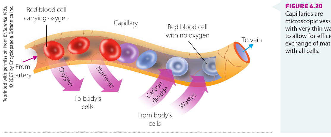
Capillaries are extremely tiny vessels that bring the blood into close contact with the tissues, enabling the exchange of chemical substances between cells and the bloodstream.
Structure
The walls of capillaries consist of only one layer of cells. Capillaries have structural features that slow down blood flow and maximise exchange:
- Thin walls: Allow for efficient diffusion of substances (substances do not have far to travel between the blood and body cells)
- Narrow internal diameter: Only slightly larger than the diameter of red blood cells, forcing them to pass through in single file
- Extensive network: Spread blood flow over a large surface area so that no cells are far from the blood supply
Function
The structure of capillaries is suited to slowing down the flow of blood, which is important because diffusion is a fairly slow, passive process.
Benefits of Capillary Structure:
- Thin walls allow efficient exchange of materials
- Red blood cells passing in single file slows down their flow
- Single-file passage increases the exposed surface area for exchange of gases, nutrients, and wastes
- Extensive branching ensures all cells are close to a blood supply
Blood remains in the capillaries at all times, but any chemical substances required by cells leave the capillaries in a dissolved form and move to the fluid that surrounds the cells (interstitial fluid).
The Heart - The Driving Force
The heart is the driving force in the circulatory system of animals. Mammals have a four-chambered heart, which pumps blood around the body.
Structure of the heart
Each side of the heart has two chambers:
- Atria (singular: atrium): The top chambers on each side
- Ventricles: The bottom chambers on each side
Key structural features
- Septum: A muscular wall that separates the left and right sides of the heart
- Cardiac muscle tissue: Composes the heart and produces the heartbeat when it contracts
- Valves: Maintain the one-way direction of blood flow in the heart
- Varying wall thickness:
- The left ventricle has much thicker walls of muscle tissue than the right ventricle
- This is because the left ventricle must pump blood to all areas of the body
- The right ventricle only has to pump deoxygenated blood to the lungs, which are in close proximity to the heart
Why Wall Thickness Matters:
The difference in wall thickness between the left and right ventricles reflects the distance blood must travel. The left ventricle needs to generate enough pressure to push blood throughout the entire body, while the right ventricle only sends blood to the nearby lungs. This is an excellent example of structure matching function.
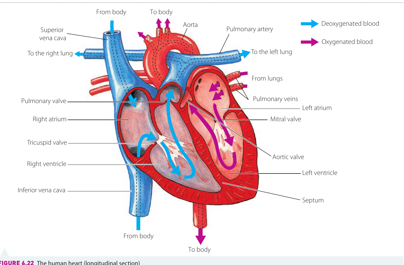
Blood flow through the heart
Right side of the heart (deoxygenated blood)
- Deoxygenated blood returns from the body to the right atrium via two large veins:
- Superior vena cava
- Inferior vena cava
- Blood moves from the right atrium to the right ventricle
- The right ventricle pumps blood via the pulmonary artery to the lungs
- In the lungs:
- Carbon dioxide diffuses from the blood into the alveoli
- Oxygen diffuses from the alveoli into the blood
Left side of the heart (oxygenated blood)
- Oxygenated blood returns to the left atrium via the pulmonary vein
- Blood moves from the left atrium to the left ventricle
- The left ventricle pumps blood via the major artery (the aorta) to all areas of the body
Circulation pathways
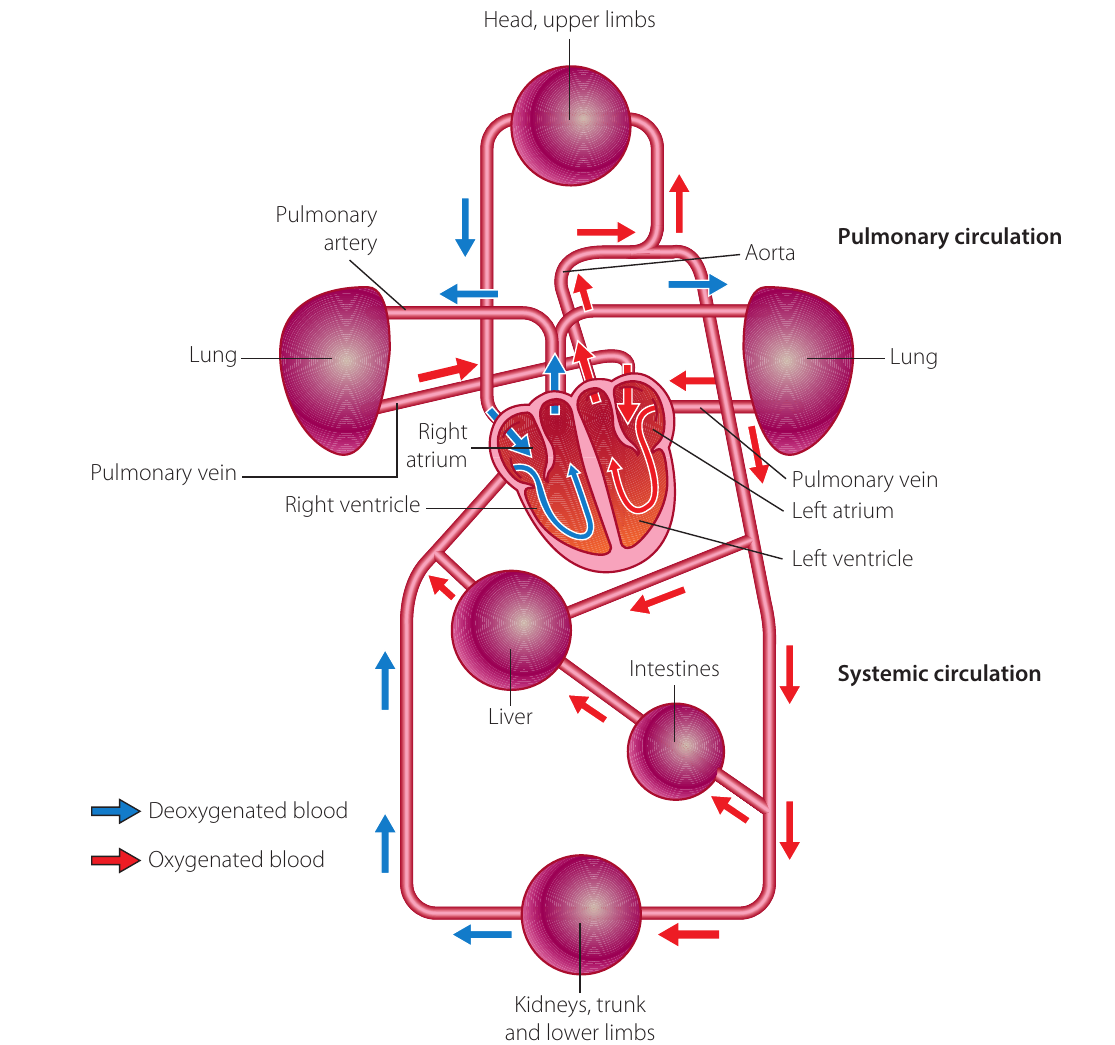
There are two main circulation pathways in the cardiovascular system:
- Systemic circulation: The pumping of oxygenated blood to all parts of the body and the return of deoxygenated blood to the heart
- Pulmonary circulation: The pathway of blood from the heart to the lungs and back to the heart
The heart is really a double pump, with each side beating almost simultaneously. This dual-pump system ensures efficient separation of oxygenated and deoxygenated blood while maintaining continuous circulation to both the lungs and the rest of the body.
Summary
Key Points to Remember:
-
Open circulatory systems (found in invertebrates) have haemolymph that leaves vessels and bathes organs directly, while closed circulatory systems (found in vertebrates) keep blood enclosed in vessels at all times.
-
The lymphatic system works alongside the cardiovascular system to return excess tissue fluid to the blood and plays a role in body defence.
-
Blood consists of plasma (55%) and blood cells (45%), including red blood cells (oxygen transport), white blood cells (defence), and platelets (clotting).
-
Blood vessels have structures suited to their functions: arteries have thick, elastic walls for high-pressure blood flow; veins have thinner walls and valves to return blood to the heart; capillaries have thin walls (one cell thick) for efficient exchange with tissues.
-
The four-chambered heart in mammals acts as a double pump, with the right side pumping deoxygenated blood to the lungs (pulmonary circulation) and the left side pumping oxygenated blood to the body (systemic circulation).