Analysis of Organic Substances (HSC SSCE Chemistry): Revision Notes
NMR Spectroscopy
Introduction to NMR spectroscopy
Nuclear magnetic resonance (NMR) spectroscopy is one of the most powerful techniques chemists use to work out the structures of organic molecules. It's so important that Richard Ernst won the Nobel Prize in Chemistry in 1991 for developing this method. Ernst described NMR as allowing nuclei to play their own "magnetic melody" - each type of atom produces a characteristic signal that chemists can detect and use to identify where atoms are located within complex molecules.
NMR spectroscopy works by observing how atomic nuclei behave when placed in a strong magnetic field. This technique can distinguish between different isomers of the same molecular formula and pinpoint the exact arrangement of atoms in a molecule.
NMR spectroscopy is so powerful that it has become an indispensable tool in chemistry, biochemistry, and medicine. The technique's ability to reveal molecular structure non-destructively has revolutionized how scientists study everything from small organic molecules to large proteins and even diagnose medical conditions through MRI (Magnetic Resonance Imaging).
Principles of NMR spectroscopy
Nuclear spin and magnetic behaviour
When you place matter in a magnetic field, certain atomic nuclei act like tiny magnetic compass needles. These nuclei are spinning charged particles, and their spin creates small magnetic fields. However, only nuclei with an odd number of nucleons (protons and neutrons combined) possess the spin property needed for NMR spectroscopy.
Critical Requirement for NMR: Only nuclei with an odd number of nucleons can be studied using NMR spectroscopy. This is because they possess the necessary spin property that creates a magnetic moment. Nuclei with even numbers of nucleons do not have this property and cannot be detected by NMR.
The two most commonly studied nuclei are:
- H (hydrogen-1, or protons)
- C (carbon-13)
These nuclei are used to produce H NMR spectra and C NMR spectra respectively.
Energy states
When nuclei are placed in a magnetic field, most align themselves with the field direction. This aligned state is the lower energy state (called the α state). Some nuclei align against the field in a higher energy state (called the β state).
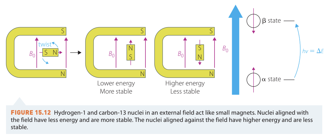
The key to NMR spectroscopy is that nuclei in the lower energy state can absorb energy from radio waves and "flip" to the higher energy state. This only happens when the radio waves have exactly the right frequency that matches the resonance of the nuclei. Think of it like pushing a child on a swing - you need to push at just the right moment for it to work.
Understanding Resonance: The concept of resonance is fundamental to NMR. Just as a musical instrument produces sound at specific frequencies, atomic nuclei respond to specific radio wave frequencies. When the frequency matches perfectly, energy is absorbed and the nucleus flips - this is called being "in resonance."
After absorbing energy and flipping to the higher energy state, nuclei will flip back down to the more stable lower energy state and emit energy. The NMR spectrometer detects this emitted energy. The energy difference between these two states depends on:
- The nature of the nucleus (H or C)
- The chemical environment surrounding the nucleus
The NMR spectrometer
An NMR spectrometer contains several key components working together:
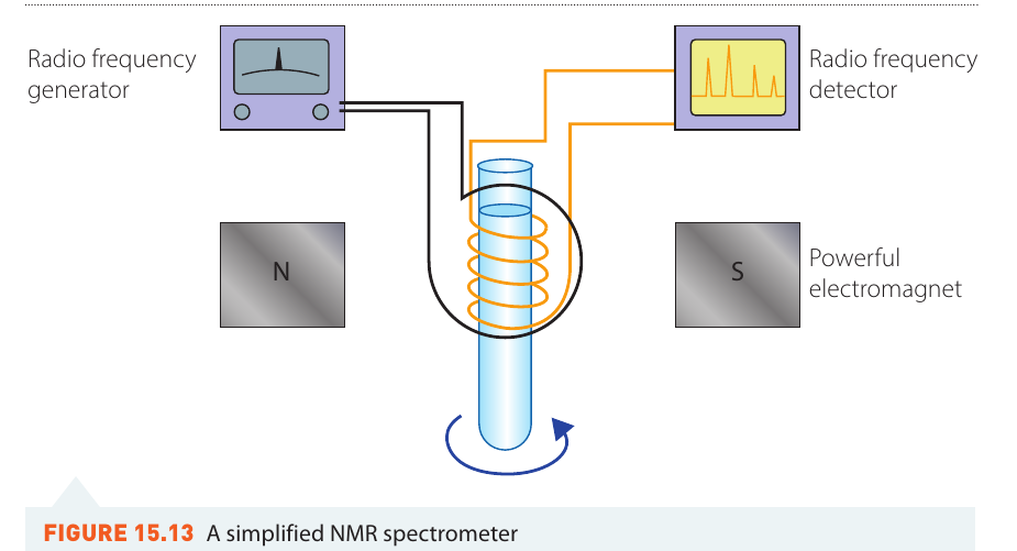
The main parts are:
- A powerful electromagnet that creates a strong magnetic field
- A sample holder where the substance being analysed sits
- A radio frequency generator that produces radio waves of different frequencies
- A radio frequency detector that records the energy emitted when nuclei flip back to the lower energy state
The sample is placed in the magnetic field and irradiated with a range of radio wave frequencies. When nuclei absorb specific frequencies and flip, the detector records the energy they emit when returning to the lower energy state. This produces an NMR spectrum showing a series of peaks that correspond to different types of nuclei in different chemical environments.
Chemical environment
The amount of energy required to flip a nucleus tells us about the nucleus's chemical environment - what atoms it's bonded to and how those bonds are arranged. When hydrogen nuclei (protons) are in identical environments, they absorb the same frequency and produce one peak on the spectrum.
Electron shielding
Electrons are also spinning charged particles, so they create small magnetic fields too. The electron clouds around nuclei can shield the nucleus from the effect of the external magnetic field. A shielded nucleus requires higher energy to flip than an unshielded nucleus.
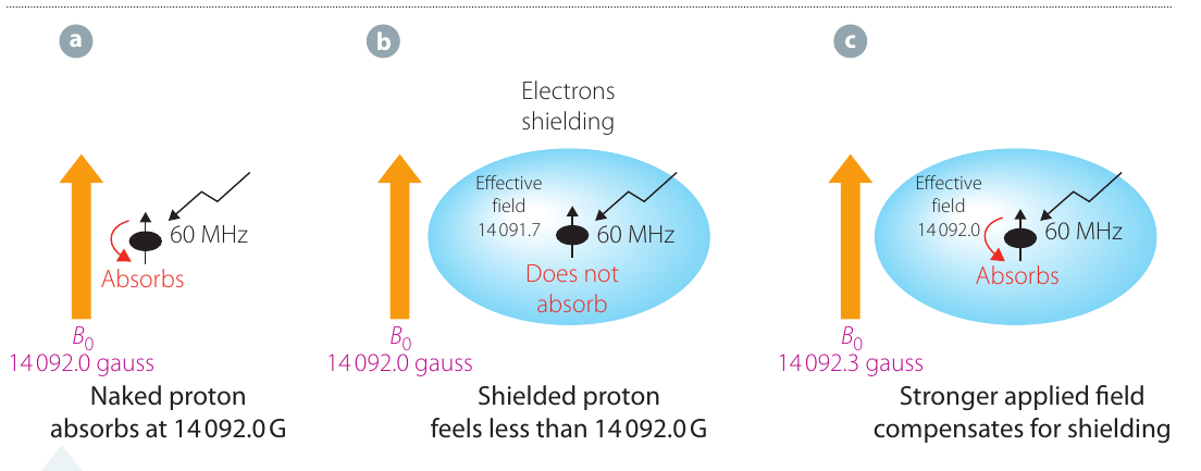
Here's how shielding works:
- In diagram (a), a "naked" proton (with no electron shielding) absorbs radio waves at a specific magnetic field strength and flips to the higher energy state
- In diagram (b), electrons shield the proton. The same amount of energy is not sufficient to flip this shielded proton
- In diagram (c), a stronger magnetic field compensates for the shielding, providing enough effective field strength to cause the proton to flip
Shielding and Chemical Shift: The shielding effect is why different protons appear at different positions on the NMR spectrum. More shielded protons require stronger magnetic fields to flip, while less shielded (deshielded) protons require weaker fields. This difference in required field strength is what creates the chemical shift values we observe.
Different chemical environments
Each atom involved in a chemical bond shares electrons. Polarity is crucial in determining the chemical environment because it affects how electrons are shared. Nuclei are in the same environment when they're bonded to the same group of atoms in the same way.
If all nuclei were in the same chemical environment, NMR wouldn't provide much useful information. Fortunately, when the bonding of nuclei differs, the chemical environment differs too. This means each different nucleus absorbs (and then emits) a different frequency, producing a different peak on the spectrum.
Examples of chemical environments
Let's look at two molecules to understand chemical environments better:
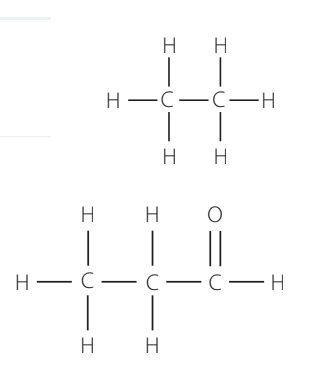
Example 1: Ethane (CH₃CH₃)
- Each carbon atom is attached to another carbon and three hydrogen atoms
- Both carbon atoms have the same chemical environment
- All six hydrogen atoms have the same chemical environment
- Result: The H spectrum would show only one peak, as would the C spectrum
Example 2: Propanal (CH₃CH₂CHO)
- There are three different carbon environments (the CH₃ carbon, the CH₂ carbon, and the CHO carbon)
- There are three different hydrogen environments (the CH₃ hydrogens, the CH₂ hydrogens, and the CHO hydrogen)
- Result: Both the H spectrum and the C spectrum will each show three peaks
Chemical shift
NMR frequencies depend on the strength of the magnetic field in the spectrometer being used. This creates a problem when comparing results from different instruments. To solve this, chemists use a standardised scale called the chemical shift (represented by the symbol δ, pronounced "delta").
The chemical shift is calculated by measuring the difference between the sample's absorption frequency and a reference compound's absorption frequency. This difference is divided by the reference absorption frequency to produce a value measured in parts per million (ppm). Because this is a ratio, it's independent of the magnetic field strength, allowing spectra from any spectrometer to be compared directly.
Tetramethylsilane (TMS) as the reference standard
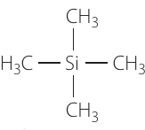
The reference compound used in NMR spectroscopy is tetramethylsilane, or TMS (Si(CH₃)₄). TMS is chosen because:
- Silicon is less electronegative than carbon, so bonding electrons spend more time around the hydrogen nuclei
- This makes TMS the most shielded compound - all other compounds will be less shielded
- In TMS, the 4 carbon atoms are in the same chemical environment and the 12 hydrogen atoms are in the same chemical environment, producing a strong, single signal
- TMS is assigned a chemical shift of zero
Why TMS is the Universal Standard: TMS produces the most shielded signal of any compound, meaning all other chemical shifts will appear to the left of TMS (at higher ppm values) on the spectrum. This ensures consistency across all NMR measurements. The chemical shift value remains the same regardless of the spectrometer's field strength - a nucleus will have the same chemical shift whether measured in a 60 MHz, 100 MHz, or 300 MHz NMR spectrometer.
Solvents in NMR spectroscopy
Molecular structures are determined in solution. Because H nuclei are very common, the signal from the compound being studied must be isolated from any signal from the solvent. Chemists solve this problem by using deuterated solvents - solvents made with deuterium (H or D) instead of regular hydrogen.
Why Deuterated Solvents Work: Deuterium has an even number of nucleons (1 proton + 1 neutron = 2 total), so it doesn't have a magnetic dipole moment and is invisible to the NMR spectrometer. This allows the solvent to dissolve the sample without interfering with the spectrum.
Common deuterated solvents include:
- D₂O (deuterium oxide) - has 10 protons and 10 neutrons (even number)
- CD₂Cl₂ (deuterated dichloromethane) - has 42 protons and 42 neutrons (even number)
Interpreting NMR spectra
NMR spectroscopy helps determine the structure of organic compounds and can distinguish between isomers. When a compound is irradiated with radio waves of different frequencies, certain frequencies are absorbed. When these frequencies are emitted, they produce peaks on the spectrum.
An NMR spectrum shows:
- A calibration peak at zero (from TMS reference)
- A series of peaks along the horizontal axis (labelled "chemical shift" in ppm)
Low-resolution H NMR spectroscopy
Low-resolution H NMR (also called proton NMR) is the most common technique used. The H nucleus is abundant, and hydrogen is present in almost all organic molecules. Low-resolution NMR can determine:
- The chemical environment of hydrogen nuclei (such as —OH or —CH₂—)
- The number of different environments
- The number of protons in each environment
Important Terminology Note: In NMR spectroscopy, the term "H" or "proton" refers to the hydrogen nucleus with its orbiting electrons. The electrons provide the shielding effect. This is different from acid-base chemistry, where "proton" refers to the H⁺ ion (a hydrogen nucleus without any electrons).
Number of peaks
The number of peaks shows how many different chemical environments are present. If protons are in the same environment, they produce a peak with the same chemical shift and appear as one peak.
Example: Ethane
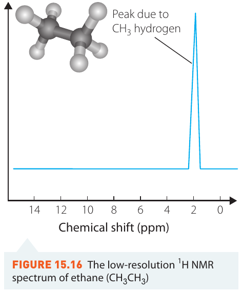
Due to the symmetry of the ethane molecule (CH₃CH₃), all hydrogen atoms are in the same chemical environment. The H spectrum shows only one peak, appearing at approximately 1 ppm.
Location of peaks
The location of each peak (its chemical shift value) shows how shielded or deshielded the proton is. Reference tables of typical proton chemical shifts help identify the types of protons present.
| Type of proton | Chemical shift (ppm) | Type of proton | Chemical shift (ppm) |
|---|---|---|---|
| Alkane CH₃ | 0.9 | R—NH₂ | Variable, about 1.5–4 |
| R—CH₂—R | 1.3 | RCH=CH₂ | 4.6–6.0 |
| R—C=OCH₃ | 2.1 | R—O—CH₃ or ROCH₂R | 3.3 |
| R—CH₂—X (X = F, Cl, Br or I) | 3–4 | R—OH | Variable, about 1–6 |
| CH₃—COOR | 2.0 | Ar—OH | Variable, about 4–7 |
| RCH₂OH | 3.3–4.5 | R—COOH | 9–13 |
| RCH=CH—CH₃ | 1.6–1.9 | R—C(O)H | 9–10 |
Example: Ethanoic acid
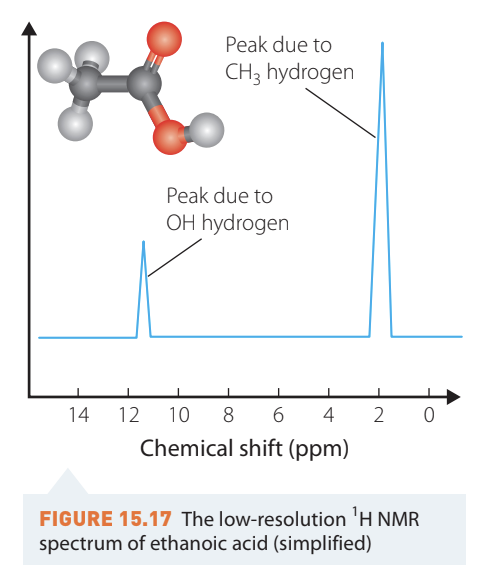
Ethanoic acid (CH₃COOH) has four hydrogen atoms:
- Three are attached to the same carbon, forming a methyl group (—CH₃)
- One is attached to oxygen, forming a hydroxyl group (—OH)
There are two hydrogen environments, so the spectrum shows two peaks:
- A peak at about 2 ppm for the methyl group hydrogens. These protons are slightly deshielded because the adjacent carbon has two oxygen atoms attached, which withdraw electrons
- A peak near 12 ppm for the —COOH group hydrogen. This large chemical shift occurs because two oxygen atoms strongly deshield this proton
Compared to ethane, the methyl protons in ethanoic acid appear further downfield (higher ppm) because the nearby oxygens reduce the electron shielding.
Intensity of the signal
The intensity of each peak shows the number of protons of that type. The area under each peak is proportional to the number of hydrogen atoms in that environment.
When peaks have similar widths at their base, you can use the height of peaks as a ratio to calculate the number of protons in each environment:
- Ethane: one peak (all 6 hydrogens are equivalent)
- Ethanoic acid: two peaks with a ratio of 3:1 (three CH₃ hydrogens to one OH hydrogen)
Example: Ethanol
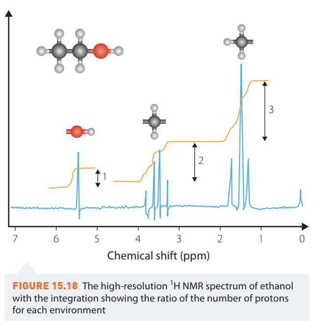
Ethanol (CH₃CH₂OH) shows three peaks with a relative intensity ratio of 3:2:1:
- The tallest peak (intensity = 3) corresponds to the three CH₃ hydrogens
- The middle peak (intensity = 2) corresponds to the two CH₂ hydrogens
- The smallest peak (intensity = 1) corresponds to the one OH hydrogen
The orange curves overlaid on the spectrum show the integration - the area under each peak, which confirms the ratio of hydrogen atoms.
Worked Example: Low-resolution H NMR
Consider the molecule CH₃COH (ethanal). Determine:
- Number of H environments
- Relative intensity of the peaks
- Expected chemical shifts
Solution:
Draw the structure first to visualise the molecule clearly.
1. Number of H environments: There are two H environments:
- The three H atoms in the CH₃ group are all equivalent
- The one H atom in the C(O)H group is different
2. Relative intensity: The ratio is 3:1 (three methyl hydrogens to one aldehyde hydrogen)
3. Chemical shifts:
- CH₃ group: approximately 0.9 ppm (typical for alkane CH₃)
- C(O)H group: 9–10 ppm (typical for aldehyde protons)
High-resolution H NMR spectroscopy
High-resolution H NMR provides one additional piece of information compared to low-resolution NMR. The extra detail shows peaks splitting into clusters of smaller peaks. This splitting pattern reveals the number of protons on adjacent atoms - the neighbouring atoms.
The n + 1 rule
The splitting pattern follows a simple rule: there is one more peak than there are hydrogen atoms attached to the neighbouring atoms.
For example:
- In low-resolution NMR, a methyl group (—CH₃) appears as a single line at about 0.9 ppm with an integration value of 3
- In high-resolution NMR, this same methyl peak may appear split into multiple peaks
The number of peaks can be calculated using the n + 1 rule, where n = the number of protons on adjacent atoms.
Example: Ethanol (CH₃CH₂OH)
- The CH₃ group will split into a triplet (three peaks) because there are 2 hydrogens on the adjacent CH₂ group: 2 + 1 = 3
- The CH₂ group will split into a quartet (four peaks) because there are 3 hydrogens on the adjacent CH₃ group: 3 + 1 = 4
- The OH group remains a singlet (one peak) despite having 2 hydrogens on the neighbouring carbon (more on this exception below)
Understanding the splitting pattern
The splitting occurs due to spin-spin coupling with neighbouring non-equivalent protons. Here's why:
The protons on the next carbon can align either with or against the magnetic field, affecting the shielding of the proton you're observing:
- When neighbouring protons align with the field, they provide extra shielding
- When neighbouring protons align against the field, they cause deshielding, shifting the peak downfield
For one neighbouring proton:
There are two possibilities - the neighbouring proton can align with or against the field. This splits the signal into two peaks (a doublet) with equal intensities (ratio 1:1).
For two neighbouring protons:
There are three possibilities:
- Both protons aligned with the field
- Both protons aligned against the field
- One proton with and one against (this can happen in two ways)
This splits the signal into three peaks (a triplet) with intensity ratio 1:2:1. The middle peak is twice as intense because there are two ways to achieve that configuration.
For three neighbouring protons:
This produces four peaks (a quartet) with intensity ratio 1:3:3:1.
The pattern follows Pascal's triangle, which predicts both the number of peaks and their relative intensities.
Common splitting patterns
| Neighbouring protons | Pattern | n + 1 = | Intensity ratio |
|---|---|---|---|
| 0 | Signal is not split | Singlet | 1 |
| 1 | Signal splits into two | Doublet | 1:1 |
| 2 | Signal splits into three | Triplet | 1:2:1 |
| 3 | Signal splits into four | Quartet | 1:3:3:1 |
| 4 or more | Multiplet |
When splitting doesn't occur
There are two situations when you don't observe signal splitting:
Exceptions to the n + 1 Rule:
-
When neighbouring nuclei have the same chemical shift: If the neighbouring hydrogen atoms are in the same chemical environment as the ones you're observing, no splitting occurs
-
For —OH group protons: The hydrogen on an —OH group shows special behaviour:
- It doesn't cause splitting in neighbouring hydrogens
- It never splits itself
- It always appears as a singlet
- Its chemical shift may vary depending on conditions (concentration, temperature, solvent), but it remains a singlet
This is why in ethanol, the —OH hydrogen appears as a singlet even though there are two hydrogens on the neighbouring carbon.
C NMR spectroscopy
The C isotope is only about 1% abundant compared to the C isotope. Because of this low natural abundance, C NMR spectra take much longer to collect and don't provide the same level of detail as H NMR spectra.
Number of peaks
C NMR spectra indicate the number of different carbon environments by showing one peak for each unique carbon environment. The type of carbon is identified by the peak's chemical shift.
Important differences from H NMR:
- Peak height does NOT relate to the number of carbons in each environment
- Splitting is NOT observed in standard C NMR
This makes C spectra much simpler to interpret than H spectra.
Location of peaks
The peak position (chemical shift) indicates how shielded or deshielded the carbon nucleus is. Reference tables help identify carbon types.
| Type of carbon | Chemical shift (ppm) |
|---|---|
| R—CH₃ | 8–25 |
| R—CH₂—R | 20–45 |
| R₃—CH | 40–60 |
| R₄—C | 36–45 |
| RCH₂—X | 15–80 |
| RCNH₂ | 35–70 |
| RCH₂—O | 50–90 |
| RC=CR | 110–150 |
| RCOOH | 160–185 |
| RC(O)H (aldehydes) | 190–200 |
| RC=O (in ketones) | 205–220 |
Example: Ethanol
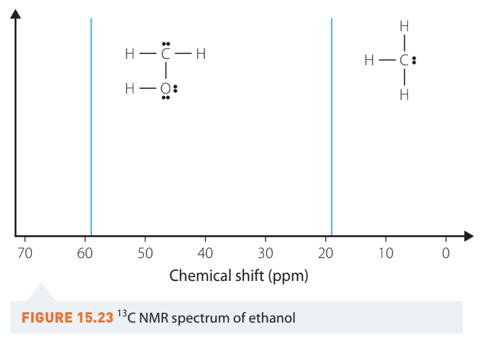
Ethanol (CH₃CH₂OH) has two carbon atoms:
- One carbon attached to three hydrogens (the methyl group)
- One carbon attached to two hydrogens and one oxygen (the CH₂OH group)
The C NMR spectrum shows two peaks:
- A peak just below 20 ppm from the methyl carbon (R—CH₃ range: 8–25 ppm)
- A peak around 59 ppm from the —CH₂OH carbon (RCH₂—O range: 50–90 ppm)
The carbon attached to oxygen appears at higher ppm because oxygen withdraws electrons, deshielding the carbon nucleus. Less shielded nuclei require less energy to flip, so they appear further downfield.
Worked Example: C NMR
Consider the molecule CH₃CHO (ethanal). Determine:
- Number of C environments
- Expected chemical shifts
Solution:
Draw the structure to visualise the molecule.
1. Number of C environments: There are two C environments:
- The carbon in the CH₃ group
- The carbon in the C(O)H group (the aldehyde carbon)
2. Chemical shifts:
- CH₃ group: 8–25 ppm (typical for methyl carbons)
- C(O)H group: 190–200 ppm (typical for aldehyde carbons)
The aldehyde carbon appears much further downfield because the carbonyl oxygen strongly deshields it.
Key terms
Chemical environment: The arrangement of atoms and bonds surrounding a nucleus, which affects how it behaves in NMR spectroscopy
Chemical shift (δ): A standardised measurement (in ppm) that indicates where a peak appears on an NMR spectrum, measured relative to TMS
Deshielding: When electron density around a nucleus is reduced, causing it to experience more of the external magnetic field and absorb at higher ppm
Doublet: A peak split into two lines with equal intensity (ratio 1:1)
High-resolution NMR: NMR spectroscopy that shows splitting patterns, providing information about neighbouring atoms
Integration: The area under an NMR peak, which is proportional to the number of nuclei producing that signal
Low-resolution NMR: NMR spectroscopy that shows the number of different environments, their chemical shifts, and relative numbers of nuclei, but not splitting patterns
Multiplet: A complex splitting pattern with four or more peaks
n + 1 rule: A rule stating that the number of peaks in a splitting pattern equals the number of neighbouring protons plus one
Quartet: A peak split into four lines with intensity ratio 1:3:3:1
Shielding: When electron density around a nucleus protects it from the external magnetic field, causing it to absorb at lower ppm
Singlet: A peak that appears as a single line (not split)
Spin-spin coupling: The interaction between neighbouring non-equivalent nuclei that causes peak splitting in high-resolution NMR
Tetramethylsilane (TMS): The reference compound (Si(CH₃)₄) assigned a chemical shift of 0 ppm in NMR spectroscopy
Triplet: A peak split into three lines with intensity ratio 1:2:1
Exam tips
Essential Exam Strategies:
- Always start by drawing the structural formula of the molecule - this helps you identify different environments and neighbouring atoms
- When counting environments, look for symmetry - equivalent atoms produce the same signal
- Remember that the n + 1 rule only applies to high-resolution H NMR, not to C NMR
- —OH and —NH protons don't follow the splitting rules - they always appear as singlets and don't split neighbouring protons
- When comparing peak intensities, use the ratio of areas (integration values), not absolute heights
- Chemical shift values from reference tables are ranges, not exact values - the solvent and conditions can cause slight variations
- In C NMR, you cannot determine how many carbons are in each environment from peak height - you only know there's at least one
- For structural determination problems, use all available information: molecular formula, chemical shifts, integration ratios, and splitting patterns
Remember!
Key Points to Remember:
-
NMR spectroscopy works because nuclei with odd numbers of nucleons (H and C) act like tiny magnets that flip between energy states when they absorb radio waves in a magnetic field
-
Chemical shift (δ, in ppm) is a standardised scale measured relative to TMS (set at 0 ppm). It's independent of the magnetic field strength, allowing comparison between different spectrometers
-
In H NMR, three key features provide structural information: the number of peaks (how many different H environments), the position of peaks (what type of H), and the area under peaks (how many H atoms in each environment)
-
The n + 1 rule predicts splitting patterns in high-resolution H NMR: the number of peaks equals the number of neighbouring protons plus one. For example, 2 neighbouring protons give a triplet (3 peaks)
-
C NMR is simpler than H NMR because it shows no splitting and peak heights don't indicate the number of carbons - you only see the number of different carbon environments and their chemical shifts