Understanding the Role of the Brain (VCE SSCE Psychology): Revision Notes
Understanding the Role of the Brain
Why is understanding the brain challenging?
Despite living in a technologically advanced world, there is still much we do not know about the brain's inner workings. The brain presents unique challenges for study due to its remarkable adaptability and the interconnected nature of its different regions. The brain can reroute and alter its functioning in response to injury, weakness or change, meaning that modifications in one area can have widespread effects throughout the entire structure. This plasticity, combined with the complex relationships between brain regions, makes it difficult to isolate and study specific functions.
Brain plasticity is the brain's ability to reorganize itself by forming new neural connections throughout life. This remarkable feature allows the brain to compensate for injury and adapt to new situations, but it also means that studying one brain region in isolation doesn't tell us the complete story of how the brain functions as a whole.
Features and functions of the brain
The human brain is an extraordinarily complex organ composed of approximately 100 billion neurons (nerve cells). These neurons connect to the rest of the body through the spinal cord and associated peripheral nerves, forming an intricate communication network. The brain also interacts with the endocrine (hormonal) system, allowing it to influence virtually all bodily processes.
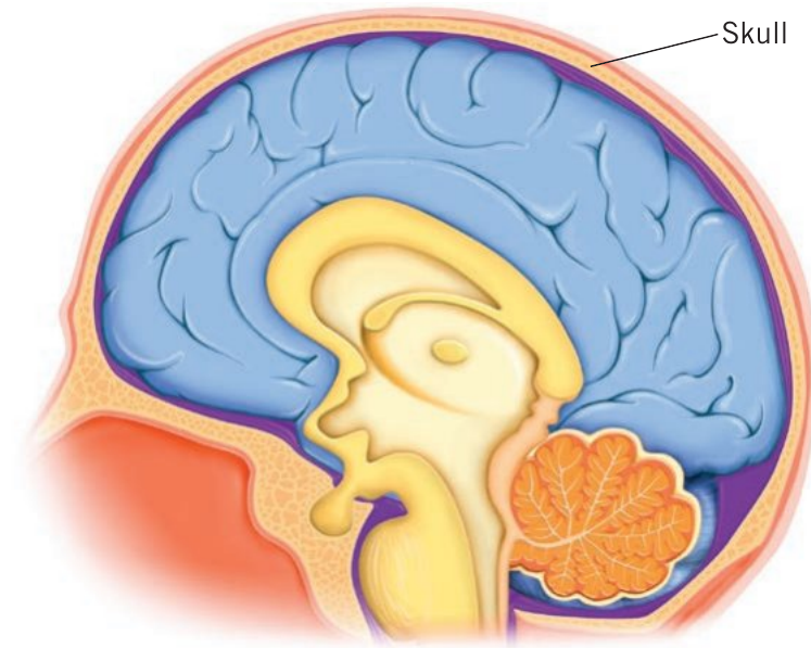
Physical characteristics and protection
The average adult brain occupies roughly 1300 cm³ of space and weighs between 1.4 and 1.5 kg. The brain is protected by multiple layers:
- The skull: A hard, bony case that encloses the brain
- The meninges: Three layers of protective membrane surrounding the soft brain tissue
- Cerebrospinal fluid: Circulates between the meninges, providing cushioning against sudden movements or impacts
- Blood supply: A rich network of arteries delivers oxygen and nutrients to billions of neurons and supporting cells
What makes us human
Beyond controlling bodily processes, the brain contributes to what distinguishes humans from other animals. Our brain enables us to:
- Imagine and create
- Develop complex languages and communication systems
- Problem-solve and reason
- Form hopes and plan for the future
- Experience complex emotions and thoughts
Historical approaches to understanding the brain
Our understanding of the brain's role has evolved considerably over time, with different approaches emerging as technology and scientific thinking advanced.
The brain-heart debate (c475 BCE – c1650 CE)
Brain-heart debate: The question of whether our thoughts, feelings and behaviours originate from our brain or our heart.
This debate may seem straightforward today, but it was hotly contested throughout ancient history. The question centered on which organ was responsible for mental processes and behaviour.
Why This Debate Mattered
Understanding which organ controlled thought and behaviour was crucial for early medicine and philosophy. The resolution of this debate laid the foundation for modern neuroscience and our understanding of how the nervous system controls human behaviour and cognition.
Arguments for the heart
Several prominent Greek philosophers argued that mental processes originated in or around the heart:
- Empedocles (approximately 494–430 BCE) believed mental processes were located in and around the heart, and that perceptions were formed in the blood
- Aristotle (384–322 BCE) supported the heart hypothesis, and his ideas dominated scientific thought in Europe until the seventeenth century
Arguments for the brain
Other ancient thinkers supported the brain hypothesis:
-
Alcmaeon (approximately 475 BCE) was among the first to identify mental processes as being housed in the brain. He concluded that sensory organs, such as the eye, were connected to the brain
-
Hippocrates (460–375 BCE) and Herophilus (335–280 BCE) believed mental processes and emotion arose from the brain, basing their conclusions on dissections of animal and human bodies
-
Galen (129–216 CE), a Greek physician, made crucial observations while treating Roman gladiators. He noted that head and brain injuries affected behaviour, whereas gladiators with heart injuries could still think and reason. Though some of his ideas about brain function were inaccurate, his work significantly advanced the brain hypothesis
Modern resolution
Today, we understand that whilst there is a bidirectional connection between the heart and brain (each influencing the other), overwhelming evidence from thousands of studies confirms that the brain houses mental processing and generates instructions for behaviour.
The mind-body problem (c1640 – present)
Mind-body problem: The extent to which the mind and the body are the same or separate things.
This philosophical question explores the relationship between mental and physical aspects of human existence.
Key distinctions
To understand this problem, it is important to distinguish between:
- The mind: A non-physical entity representing our conscious, thinking self that experiences thoughts
- The body: The physical entity, including the brain, that carries out biological processes
Historical perspectives
Aristotle's view: Before the seventeenth century, most Western philosophers, influenced by Aristotle, considered the mind and body to be unified. Aristotle believed the soul (comparable to what we call the mind) was the living essence of a person's physical body.
Descartes' dualism: In the seventeenth century, French philosopher René Descartes proposed a theory of 'dualism'. He suggested that mind and body were separate entities—one physical, one non-physical—that interacted through the pineal gland (a small structure deep in the brain). Descartes believed this two-way interaction between body and mind contributed to our thoughts, sensations and bodily emotions.
Whilst Descartes was incorrect about the pineal gland's role in mind-body interaction (it actually regulates the body clock through melatonin production), his ideas were revolutionary for their time and advanced scientific understanding of mind-body interactions by encouraging systematic investigation of brain structures.
Modern understanding
Contemporary science has revealed that mind and body influence each other through countless pathways, with no single central structure linking them. Research continues to explore:
- The relationship between conscious experience and brain activity
- Whether the mind can be aware of brain activity
- Whether conscious awareness can exist without brain activity
An Active Research Question
The mind-body problem remains an active and compelling area of scientific investigation. Modern neuroscience is uncovering new insights into how physical brain processes relate to subjective conscious experience, but many fundamental questions remain unanswered.
Phrenology (1796 – c1840s)
Phrenology: The study of the shape of the skull as an indicator of the extent of one's mental faculties and character traits.
Though now discredited, phrenology represented an important step in understanding brain function.
Gall's theory
German doctor Franz Joseph Gall (1758–1828) developed phrenology through comparative studies of animal and human skulls and brains. He proposed that:
- Each part of the cerebral cortex enlarged or reduced depending on an individual's mental faculties, traits and personality
- These differences created subtle changes in skull contours that could be felt externally
- Specific brain areas were responsible for specific functions
- Bumps and indentations on the head reflected personality, character and abilities
The 27 faculties
Working with colleague Johann Spurzheim (1776–1832), Gall examined skulls in prisons and hospitals, developing a system of 27 'faculties', each supposedly located in different head regions. These included:
- Reproductive instincts and love of offspring
- Affection, friendship and compassion
- Self-defence, courage and fighting ability
- Pride, vanity and ambition
- Memory (verbal memory, recollection of people)
- Sense of locality and place
- Language ability and sense of colours
- Musical, mathematical and mechanical talents
- Wit, poetic talent and metaphysics
- Religiosity and perseverance
(The full list included all 27 faculties, ranging from basic instincts to complex cognitive abilities.)
Decline and legacy
Spurzheim promoted phrenology in the United States and United Kingdom, where it became popular. However, by the 1850s, the method had become discredited as it was increasingly exploited for making unfounded behavioural and personality assessments.
Phrenology's Lasting Contribution
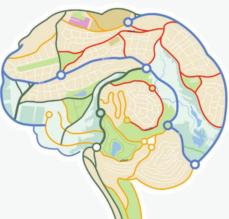
Despite phrenology's rejection as a science, Gall's core idea—that particular brain areas play specific roles in body functioning—was accurate. This concept is now called localisation of function, though we now understand that many brain areas typically work together when we think, feel and act.
The first brain experiments (c1800 – present)
Many early brain experiments would be considered unethical by modern standards, but they provided valuable insights into brain function.
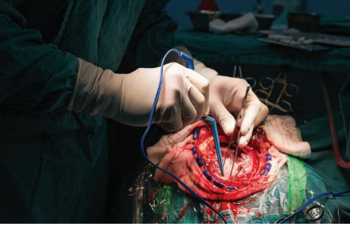
Ethical Considerations
Research using human subjects for invasive brain procedures is now virtually non-existent due to high risk and ethical concerns. Modern neuroscience relies on less invasive techniques, and when ablation, lesioning or electrical stimulation are used, they are only employed in brain surgery to treat serious disorders such as epilepsy, under strict controls and ethical oversight.
Ablation and lesioning
Ablation: The surgical removal or destruction of tissue (e.g. brain tissue) by lesioning or using electrodes.
Lesioning: The creation of small areas of damage (lesions) in the brain.
Some of the earliest brain experiments involved removing brain tissue to observe behavioural or functional changes.
Pierre Flourens (1794–1867), a French physiologist, performed brain ablation by lesioning in animals such as rabbits and pigeons. His work:
- Challenged phrenology's assumptions
- Established the concept of 'holistic' brain function
- Revealed that removing small cortex sections initially caused movement loss, but this could recover over time
- Suggested that remaining cortex areas could assume lost functions
Karl Lashley, an American psychologist, built upon Flourens' work using ablation on chimpanzees, monkeys and rats. He attempted to locate brain areas responsible for learning and memory by:
- Teaching animals specific tasks
- Using brain lesioning to test for memory loss
- Concluding that learning and memory are distributed throughout the brain rather than concentrated in one location
Lashley's experiments led to two important brain function principles:
Equipotentiality: The ability of healthy cortex areas to take over the functions of injured parts.
Mass action: The involvement of large brain areas functioning as a whole to carry out complex functions. If part of the brain is destroyed, functional impairments depend on the amount of tissue lost.
Brain Plasticity in Action
The concepts of equipotentiality and mass action demonstrated that the brain is not simply a collection of isolated functional units. Instead, these principles revealed the brain's remarkable plasticity and its ability to reorganize and compensate for damage—a finding that has profound implications for rehabilitation after brain injury.
Electrical stimulation
Electrodes: Small wires used to electrically stimulate biological tissues or measure electrical activity in these tissues.
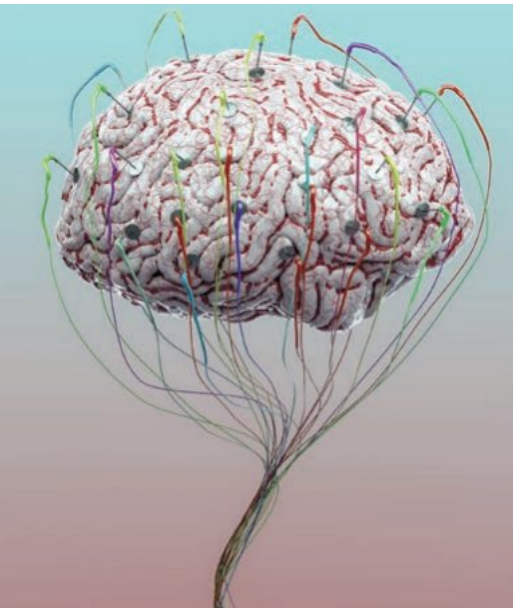
Brain neurons generate weak electrical signals that can be detected by electrodes. If placing an electrode on or in a specific brain area and stimulating it produces a bodily response (e.g. muscle activation), researchers can infer that brain area's responsibility for that action.
Gustav Fritsch (1838–1927) and Eduard Hitzig (1838–1907), German doctors, were among the first to use electrical brain stimulation. Using electrodes on a dog's brain, they discovered that stimulating motor cortex areas produced movements on the opposite side of the dog's body.
Wilder Penfield (1891–1976), a Canadian neurosurgeon, used electrical stimulation on human patients to treat epilepsy. He needed to locate abnormally and normally functioning brain tissue before removing sections contributing to seizures. Penfield:
- Electrically stimulated different brain areas using electrodes
- Recorded responses from patients (most were conscious during the procedure)
- Pooled data collected over more than 20 years
- Created 'brain maps' linking brain areas and functions that remain in use today
Penfield's Brain Mapping Technique
When Penfield electrically stimulated a specific area of a patient's motor cortex, the patient's hand might suddenly move. By systematically stimulating different areas and recording the responses, Penfield could create detailed maps showing which brain regions controlled which body parts. This technique revealed the famous "motor homunculus"—a distorted representation of the human body based on the proportion of motor cortex dedicated to controlling each body part.
Neuroimaging techniques (1890s – present)
Neuroimaging techniques capture images of the brain and are considerably less invasive than earlier methods that required exposing the brain. In research, participants typically think, feel or behave in specific ways whilst concurrent brain images are obtained.
Neuroimaging techniques can be classified as:
- Structural: Obtain images of brain anatomy (e.g. MRI, CT)
- Functional: View the brain 'live' during responses, providing information about both function and structure (e.g. PET, fMRI)
Computerised tomography (CT)
Computerised tomography (CT): An imaging technique that combines a series of x-ray images taken from different angles to create cross-sectional images of the body.
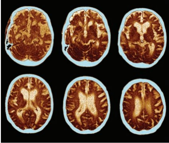
CT scanning involves:
- Taking multiple x-ray images from different angles
- Sometimes requiring patients to take or receive injections of 'contrast' (a dye making certain structures more visible)
- Creating cross-sectional body images
- Helping interpret and diagnose brain conditions
CT is used to:
- Locate brain tumours
- Observe brain changes from conditions like Alzheimer's or Parkinson's disease
- Determine brain injury extent following trauma or stroke
CT scans are particularly useful in emergency situations because they are quick to perform and can rapidly identify life-threatening conditions such as brain haemorrhages, skull fractures, or stroke. However, they expose patients to radiation, so they are used judiciously.
Magnetic resonance imaging (MRI)
Magnetic resonance imaging (MRI): An imaging technique that uses magnetic fields to activate atoms in the brain, which then allows a computer to generate an image of the brain.
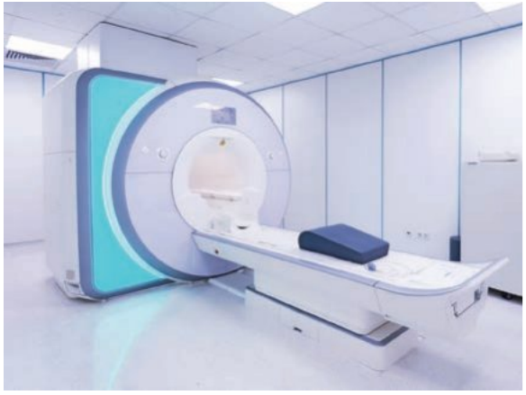
MRI produces more detailed and clearer images than CT, making it useful for:
- Diagnosing structural brain abnormalities
- Identifying cancerous tissue
- Detecting stroke signs
- Revealing subtle abnormalities in conditions like multiple sclerosis and other neurological disorders
MRI Advantages Over CT
MRI does not expose patients to ionizing radiation like CT scans do, making it safer for repeated use. The magnetic fields used in MRI provide superior soft tissue contrast, allowing for more detailed visualization of brain structures. However, MRI scans take longer to perform and cannot be used for patients with certain metal implants.
Positron emission tomography (PET)
Positron emission tomography (PET): An imaging technique that provides information not only about brain structure but also activity and function by recording the use of glucose by cells in the brain.
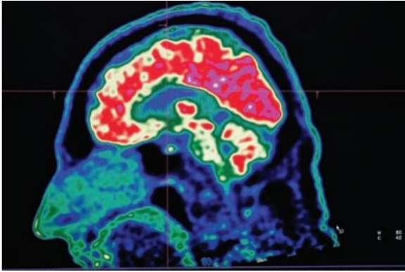
PET scanning procedure:
- Participants are injected with a glucose solution containing a radioactive tracer
- Participants engage in specific activities whilst images are taken
- Active neurons show increased blood flow and glucose consumption
- Colour codes indicate areas of high (warm colours) and low (cool colours) brain activity
- Researchers can determine which brain areas align with certain tasks
PET provides full-colour images of both brain structure and the 'live brain' at work.
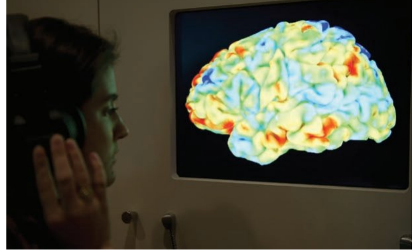
Functional magnetic resonance imaging (fMRI)
Functional magnetic resonance imaging (fMRI): A type of MRI that shows brain activity by measuring oxygen consumption in the brain, with the assumption that active areas consume more oxygen.
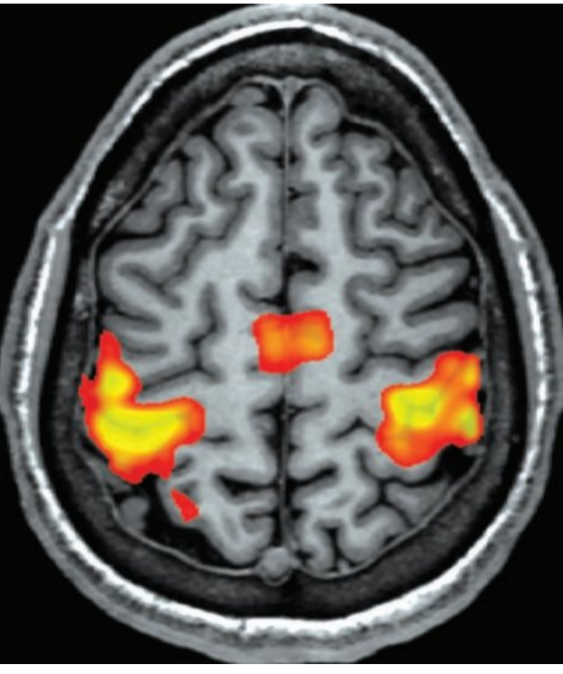
fMRI characteristics:
- Available since the 1990s
- Measures oxygen consumption in the brain
- Assumes blood is more oxygenated in active brain areas
- Does not expose participants to radioactive tracers (advantage over PET)
- Produces coloured images showing higher (red) and lower (blue) activity areas
- Creates more detailed and accurate pictures than PET
- Can produce more images in rapid succession
- Currently the preferred imaging technique in psychological research
Why fMRI is the Preferred Technique
fMRI has become the gold standard in brain imaging research because it combines several crucial advantages: it provides excellent spatial and temporal resolution, does not require radioactive substances, is non-invasive, and can capture dynamic brain activity as participants perform tasks. This makes it ideal for understanding how different brain regions work together during cognitive processes, emotional responses, and behavioural tasks.
Key Points to Remember:
-
The brain is protected by three layers: the skull, meninges, and cerebrospinal fluid, which together safeguard approximately 100 billion neurons.
-
Historical understanding of the brain evolved from the brain-heart debate (resolved in favour of the brain) through philosophical questions about mind-body relationships to scientific experimentation and modern neuroimaging.
-
Early experimental methods included ablation (tissue removal), lesioning (creating small areas of damage), and electrical stimulation, leading to important concepts like equipotentiality (healthy areas taking over injured functions) and mass action (large brain areas working together).
-
Modern neuroimaging techniques are far less invasive than early methods. Structural techniques (CT and MRI) show brain anatomy, whilst functional techniques (PET and fMRI) reveal both structure and activity during tasks.
-
fMRI is currently the preferred research technique because it provides detailed, accurate images of brain activity without exposing participants to radioactive substances, measuring oxygen consumption to indicate which brain areas are most active during specific tasks.