Microscopy (AQA GCSE Biology Combined Science): Revision Notes
📚 Revision Notes
1.1.5 Microscopy
infoNote
Extremely small structures, like cells, cannot be seen without microscopes, which enlarge the image.
Light Microscopes
- First Observations: Robert Hooke observed the first cells of a cork in 1665 using a light microscope.
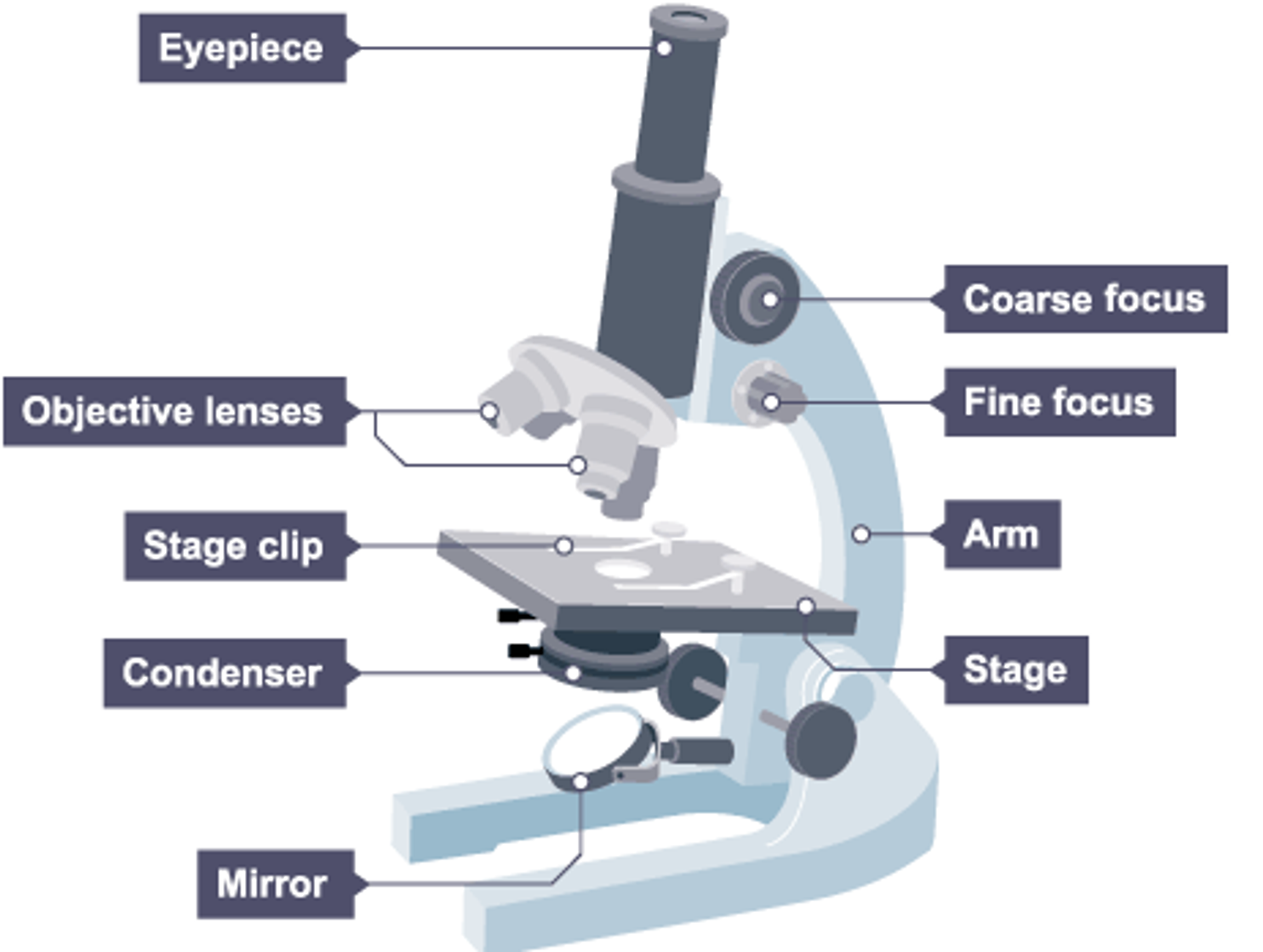
- Structure:
- Two lenses: objective lens and eyepiece lens.
- Objective Lens: Produces a magnified image.
- Eyepiece Lens: Magnifies the image further for viewing.
- Illuminated from underneath.
- Capabilities:
- Maximum magnification: ×2000
- Resolving power: 200nm (ability to distinguish between two points)
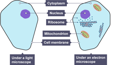
- Used to view tissues, cells, and large sub-cellular structures.
Electron Microscopes
- Development: In the 1930s, electron microscopes were developed to view detailed sub-cellular structures like mitochondria, ribosomes, chloroplasts, and plasmids.
- How They Work:
- Use electrons instead of light to form an image, allowing for a smaller wavelength.
- Types:
- Scanning Electron Microscope (SEM): Creates 3D images.
- Transmission Electron Microscope (TEM): Creates 2D images detailing organelles.
- Capabilities:
- Magnification: up to ×2,000,000
- Resolving power: 10nm (SEM) and 0.2nm (TEM)
Common Calculations
- Magnification of a Light Microscope:
- Magnification of eyepiece lens × Magnification of objective lens.
- Size of an Object:
- Size of image / Magnification = Size of object (Ensure units are consistent).
Standard Form
- Usage: Helpful for working with very large or small numbers in microscopy.
- Format: A number multiplied by a power of 10, where the number is between 1 and 10.
lightbulbExample
EXAMPLES: