The Cell (Junior Cert Science): Revision Notes
The Cell
What is a cell?
A cell is the smallest working unit of a living thing. Every organism, from tiny bacteria to humans, is made up of cells. Some organisms are made of just one cell (unicellular), like bacteria, while others are made of thousands or millions of cells (multicellular), like plants and animals.
The word "cell" was first used by scientist Robert Hooke in the 1600s. While looking at a piece of cork under his microscope, he noticed it was made up of tiny box-like compartments. These reminded him of the small rooms that monks lived in, which were called cells. This is how cells got their name.
Another important scientist, Antonie van Leeuwenhoek, made many improvements to microscopes. He was the first person to observe and draw bacteria. Robert Hooke used van Leeuwenhoek's microscope to study cells in more detail.
The microscope
A microscope is a scientific instrument used to observe cells and other tiny objects that are too small to see with the naked eye. The microscopes used in school laboratories are called light microscopes because they use light to help you see the specimen.
Parts of a microscope
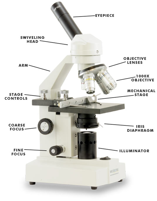
A microscope has several important parts:
- Eyepiece: This is the lens you look through at the top of the microscope
- Objective lens: These are the lenses close to the specimen that magnify the image. Most microscopes have several objective lenses of different strengths
- Stage: This is the flat platform where you place your microscope slide
- Diaphragm: Controls the amount of light passing through the specimen
- Light source: Provides illumination for viewing the specimen
- Coarse focus: Used to bring the object into general focus
- Fine focus: Used to bring the object into sharp, clear focus
- Arm: Connects the tube to the base and is used to carry the microscope
Using a microscope
To use a light microscope properly, follow these steps:
- Click the objective lens into place, starting with the lowest magnification
- Look through the eyepiece
- Magnify the object by using different objective lenses
- Revolving nosepiece allows you to switch between objective lenses
- Adjust the light coming through using the diaphragm
- Bring the object into focus using the coarse focus first, then the fine focus
- Place the slide on the stage
- Plug the microscope in and switch on the light
Always start with the lowest magnification objective lens and work your way up. This makes it easier to locate and focus on your specimen. Use the coarse focus first, then fine-tune with the fine focus knob.
Calculating magnification
The magnification of a microscope tells you how many times bigger the object appears compared to its actual size. Light microscopes in school laboratories usually magnify objects by a maximum of times.
To calculate the total magnification, you multiply the eyepiece lens magnification by the objective lens magnification:
Worked Example: Calculating Total Magnification
If the eyepiece magnification is and the objective lens magnification is :
This means the object appears times larger than its actual size.
Animal cell structure
Animal cells are complex structures containing several different parts. The individual parts of a cell are called cell organelles. Each organelle has a specific job to help the cell function properly.
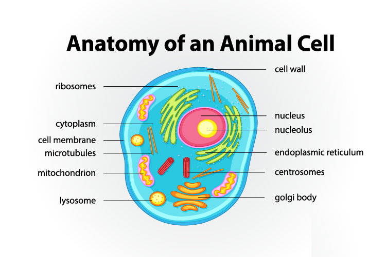
Key organelles in animal cells
Cell membrane: This is the outer boundary of the cell. It controls what substances can move in and out of the cell, acting like a selective barrier.
Cytoplasm: This is the jelly-like substance that fills the cell. It is where many chemical reactions take place.
Nucleus: This is the control centre of the cell. It contains DNA, which carries the genetic information that controls all the cell's activities.
Mitochondria (singular: mitochondrion): These are the powerhouses of the cell. Energy is released from food during a process called respiration, which happens in the mitochondria.
Ribosomes: These tiny structures are where proteins are made. Proteins are essential molecules needed for growth and repair.
Memory Aid: Think of mitochondria as "mighty mitochondria" that make energy for the cell! The nucleus is like the "brain" of the cell, controlling everything that happens.
Preparing animal cells for observation
To observe animal cells under a microscope, you can prepare a slide of cheek cells. Here's a summary of the process:
- Gently scrape the inside of your cheek with a clean lollipop stick
- Spread the sample onto a clean glass slide
- Add a drop of methylene blue stain to make the cells easier to see
- Carefully place a cover slip over the sample
- Use a tissue or bottle to gently remove excess stain
- The slide is now ready to view under the microscope
The methylene blue stain helps the cheek cells show up more clearly because it colours certain parts of the cell.
Plant cell structure
Plant cells have a similar basic structure to animal cells, but they also contain some additional organelles that animal cells don't have. These extra structures help plants carry out photosynthesis and maintain their shape.
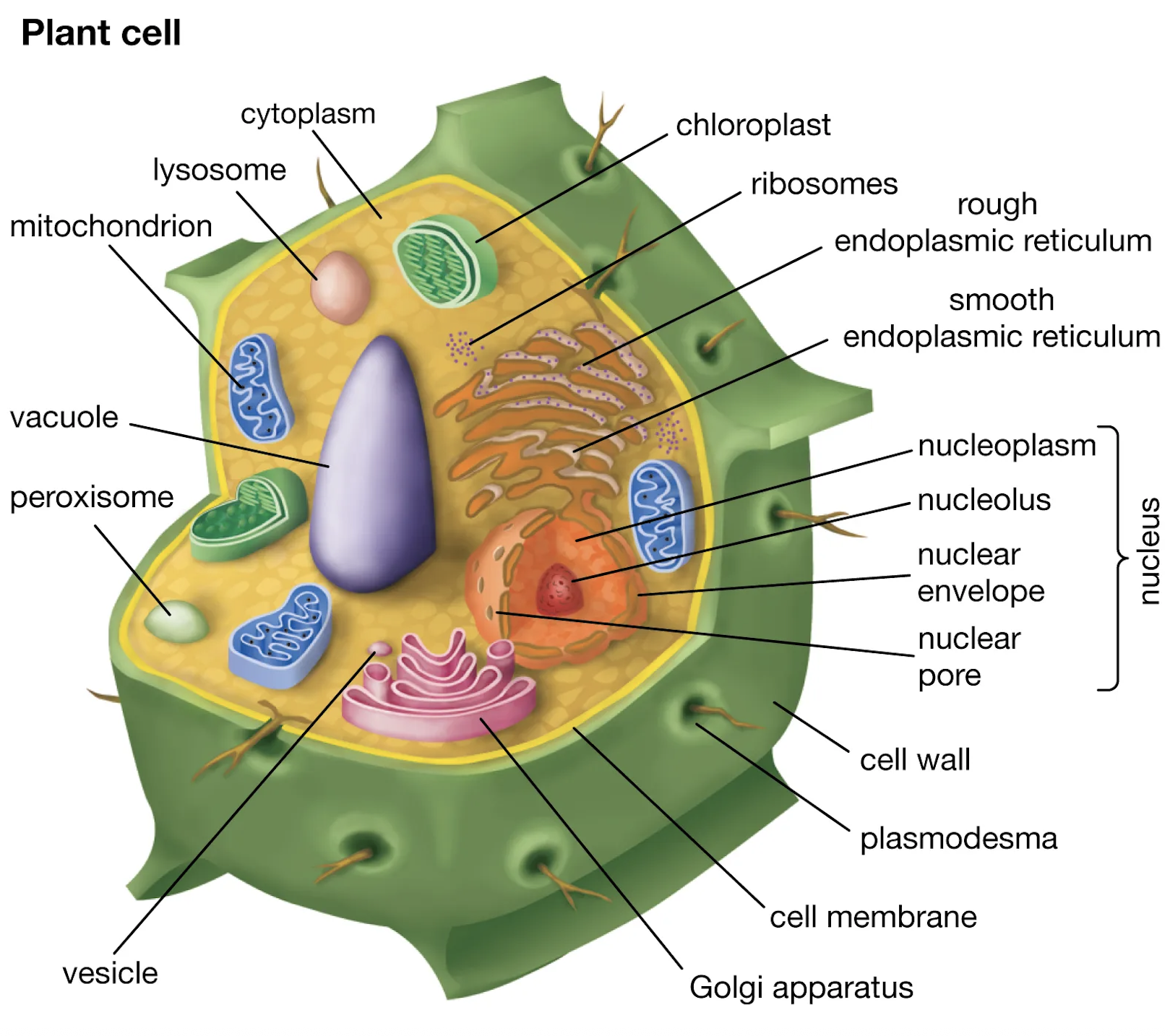
Key organelles in plant cells
Plant cells contain all the organelles found in animal cells (cell membrane, cytoplasm, nucleus, mitochondria, and ribosomes), plus three additional structures:
Cell wall: This rigid outer layer provides extra protection and support for the cell. It helps the plant maintain its shape. The cell wall is outside the cell membrane.
Vacuole: Plant cells have a large central vacuole filled with a watery fluid called cell sap. This helps keep the cell firm and stores various substances.
Chloroplasts: These green structures are where photosynthesis takes place. They contain a green pigment called chlorophyll, which captures light energy to make food for the plant.
Photosynthesis
Photosynthesis is the process by which green plants make their own food. This happens in the chloroplasts. Plants use light energy, carbon dioxide, and water to produce glucose (a type of sugar) and oxygen. This is why plants are often called producers – they can produce their own food rather than having to consume other organisms.
Photosynthesis is the key process that makes plants different from animals. Only cells with chloroplasts can carry out photosynthesis. This is why plants are green and can make their own food!
Preparing plant cells for observation
To observe plant cells under a microscope, you can prepare a slide of onion cells. Here's a summary of the process:
- Cut an onion lengthways down the centre
- Scoop out the centre portion using tweezers to separate the layers
- Peel off a very thin, transparent skin from the onion layer
- Place the onion skin on a clean, dry glass slide
- Add a drop of iodine stain to make the cells more visible
- Place a cover slip onto the slide at a angle to avoid air bubbles
- The slide is now ready to view under the microscope
Comparing plant and animal cells
While plant and animal cells share many similarities, there are three key differences:
| Organelle/Cell structure | Plant cell | Animal cell |
|---|---|---|
| Cell wall | Present | Absent |
| Vacuole | Present (large) | Absent (or very small) |
| Chloroplast | Present | Absent |
Cell wall: Only plant cells have a rigid cell wall. This gives plant cells their regular, box-like shape and provides structural support. Animal cells only have a cell membrane, so they tend to have more irregular shapes.
Vacuole: Plant cells have a large central vacuole that takes up most of the cell's volume. Animal cells may have small temporary vacuoles, but nothing like the large permanent vacuole in plant cells.
Chloroplast: Only plant cells have chloroplasts, which contain the green pigment chlorophyll. This is what makes plants green and allows them to carry out photosynthesis. Animal cells cannot make their own food, so they don't need chloroplasts.
Exam tip: A common exam question asks you to identify differences between plant and animal cells. Remember the three key structures that plant cells have but animal cells don't: cell wall, large vacuole, and chloroplasts. You can use the mnemonic "VCW" to remember them!
Key Points to Remember:
- A cell is the smallest working unit of a living organism, first observed by Robert Hooke
- Microscopes are essential tools for observing cells. Total magnification = eyepiece magnification × objective lens magnification
- Animal cells contain organelles including the nucleus (control centre), mitochondria (energy production), ribosomes (protein synthesis), cell membrane, and cytoplasm
- Plant cells have all the organelles of animal cells, plus three additional structures: cell wall (support), large vacuole (storage and firmness), and chloroplasts (photosynthesis)
- Photosynthesis is the process by which green plants make their own food in chloroplasts using light energy
- Remember "VCW" for the three structures unique to plant cells: Vacuole, Chloroplasts, and cell Wall