The Breathing System (LC 2027) (Leaving Cert Biology): Revision Notes
The Breathing System
The human breathing system is a complex network of organs and tissues that work together to supply your body with oxygen and remove carbon dioxide. This system is essential for cellular respiration and maintaining life.
Understanding the breathing system requires knowledge of both anatomy and physiology.
Anatomy refers to the study of the structure of an organism or its parts, while physiology focuses on the functions and processes of an organism or its parts.
Anatomy of the breathing system
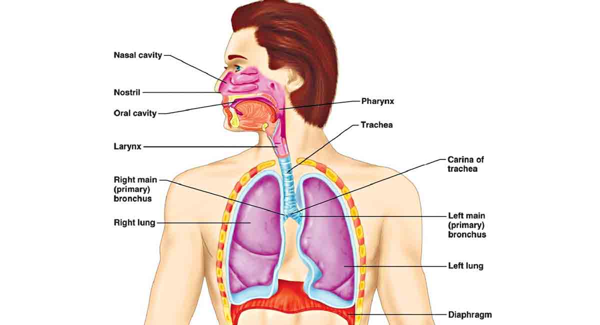
The breathing system consists of two main parts: the lungs and a series of tubes that transport air to and from the lungs. The entire system is located within the thoracic cavity (chest area), which is separated from the abdominal cavity by a sheet of muscle called the diaphragm.
Upper respiratory tract
The nose serves as the primary entrance for air into your respiratory system. Air enters through two openings called nostrils, which are separated by cartilage called the septum. Each nostril leads into nasal chambers or passages.
Breathing through your nose provides several important benefits:
Benefits of nasal breathing:
- Filtration: Hairs and mucus in the nostrils trap dust, pollen, bacteria and other particles
- Moistening: Dry air becomes humidified as it passes through the nasal passages
- Warming: Cold air is heated to body temperature, making it easier for the lungs to process
The pharynx and larynx are located in your throat area. The pharynx (throat) connects your nasal passages to your windpipe. A flap of tissue called the epiglottis covers the trachea when you swallow, preventing food and drink from entering your airways.
Just below the epiglottis sits the larynx, commonly known as your voice box. The larynx contains two vocal cords that vibrate to produce sound when air passes through them. Your tongue and lips then shape these sounds into speech.
Lower respiratory tract
The trachea (windpipe) is a tube made of cartilage and elastic fibres that extends from your throat down into your chest. The cartilage rings prevent the trachea from collapsing when air is drawn through it during breathing.
Bronchi and bronchioles form the branching airways within your lungs. The trachea divides into two main bronchi, with each leading to one lung. Inside the lungs, each bronchus subdivides repeatedly to form many smaller bronchioles, creating a tree-like structure.
The walls of the smaller bronchioles contain muscle and elastic fibres but lack cartilage. During an asthma attack, these muscles can contract and narrow the airways, making breathing difficult.
All tubes in the respiratory system are lined with mucus and cilia (tiny hairs). The mucus traps particles like dust, pollen, bacteria and viruses, while the cilia beat in coordinated waves to move this trapped material upward and out of the respiratory system.
Lungs and alveoli
The lungs are large, pink, spongy organs where gas exchange occurs. Each lung is surrounded by two pleural membranes that create a pleural cavity between them. This cavity contains a small amount of fluid that lubricates the membranes and reduces friction during breathing.
Alveoli are tiny, balloon-like air sacs found at the ends of the smallest bronchioles. Each bronchus eventually subdivides into approximately one million bronchioles, and each bronchiole ends in clusters of alveoli.
Alveolar adaptations for gas exchange:
The alveoli are perfectly designed for gas exchange because they have:
- Enormous surface area: Over 700 million alveoli provide a huge area for gas exchange
- Thin walls: Only one cell thick, allowing gases to pass through easily
- Moist surfaces: Essential for efficient gas exchange
- Rich blood supply: Surrounded by a dense network of tiny blood vessels called capillaries
Gas exchange process
Gas exchange is the process by which oxygen enters your bloodstream and carbon dioxide is removed. This occurs in two main locations: in the lungs between alveoli and blood, and in body tissues between blood and cells.
Gas exchange in the alveoli
When you breathe in, air rich in oxygen reaches the alveoli. The oxygen dissolves in the moist alveolar walls and then diffuses across the thin membrane into the surrounding capillaries. At the same time, carbon dioxide and water move in the opposite direction - from the blood into the alveolar air space.
Worked Example: Gas Exchange Process
Step 1: Oxygen movement
- High oxygen concentration in alveolar air (21%)
- Low oxygen concentration in blood arriving at lungs
- Oxygen diffuses from alveoli → blood
Step 2: Carbon dioxide movement
- High carbon dioxide concentration in blood from tissues
- Low carbon dioxide concentration in alveolar air (0.04%)
- Carbon dioxide diffuses from blood → alveoli
Step 3: Result
- Blood becomes oxygen-rich (oxygenated)
- Alveolar air becomes carbon dioxide-rich for exhalation
This process works because gases naturally move from areas of high concentration to areas of low concentration through diffusion. The blood arriving at the alveoli has a low oxygen concentration and high carbon dioxide concentration, while the alveolar air has high oxygen and low carbon dioxide.
Gas exchange in body tissues
A similar exchange occurs at the tissue level throughout your body. Oxygen-rich blood from the lungs is pumped by your heart to all body cells. The oxygen diffuses from the blood into the cells where it's needed for cellular respiration. Meanwhile, carbon dioxide and water (waste products from cellular respiration) diffuse from the cells into the bloodstream to be transported back to the lungs.
Transport of gases
Oxygen transport: Most oxygen in your blood is carried by combining with haemoglobin in red blood cells to form oxyhaemoglobin. Only about 3% of oxygen dissolves directly in blood plasma.
Carbon dioxide transport: Carbon dioxide is transported in blood plasma as dissolved gas and as carbonic acid formed when carbon dioxide reacts with water.
Mechanism of breathing
Breathing involves two main processes: inhalation (breathing in) and exhalation (breathing out). These processes are controlled by the coordinated movement of your diaphragm and intercostal muscles.
Inhalation (active process)
Inhalation requires energy and active muscle contraction - it is an active process.
Inhalation requires energy and active muscle contraction:
- Brain control: Your brain sends signals to the intercostal muscles and diaphragm
- Muscle contraction: The intercostal muscles contract, pulling your ribs up and outward
- Diaphragm movement: The diaphragm contracts and curves downward
- Chest expansion: The volume of your chest cavity increases
- Pressure decrease: As the chest expands, air pressure inside decreases
- Air inflow: External air pressure is now higher than internal pressure, forcing air into your lungs
Exhalation (passive process)
Exhalation is normally a passive process that doesn't require energy:
- Muscle relaxation: The intercostal muscles and diaphragm relax
- Chest contraction: Your ribs move down and inward, and the diaphragm curves upward
- Volume decrease: The chest cavity volume decreases
- Pressure increase: Air pressure inside your chest increases
- Air outflow: Higher internal pressure forces air out of your lungs
Composition of inhaled and exhaled air
The composition of air changes as it passes through your respiratory system:
Air composition changes during breathing:
Inhaled air contains:
- 21% oxygen
- 0.04% carbon dioxide
- Low water concentration
Exhaled air contains:
- 14% oxygen (decreased)
- 4.4% carbon dioxide (increased)
- High water concentration (increased)
This difference demonstrates that your body uses oxygen for cellular processes and produces carbon dioxide and water as waste products.
Control of breathing
Your breathing rate is automatically controlled by the medulla oblongata, a region in your brainstem. This control system monitors the level of carbon dioxide in your blood rather than oxygen levels.
How breathing control works:
- Detection: The medulla oblongata detects increases in blood carbon dioxide levels
- pH change: Carbon dioxide dissolves in water to form weak carbonic acid, slightly lowering blood pH
- Response: When carbon dioxide levels rise, the medulla oblongata sends signals to increase breathing rate and depth
- Correction: Faster, deeper breathing removes excess carbon dioxide and returns blood chemistry to normal
During exercise, your muscle cells produce more carbon dioxide through increased cellular respiration. The medulla oblongata detects this rise and automatically increases your breathing rate to remove the excess carbon dioxide.
Key misconception to avoid:
Your brain doesn't normally respond to low oxygen levels. Instead, it responds to high carbon dioxide levels, which is why you breathe faster when carbon dioxide builds up rather than when oxygen runs low.
Remember!
Key Points to Remember:
-
The breathing system consists of airways (nose to bronchioles) and gas exchange surfaces (alveoli) that work together to supply oxygen and remove carbon dioxide
-
Alveoli are perfectly adapted for gas exchange with their enormous surface area, thin walls, moist surfaces, and rich blood supply
-
Inhalation is an active process requiring muscle contraction, while exhalation is normally passive, relying on muscle relaxation and elastic recoil
-
Gas exchange occurs by diffusion - oxygen moves from high concentration in alveolar air to low concentration in blood, while carbon dioxide moves in the opposite direction
-
Breathing rate is controlled by the medulla oblongata, which responds to carbon dioxide levels in blood rather than oxygen levels, automatically adjusting breathing to maintain proper blood chemistry