The Musculoskeletal System (LC 2027) (Leaving Cert Biology): Revision Notes
The Musculoskeletal System
The musculoskeletal system is formed when the skeletal and muscular systems work together in most animals. This integrated system is controlled by the nervous system and provides the structural foundation for movement and support in the human body.
Functions of the musculoskeletal system
The musculoskeletal system performs several vital functions that keep our bodies functioning properly:
Support: The bones of the skeleton create a rigid framework that holds the body upright and maintains our shape. Without this bony structure, we would collapse under our own weight.
Protection: Different parts of the skeleton protect vital organs from damage. The skull protects the brain, the vertebrae safeguard the nerves of the spinal cord, and the ribs shield the heart and lungs from injury.
Movement: Bones provide anchor points for muscles to attach to. When muscles contract, they pull against these rigid bones, creating movement. Without solid bones, effective movement would be impossible.
Shape: The skeletal framework determines body proportions and overall appearance. The shape of bones, particularly the long bones in the arms and legs, influences how tall or wide a person appears.
Manufacture of blood components: Bone marrow inside certain bones produces red blood cells, white blood cells, and platelets through a process that continues throughout life.
The musculoskeletal system's five key functions - support, protection, movement, shape, and blood cell production - work together to maintain life and enable daily activities.
Structure of the human skeleton
The adult human skeleton contains 206 bones organised into two main divisions: the axial skeleton and the appendicular skeleton.

Axial skeleton
The axial skeleton forms the central axis of the body and consists of the skull, backbone (vertebral column), and ribs. This central framework provides the main structural support for the body.
Skull: The skull, also called the cranium, consists of over 20 bones fused together. Its primary function is protecting the brain from injury.
Vertebral column: The backbone or spine contains 33 bones called vertebrae that protect and support the nerves in the spinal cord and allow for movement.
Ribs: The rib cage consists of the sternum (breastbone) and 12 pairs of ribs. These bones protect the organs in the chest and help with breathing. All ribs attach to the vertebrae of the spine.
Appendicular skeleton
The appendicular skeleton includes the limbs (arms and legs), the pectoral girdle (shoulder area), and the pelvic girdle (hip area). These structures allow for a wide range of movement and locomotion.
Remember the distinction: Axial = central axis (skull, spine, ribs), Appendicular = appendages/limbs (arms, legs, and their connecting structures).
The vertebral column
The vertebral column, commonly known as the spine, is made up of 33 bones called vertebrae. These bones protect and support the nerves within the spinal cord and allow for flexible movement of the back.
Regions of the spine
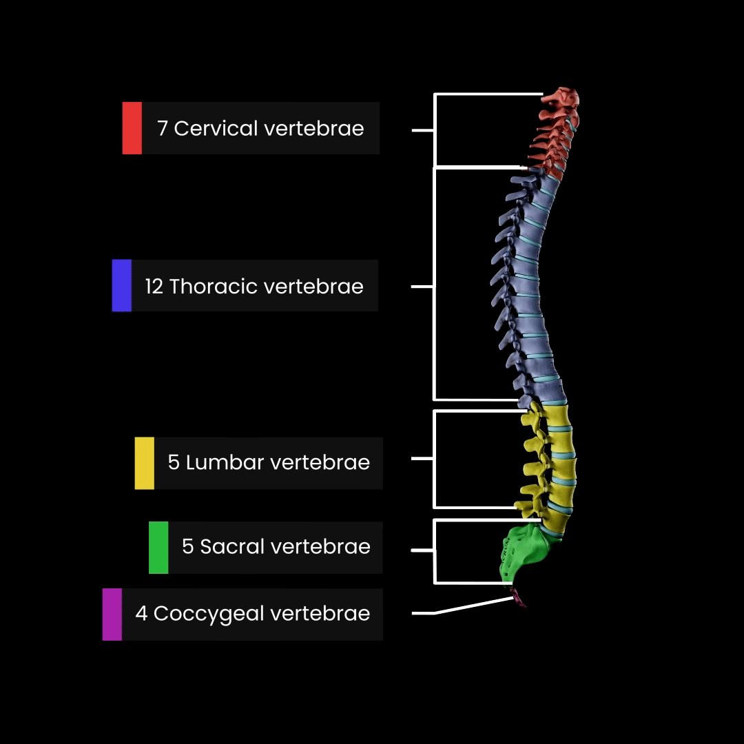
The spine is divided into five distinct regions, each with specific characteristics:
- Cervical region (neck): Contains 7 vertebrae that support the head and allow neck movement
- Thoracic region (chest): Contains 12 vertebrae that connect to the ribs
- Lumbar region (lower back): Contains 5 vertebrae that bear much of the body's weight
- Sacrum (hip area): Contains 5 fused vertebrae that form part of the pelvis
- Coccyx (tailbone): Contains 4 fused vertebrae at the bottom of the spine
The first 24 vertebrae (cervical, thoracic, and lumbar) are held together by ligaments and can move slightly in relation to each other. The last 9 vertebrae are fused together with no movement between them.
Structure of vertebrae
Each vertebra has a similar basic structure designed for both strength and flexibility.
Vertebrae have different shapes depending on their location in the spine, but they share common features:
- Neural spine: Projects backwards for muscle attachment
- Transverse processes: Extend sideways for muscle attachment
- Neural canal: Contains and protects the spinal cord
- Centrum (vertebral body): Provides structural strength
Intervertebral discs
The vertebrae are separated by intervertebral discs made of cartilage. These discs have a hard outer layer and a soft, jelly-like centre. They act as shock absorbers and protect the vertebrae from rubbing against each other during movement.
Did you know? Sometimes the soft centre of a disc bulges out and presses on spinal nerves, causing pain in the back or leg. This condition is called a 'slipped disc'. People are often taller in the mornings when their discs are fully expanded, but become slightly shorter during the day as the discs compress due to gravity.
Bones of the limbs
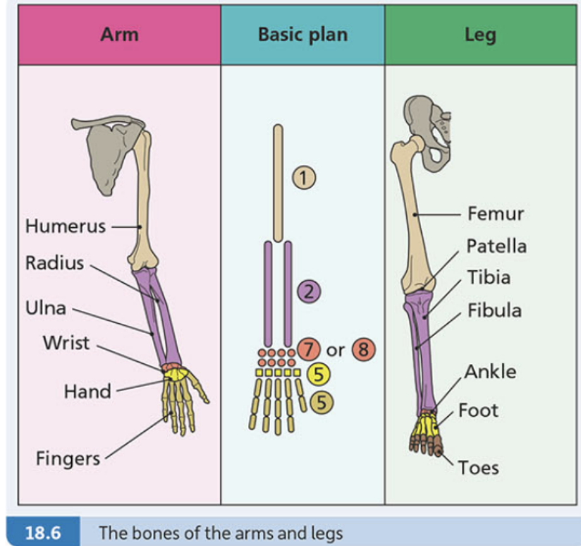
The arms and legs follow a similar basic structural plan, though they are adapted for different functions.
Pectoral girdle
The pectoral girdle consists of the collarbone (clavicle) and the shoulder blade (scapula). Its functions include connecting the arms to the axial skeleton and allowing a wide range of arm movements.
Pelvic girdle
The pelvic girdle is composed of two halves that join to form the sacrum. Each half consists of three fused bones called innominate bones, connected by flexible cartilage.
The pelvic girdle is more robust than the pectoral girdle because it must support the weight of the upper body and connect to the legs. It also protects organs in the lower abdomen.
The hollow cavity where the hip bones attach to the sacrum is called the pelvis.
Major bones of the arms
- Humerus: The upper arm bone that allows for a wide variety of arm movements due to its connections at the shoulder and elbow
- Radius: Located on the thumb-side of the lower arm, it helps the forearm move and rotate
- Ulna: Provides stability and support to the lower arm when it moves
Major bones of the legs
- Femur: The thigh bone is the longest and strongest bone in the body. Its functions include supporting body weight and allowing leg movement. The head of the femur forms a highly moveable joint with the pelvic girdle
- Patella: The kneecap protects the knee joint and allows for smooth movement of the knee
- Tibia: The main bone in the shin that supports the body when standing and moving
- Fibula: A smaller bone in the shin that is not weight-bearing but supports the ancle and provides attachment points for muscles and tendons in the legs and ankles
Interesting fact: An important feature of great apes and humans is that we have opposable thumbs. This means the thumb can be pushed against all the other fingers, giving us much greater power of grip and manipulation compared to other animals.
Remember!
Key Points to Remember:
-
The musculoskeletal system combines bones and muscles to provide support, protection, movement, shape, and blood cell production
-
The skeleton is divided into the axial skeleton (skull, spine, ribs) and appendicular skeleton (limbs and girdles)
-
The spine has 33 vertebrae arranged in 5 regions: cervical, thoracic, lumbar, sacrum, and coccyx
-
Intervertebral discs act as shock absorbers between vertebrae and can compress throughout the day
-
Arms and legs follow a similar basic plan but are adapted for different functions - arms for manipulation and legs for weight-bearing and locomotion