The Basic Units of Life (Grade 10 NSC Matric Life Sciences): Revision Notes
Cell Organelles
Cell organelles are specialised structures found within eukaryotic cells that carry out specific functions essential for life. Think of organelles as the "organs" of a cell - each one has a particular job that helps the cell survive and function properly. Understanding the relationship between structure and function is crucial when studying organelles, as their unique shapes and arrangements directly relate to their roles.
Cytoplasm
The cytoplasm is the gel-like substance that fills the space inside a cell membrane. It consists of approximately 90% water and contains dissolved nutrients, salts, and waste products. This jelly-like medium serves as the cellular environment where most life processes occur.
Functions of cytoplasm
The cytoplasm performs several vital roles in maintaining cell health and function:
- Mechanical support: The cytoplasm creates turgor pressure against the cell membrane, helping to maintain the cell's shape and structure
- Metabolic activity centre: Most cellular processes, including metabolism, cell division, and protein synthesis, take place within the cytoplasm
- Ribosome housing: The cytoplasm contains ribosomes that manufacture proteins for the cell
- Storage facility: Small molecules like carbohydrates, lipids, and proteins are stored temporarily in the cytoplasm
- Transport medium: The cytoplasm allows organelles to move around the cell and facilitates the transport of materials
In prokaryotic cells (like bacteria), all cellular contents are contained within the cytoplasm since these cells lack a membrane-bound nucleus. In eukaryotic cells, the cytoplasm surrounds the nucleus and other organelles.
Nucleus
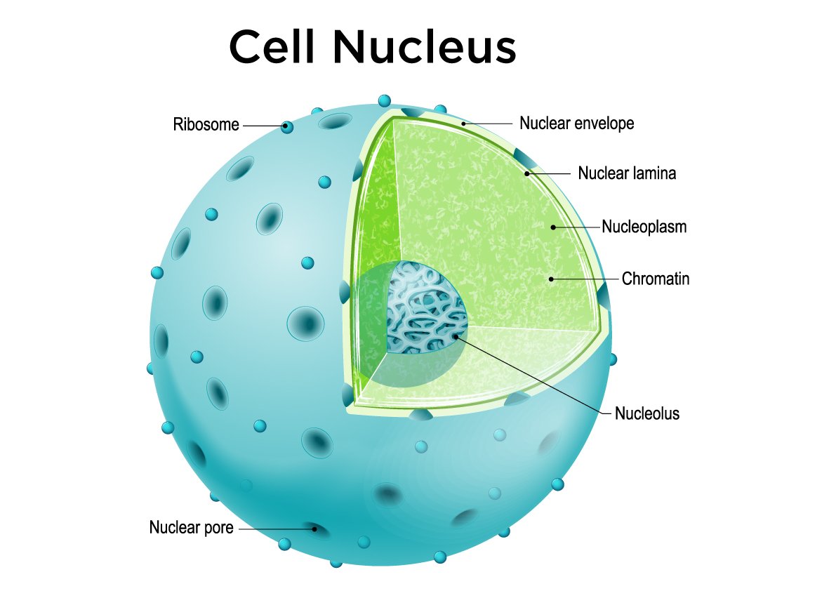
The nucleus is the largest organelle in eukaryotic cells and serves as the cell's control centre. It contains the cell's genetic information in the form of DNA and regulates all cellular activities. The presence of a nucleus is what distinguishes eukaryotic cells from prokaryotic cells.
Nuclear structure
The nucleus consists of several important components:
- Nuclear envelope: A double membrane system studded with special proteins that separates the nucleus from the cytoplasm
- Nuclear pores: Tiny openings in the nuclear envelope that control the exchange of materials (such as RNA and proteins) between the nucleus and cytoplasm
- Chromatin: Long, thin strands composed of DNA and proteins that contain the cell's genetic information
- Nucleolus: A dense region within the nucleus responsible for making ribosomal RNA (rRNA), which is essential for ribosome construction
The nuclear envelope is selectively permeable, meaning it carefully controls what enters and exits the nucleus. This regulation is essential for protecting the cell's genetic material and controlling gene expression.
Functions of the nucleus
The nucleus performs several critical functions:
- Gene expression control: The nucleus regulates which genes are turned on or off, controlling the cell's characteristics and behaviour
- DNA replication: During cell division, the nucleus coordinates the copying of genetic material
- Metabolic regulation: The nucleus produces messenger RNA (mRNA) that carries instructions for making enzymes and other proteins
- Structural protein production: The nucleus controls the synthesis of structural proteins like actin and keratin that give cells their shape
- Ribosome assembly: The nucleolus manufactures ribosomal RNA, which is crucial for protein synthesis
- Inheritance: Genetic characteristics are passed from parent to offspring through the DNA stored in the nucleus
Mitochondria
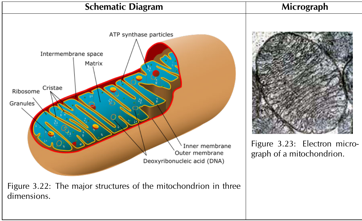
Mitochondria are often called the "powerhouses" of the cell because they generate most of the cell's chemical energy in the form of ATP (adenosine triphosphate). These organelles are found in eukaryotic cells and have a unique double-membrane structure that enables them to carry out cellular respiration efficiently.
Mitochondrial structure
Mitochondria have a complex internal structure perfectly adapted for energy production:
- Outer mitochondrial membrane: A smooth membrane that allows small molecules to pass through easily
- Intermembrane space: The narrow gap between the outer and inner membranes
- Inner mitochondrial membrane: A highly folded membrane containing specialised proteins for ATP synthesis
- Cristae: Folds in the inner membrane that dramatically increase the surface area available for energy production
- Matrix: The gel-like substance inside the inner membrane containing enzymes needed for cellular respiration
The table below shows how each mitochondrial structure relates to its function:
| Structure | Function | Adaptation to function |
|---|---|---|
| Outer mitochondrial membrane | Transfer of nutrients (e.g. lipids) to mitochondrion | Has large number of channels to facilitate transfer of molecules |
| Intermembrane space | Stores large proteins allowing for cellular respiration | Its position between two selectively permeable membranes allows it to have a unique composition compared to the cytoplasm and the matrix |
| Inner membrane | Stores membrane proteins that allow for energy production | Contains folds known as cristae which provide increased surface area, thus enabling production of ATP (chemical potential energy) |
| Matrix | Contains enzymes that allow for the production of ATP (energy) | The matrix contains a high quantity of protein enzymes which allow for ATP production |
Mitochondria are unique because they contain their own DNA, separate from the nuclear DNA. This mitochondrial DNA is inherited exclusively from the mother's side and makes up only a small percentage of the cell's total genetic material.
Energy Production Example
A single glucose molecule can produce approximately 38 ATP molecules through cellular respiration in the mitochondria. This process involves:
- Glycolysis (in cytoplasm): 2 ATP produced
- Krebs cycle (in matrix): 2 ATP produced
- Electron transport (on cristae): 34 ATP produced
The extensive folding of cristae provides the massive surface area needed for this efficient energy production.
Endoplasmic reticulum
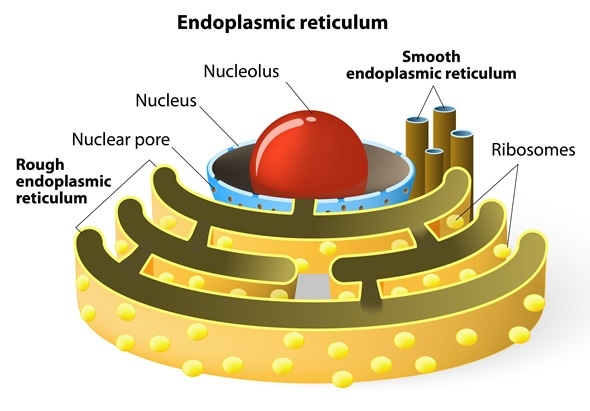
The endoplasmic reticulum (ER) is a complex network of flattened sheets, tubes, and sacs found only in eukaryotic cells. Connected to the nuclear envelope, the ER serves as the cell's manufacturing and transport system. There are two distinct types of ER, each with different functions.
Smooth endoplasmic reticulum (SER)
Smooth ER appears smooth because it lacks ribosomes on its surface. This organelle specialises in:
- Lipid synthesis: Manufacturing oils, phospholipids, and steroids
- Carbohydrate metabolism: Processing sugars and other carbohydrates
- Calcium regulation: Controlling calcium ion concentrations within the cell
- Detoxification: Breaking down harmful substances, particularly in liver cells
Rough endoplasmic reticulum (RER)
Rough ER gets its bumpy appearance from the numerous ribosomes attached to its surface. The RER is primarily responsible for:
- Protein synthesis: Manufacturing proteins, especially those destined for secretion or membrane insertion
- Membrane production: Creating new membrane material for the cell
- Quality control: Ensuring proteins are properly folded before transport
The extensive folding of ER membranes increases the surface area, allowing more ribosomes to attach and enabling greater protein production capacity. Think of it as having more "workbenches" for cellular manufacturing.
Ribosomes
Ribosomes are small but essential organelles composed of RNA and protein. They serve as the cell's protein factories, translating genetic information into functional proteins. Ribosomes can be found either floating freely in the cytoplasm or attached to the rough endoplasmic reticulum.
Types of ribosomes
- Free ribosomes: These ribosomes float independently in the cytoplasm and typically produce proteins used within the cell itself
- Polyribosomes: Multiple ribosomes working together on a single strand of messenger RNA (mRNA) to produce many copies of the same protein efficiently
Ribosomes attached to the rough ER generally manufacture proteins that will be secreted from the cell or incorporated into cell membranes. The location of ribosomes determines the destination of the proteins they produce.
Golgi body
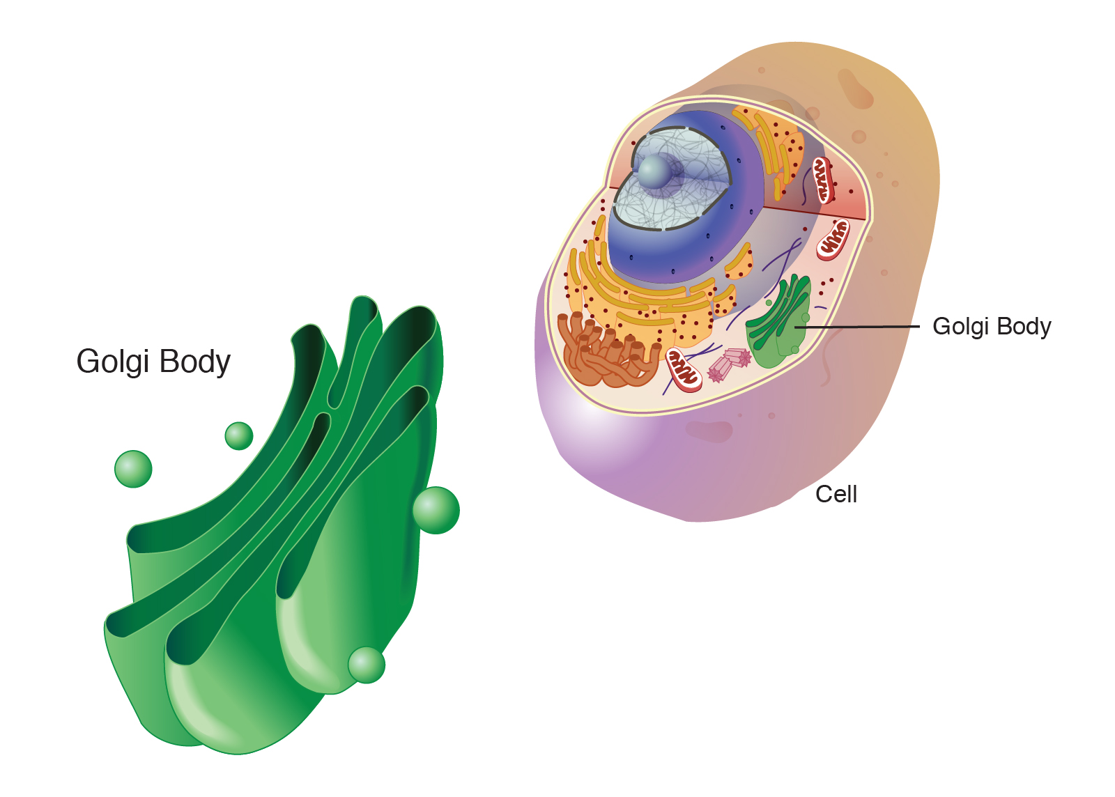
The Golgi body, sometimes called the Golgi apparatus, functions as the cell's "post office". Located near the nucleus and endoplasmic reticulum, this organelle consists of flattened membrane sacs called cisternae that work together to process and package proteins.
Functions of the Golgi body
The Golgi body plays a crucial role in protein processing and distribution:
- Protein modification: Enzymes within the Golgi cisternae modify proteins received from the rough ER, often adding sugar groups or other chemical tags
- Packaging and sorting: The Golgi body packages modified proteins into vesicles and determines where each protein should be sent within the cell
- Transport coordination: The Golgi body serves as a distribution centre, directing proteins to their final destinations, whether inside the cell or for secretion outside
- Quality control: The Golgi body ensures proteins are properly processed before they reach their final locations
Protein Processing Example
Consider a digestive enzyme being produced:
- Rough ER: Ribosome produces the basic protein
- Golgi body: Adds sugar tags and modifies the protein for stability
- Vesicle formation: Packages the finished enzyme into a secretory vesicle
- Transport: Vesicle carries enzyme to cell membrane for release
This is why the Golgi body is often compared to a postal sorting office!
Vesicles and lysosomes
Vesicles are small, membrane-bound spherical sacs that facilitate the transport, storage, and metabolism of molecules within cells. These versatile organelles are produced by the Golgi body, endoplasmic reticulum, or cell membrane and can be classified according to their contents and specific functions.
Vesicle functions
- Molecular transport: Transport vesicles carry molecules from one location to another within the cell
- Storage: Some vesicles store materials temporarily until they are needed
- Secretion: Secretory vesicles transport materials from inside the cell to the outside environment
Lysosomes
Lysosomes are specialised vesicles containing powerful digestive enzymes. These organelles are often called the cell's "recycling centres" because they break down worn-out cellular components and digest large molecules.
Key lysosome functions:
- Cellular digestion: Breaking down large molecules like carbohydrates and proteins
- Waste disposal: Digesting worn-out organelles and cellular debris
- Food digestion: In single-celled organisms, lysosomes digest food particles brought into the cell
- Cell death: When a cell dies, lysosomes release their enzymes to break down the entire cell
Lysosomes are particularly abundant in animal cells that need to process large amounts of material, such as white blood cells that digest bacteria and other foreign substances.
Vacuoles
Vacuoles are membrane-bound, fluid-filled organelles found in the cytoplasm of most plant cells, though they are very small or absent in animal cells. Plant cells typically have one large central vacuole that occupies most of the cell's volume.
Vacuole structure
- Tonoplast: The selectively permeable membrane surrounding the vacuole
- Cell sap: The liquid inside the vacuole, consisting of water, mineral salts, sugars, and amino acids
Functions of vacuoles
Vacuoles perform several important functions, particularly in plant cells:
- Storage facility: Vacuoles store water, organic compounds, and inorganic substances for later use
- Osmotic regulation: Vacuoles take in and release water through osmosis, helping maintain proper water balance
- Structural support: When full of water, vacuoles create turgor pressure that pushes against the cell wall, helping maintain plant shape and rigidity
- Waste management: Vacuoles help remove cellular waste products and excess materials
When plants lack sufficient water, vacuoles lose pressure, cells become flaccid, and the plant wilts. This demonstrates the crucial role vacuoles play in plant structure and health. Without adequate turgor pressure, plants cannot maintain their upright structure.
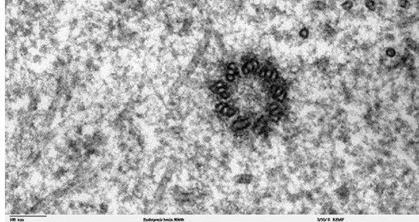
Centrioles
Centrioles are specialised organelles found only in animal cells. These cylindrical, tube-like structures are composed of nine microtubules arranged in a very specific pattern and play a vital role in cell division.
Centriole structure and function
- Centrosome: Two centrioles arranged perpendicular to each other form a structure called a centrosome
- Cell division role: Centrioles help organise the microtubules that separate chromosomes during cell division
- Chromosome positioning: They ensure chromosomes are positioned correctly so that each new cell receives the proper genetic material
The absence of centrioles in plant cells is one of the key differences between plant and animal cell organisation. Plant cells have developed alternative methods for organising cell division without these structures.
Plastids
Plastids are organelles found exclusively in plant cells and some algae. These structures are responsible for manufacturing and storing important compounds, particularly those involved in photosynthesis. There are three main types of plastids, each with distinct functions and appearances.

Three Types of Plastids
-
Leucoplasts (colourless): Found in roots and storage organs
- Store starch, oils, and proteins
- Example: Potato tubers contain starch-storing leucoplasts
-
Chloroplasts (green): Found in leaves and stems
- Contain chlorophyll for photosynthesis
- Convert sunlight into chemical energy
-
Chromoplasts (coloured): Found in fruits and flowers
- Contain red, orange, or yellow pigments
- Example: The red colour in tomatoes comes from chromoplasts
Chloroplast structure
Chloroplasts are the most important plastids because they enable plants to convert sunlight into chemical energy through photosynthesis. These double-membraned organelles have a complex internal structure perfectly adapted for capturing and using light energy.
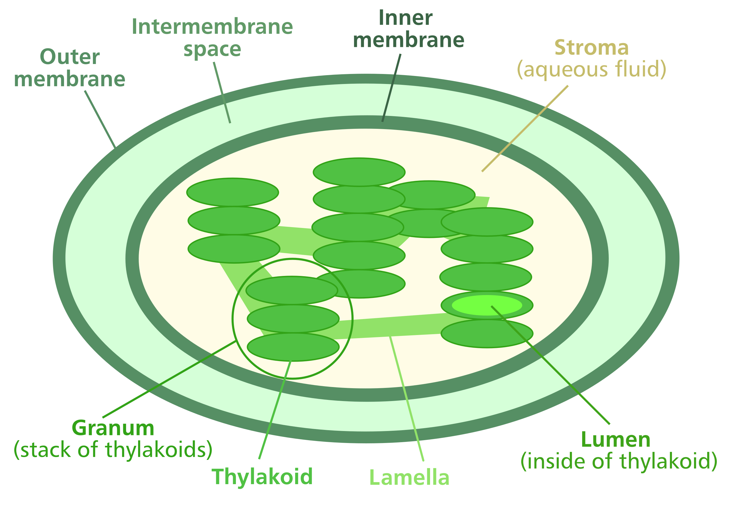
Key chloroplast components:
- Outer membrane: Controls entry of materials into the chloroplast
- Inner membrane: Contains transport proteins for moving materials
- Intermembrane space: Narrow gap between the two membranes
- Stroma: Gel-like fluid containing enzymes for photosynthesis reactions
- Thylakoids: Flattened disc-like structures containing chlorophyll
- Grana: Stacks of thylakoids that maximise light absorption
- Lamella: Connections between grana that keep thylakoid stacks separated
The structure of chloroplasts is brilliantly designed for their function. The high density of thylakoid discs and numerous grana create an enormous surface area for chlorophyll molecules, enabling maximum light absorption. The lamellae ensure efficient spacing between grana, allowing light to reach all chlorophyll molecules effectively.
Differences between plant and animal cells
Understanding the differences between plant and animal cells helps explain how these organisms have adapted to their different lifestyles. While both are eukaryotic cells sharing many common organelles, several key differences reflect their distinct functions and environments.
| Animal Cells | Plant Cells |
|---|---|
| Do not contain plastids | Almost all plant cells contain plastids such as chloroplasts, chromoplasts and leucoplasts |
| No cell wall | Have a rigid cellulose cell wall in addition to the cell membrane |
| Contain centrioles | Do not contain centrioles |
| Animals do not have plasmodesmata or pits | Contain plasmodesmata and pits |
| Few vacuoles (if any) | Large central vacuole filled with cell sap in mature cells |
| Nucleus is generally found at the centre of the cytoplasm | Nucleus is found near the edge of the cell |
| No intercellular spaces found between the cells | Large intercellular air spaces found between some cells |
Key differences explained
Cell walls: Plant cells have rigid cellulose cell walls that provide structural support and protection. This allows plants to grow tall and maintain their shape without a skeleton. Animal cells rely on internal cytoskeleton and external support systems instead.
Plastids: Only plant cells contain plastids, especially chloroplasts for photosynthesis. This fundamental difference allows plants to make their own food from sunlight, while animals must consume other organisms for energy.
Vacuoles: Plant cells typically have one large central vacuole that maintains turgor pressure and provides structural support. Animal cells may have small vacuoles but rely on other mechanisms for maintaining cell shape.
Centrioles: Animal cells use centrioles to organise cell division, while plant cells have developed alternative methods for chromosome separation during cell division.
Key Points to Remember:
-
Organelles are specialised cellular structures - each organelle has a specific function that contributes to overall cell survival and operation
-
Structure relates to function - the shape, size, and internal organisation of organelles directly support their roles (e.g., cristae in mitochondria increase surface area for ATP production)
-
Plant and animal cells have different organelles - plastids, large vacuoles, and cell walls are found only in plants, while centrioles are found only in animals
-
The nucleus controls cellular activities - all cell functions are regulated by genetic information stored in the nucleus
-
Energy production occurs in mitochondria - these "powerhouses" generate ATP through cellular respiration to fuel all cellular processes