Excretory Organs and the Urinary System (Grade 11 NSC Matric Life Sciences): Revision Notes
Excretory Organs and the Urinary System
Introduction to excretion
The removal of waste from our bodies is essential for survival. When waste builds up, it becomes dangerous to cells, tissues, organs and body systems. The human body has evolved efficient ways to remove different types of waste products.
Understanding the key terminology below is crucial for mastering this topic. These terms will appear throughout your study of the excretory system.
Key terms you need to know:
| Term | Definition |
|---|---|
| Excretion | The removal or elimination of metabolic waste from an organism |
| Secretion | The release of useful substances (like enzymes or saliva) from cells or glands |
| Egestion | The removal of undigested food waste from the digestive tract as faeces |
| Metabolism | Chemical reactions happening in every cell - can be building up (anabolic) or breaking down (catabolic) |
| Renal | Anything relating to the kidney |
| Deamination | Removal of amino groups from amino acids |
Excretory organs and their functions
Different organs in your body remove different types of waste products. The human body uses a coordinated system of specialised organs to eliminate various waste materials efficiently.
| Organ | Waste Products | Origin | How It's Removed |
|---|---|---|---|
| Lungs | Carbon dioxide and water vapour | Cellular respiration | Exhaled air |
| Skin | Mineral salts, traces of urea, water | Extracted from blood | Perspiration (sweat) |
| Liver | Urea, bile pigments | Deamination of amino acids, breakdown of haemoglobin | Via faeces |
| Colon | Bile pigments, excess mineral salts | Breakdown of haemoglobin in liver | Faeces |
| Kidneys | Urea, mineral salts, water | Deamination in liver, excess from food/drink | Urine |
Notice how each organ is specialised for removing specific types of waste. This specialisation prevents any single organ from becoming overwhelmed and ensures efficient waste removal throughout the body.
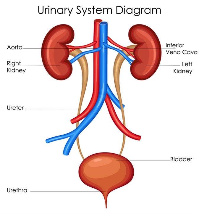
The urinary system
The urinary system is the body's primary filtration and waste removal system, working continuously to maintain internal balance.
Key terminology:
| Term | Definition |
|---|---|
| Osmoregulation | The control of water levels in the body |
| Adipose | Fat tissue |
| Aorta | The main artery leaving the heart, supplying the body with blood |
| Renal artery | Brings oxygenated, unfiltered blood to the kidneys |
| Renal vein | Carries deoxygenated, filtered blood away from the kidneys |
| Renal capsule | Outer membrane covering the kidney |
The urinary system consists of two kidneys, two ureters, one bladder and one urethra. These work together as a team to filter blood and remove waste.
The four main functions of the kidneys
The kidneys perform these essential jobs that are vital for maintaining life:
- Osmoregulation - controlling water levels in body fluids
- Excretion - removing nitrogenous waste like urea
- pH regulation - keeping body fluids at the right acidity level
- Salt regulation - controlling mineral salt concentration in body fluids
These four functions work simultaneously and are interconnected. Problems with any one function can affect the others, which is why kidney health is so important for overall wellbeing.
Structure of the kidney
Kidneys are remarkable organs with a complex internal structure designed for efficient filtering. Understanding their anatomy helps explain how they perform their vital functions.
External features
- Bean-shaped organs located halfway down your back, just under the ribcage
- Each kidney weighs between 115-170 grammes depending on age and gender
- About 11 cm long
- Protected by adipose (fat) tissue and covered by a renal capsule for protection from infections
Internal structure
The kidney has three main regions that you can see when it's cut in half:
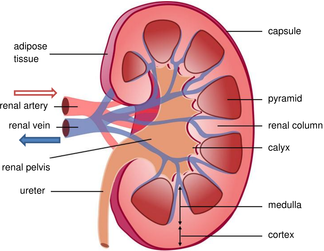
- Cortex - the outer region (dark reddish-brown colour)
- Medulla - the middle region (lighter pinkish colour)
- Pelvis - the central hollow region (creamy white) where the three tubes (renal artery, renal vein, ureter) connect
The different colours you observe in kidney cross-sections reflect the different functions of each region. The darker cortex contains most of the filtering units, while the lighter medulla focuses on concentrating urine.
Blood supply
- Renal artery brings oxygen-rich blood carrying waste products to the kidney
- Blood gets filtered by the kidney
- Renal vein carries filtered, deoxygenated blood away from the kidney
The nephron - functional unit of the kidney
Each kidney contains approximately one million tiny filtering units called nephrons. These microscopic structures do the actual work of filtering blood and making urine.
Key nephron terminology
| Term | Definition |
|---|---|
| Nephron | The microscopic functional unit of the kidney |
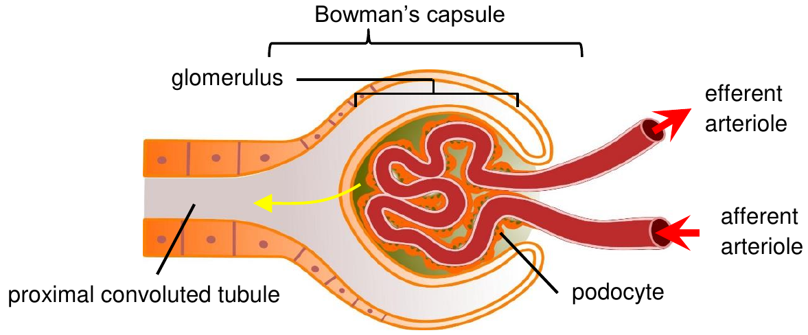
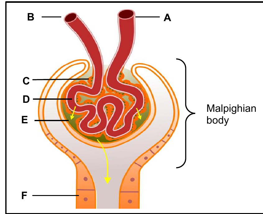
| Podocytes | Specialised cells lining Bowman's capsule |
| Afferent arteriole | Blood vessel bringing blood to the glomerulus |
| Efferent arteriole | Blood vessel taking blood away from the glomerulus |
| Glomerulus | Dense network of capillaries in Bowman's capsule |
| Bowman's capsule | Cup-shaped structure surrounding the glomerulus |
| Malpighian body | Made up of glomerulus plus Bowman's capsule |
| Proximal convoluted tubule | First coiled section after Bowman's capsule |
| Distal convoluted tubule | Second coiled section before collecting tubule |
| Peritubular capillaries | Tiny blood vessels enabling exchange between blood and nephron |
Nephron structure
The nephron has two main sections that work together to filter blood and produce urine:
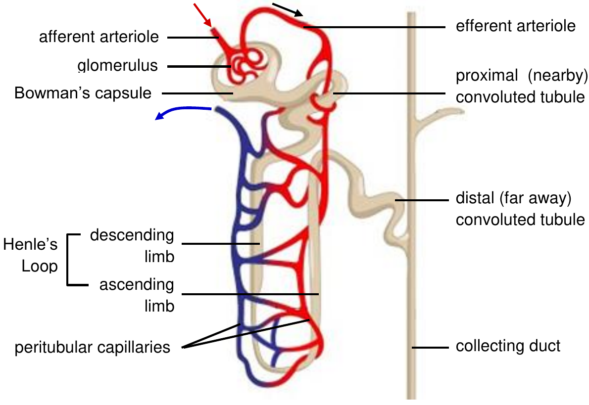
- Malpighian body (renal corpuscle) - located in the cortex
- Contains the cup-shaped Bowman's capsule
- Houses the glomerulus (cluster of capillaries)
- Special podocytes cells line the capsule with finger-like extensions and slits for filtration
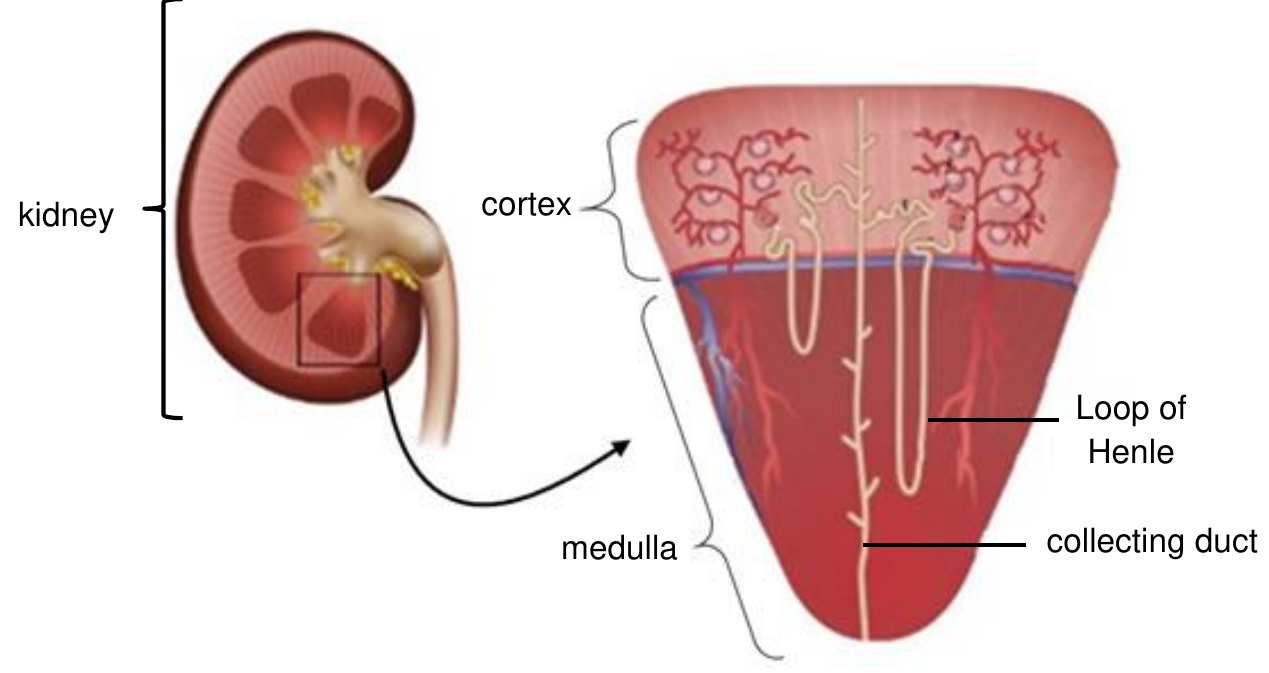
- Renal tubule - the processing pipeline
- Proximal convoluted tubule (in cortex)
- Loop of Henle (extends into medulla)
- Distal convoluted tubule (back in cortex)
- Surrounded by peritubular capillaries for reabsorption and secretion
The tubules are lined with cuboidal epithelial cells that have microvilli to increase surface area. These cells are packed with mitochondria to provide energy for active transport processes - this shows how energy-intensive kidney function really is!
Kidney functions performed by the nephron
Understanding how nephrons create urine requires knowing key concepts about water and salt concentrations:
Important concepts:
| Term | Definition |
|---|---|
| Hypertonic | Low water concentration, high salt concentration |
| Hypotonic | High water concentration, low salt concentration |
| Permeable | Allows substances to flow through easily |
| Dehydration | Loss of water |
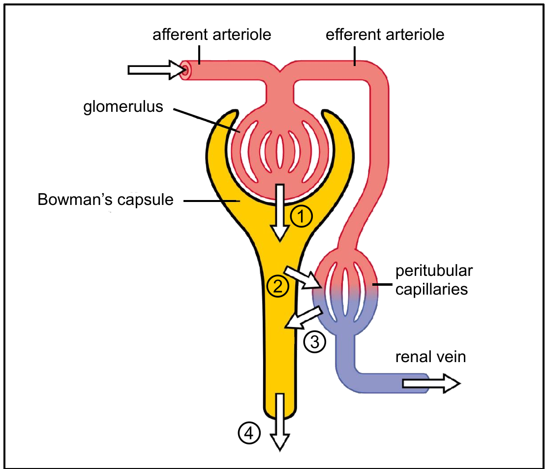
Urine formation involves four main processes working together in a coordinated sequence:
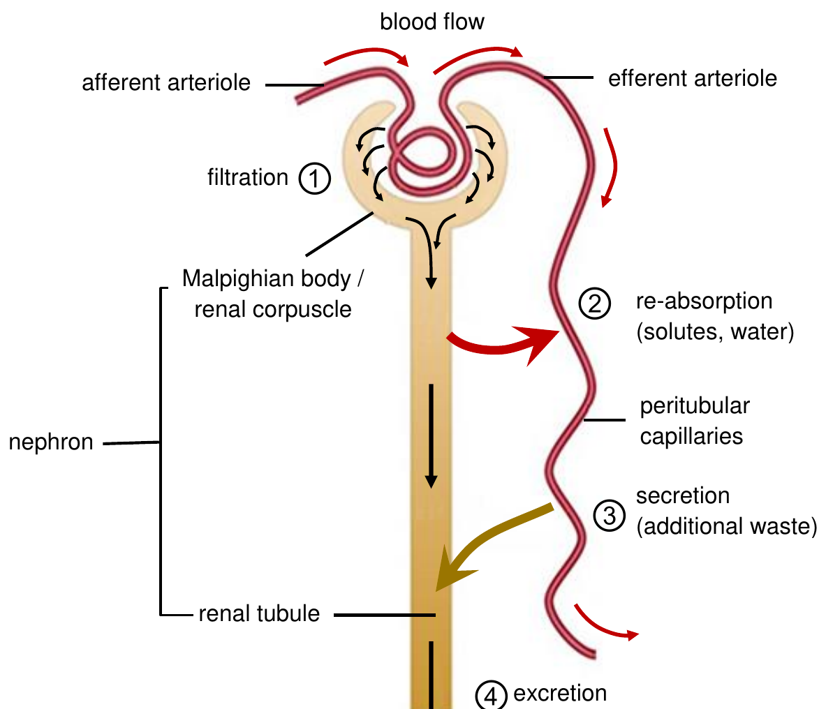
1. Glomerular filtration
This happens in the Malpighian body where blood gets filtered through a high-pressure filtration system.
How Filtration Works - Step by Step:
Step 1: Blood enters the glomerulus through the afferent arteriole (wider vessel)
Step 2: Blood exits through the efferent arteriole (narrower vessel)
Step 3: This size difference creates high pressure, forcing liquid and small dissolved substances through the filtration barrier
Step 4: Small molecules pass through podocyte slits while larger proteins remain in blood
Result: Both useful and waste substances are filtered based on size alone
Key filtration features:
- The glomerulus capillaries have thin walls (single layer of cells)
- Podocytes in Bowman's capsule have slits that allow small molecules through but keep larger proteins in the blood
- The cup shape of Bowman's capsule increases contact area for better filtration
What gets filtered: Both useful substances (glucose, amino acids, vitamins, minerals, water) and waste substances (urea, uric acid) pass through. This is a non-selective process - size matters, not whether the substance is useful or not.
2. Tubular reabsorption
This occurs mainly in the proximal convoluted tubule and is crucial for preventing dehydration.
What gets reabsorbed:
- About 65% of water returns to blood through osmosis
- All glucose, amino acids, and vitamins are actively reabsorbed
- Most mineral salts are recovered
Why reabsorption is so efficient:
- Cuboidal epithelial cells have many mitochondria for energy
- Microvilli increase surface area for maximum reabsorption
- The fluid is now called tubular filtrate
The Loop of Henle's Special Role:
- Cells in the ascending limb actively pump salt into the medulla tissue
- This makes the medulla hypertonic (very salty) with low water potential
- Creates a steep gradient that helps conserve water - essential for survival!
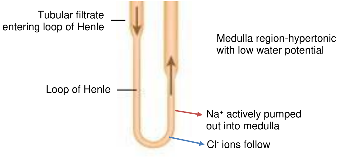
When tubular filtrate reaches the distal convoluted tubule and collecting ducts, these areas are very permeable to water. Water flows out by osmosis into the salty medulla and back into peritubular capillaries. The amount of water removed is controlled by antidiuretic hormone (ADH).
3. Tubular secretion
This is the active removal of additional unwanted substances from blood into the tubular filtrate in the distal convoluted tubule.
Substances secreted include:
- Creatinine
- Ammonia
- Potassium ions (K⁺)
- Hydrogen ions (H⁺)
- Sodium ions (Na⁺)
- Bicarbonate ions
- Some drugs (like penicillin)
Tubular secretion is like a "cleanup" process - it removes substances that weren't filtered out initially or that the body needs to eliminate to maintain proper balance.
4. Excretion of urine
The final filtrate is now called urine. It contains:
- Urea (the main nitrogenous waste)
- Excess water
- Excess mineral salts
- Other waste substances
Healthy Urine Check: Useful substances like glucose and amino acids should NOT be present in healthy urine. If they are, it indicates a problem with kidney function.
The urine flows from the collecting ducts in the medulla into the pelvis region, then down the ureter to the bladder for storage.
Homeostatic control of blood pH
One of the kidney's most important jobs is maintaining blood pH at exactly 7.4. The distal convoluted tubule plays a key role in this process.
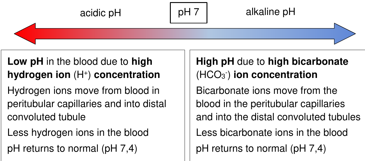
How pH Regulation Works:
Scenario 1 - Blood too acidic (low pH):
- Excess hydrogen ions (H⁺) are actively removed from blood in peritubular capillaries
- These H⁺ ions are secreted into the distal convoluted tubule
- Result: Blood pH rises back to 7.4
Scenario 2 - Blood too alkaline (high pH):
- Excess bicarbonate ions (HCO₃⁻) are removed from blood
- These ions are secreted into the tubule
- Result: Blood pH lowers back to 7.4
Both processes help restore normal blood pH to 7.4, maintaining homeostasis.
Blood composition and filtration
Understanding what's in blood helps explain how the kidneys work so effectively:
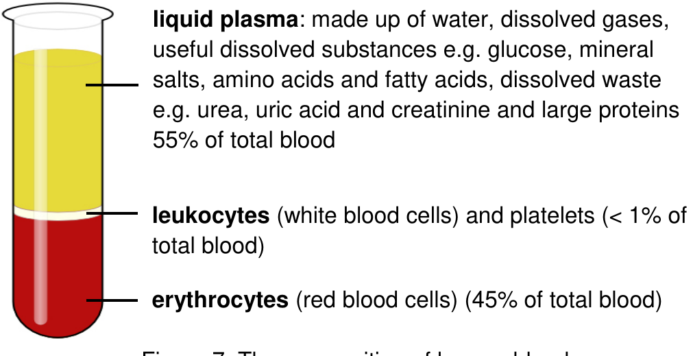
Blood consists of:
- Liquid plasma (55%) - contains water, dissolved gases, nutrients (glucose), mineral salts, amino acids, fatty acids, waste products (urea, creatinine), and large proteins
- White blood cells and platelets (<1%)
- Red blood cells (45%)
The kidneys filter the plasma portion, removing waste while keeping essential proteins and blood cells in circulation. This selective filtering is what makes kidney function so remarkable.
Key Points to Remember:
- Excretion removes metabolic waste - it's different from egestion (removing undigested food) and secretion (releasing useful substances)
- Multiple organs work as excretory organs - lungs (CO₂), skin (salts), liver (urea production), and kidneys (urea removal) all play important roles
- The nephron is the kidney's functional unit - each kidney contains about 1 million nephrons that filter blood and produce urine
- Four processes create urine: filtration (non-selective), reabsorption (recovers useful substances), secretion (removes additional waste), and excretion (final urine formation)
- Kidneys maintain homeostasis - they control water balance, salt levels, pH, and remove nitrogenous waste to keep your internal environment stable