Cell Structure and Technologies (HSC SSCE Biology): Revision Notes
Organelles
Introduction to organelles
Eukaryotic cells contain specialised internal structures known as organelles. Each organelle is enclosed by its own membrane and performs a distinct function within the cell. These structures work together as a coordinated system to ensure the cell functions efficiently as a complete unit.
Key features of organelles include:
- All organelles are membrane-bound structures (surrounded by membranes)
- Membranes can be either single or double-layered
- Many organelles have evolved structures that maximise their surface area to enhance their specific functions
- Different organelles vary significantly in size
The visibility of organelles depends on the type of microscope used. Larger structures such as the nucleus, vacuoles and chloroplasts can be observed with a basic light microscope. However, smaller organelles like mitochondria and ribosomes require the higher magnification and resolution of an electron microscope to see their detailed structure.
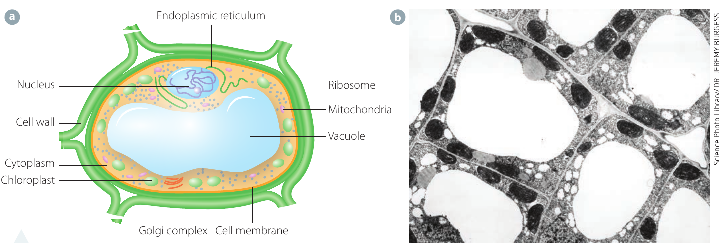
Comparing plant and animal cells
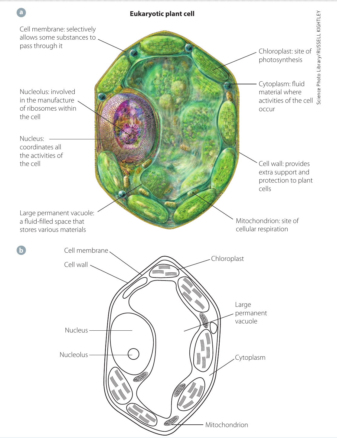
Both plant and animal cells are eukaryotic and share many common organelles. However, there are some key differences between them. Plant cells possess three distinctive structures that animal cells lack:
- Cell wall - provides rigid support and protection
- Chloroplasts - carry out photosynthesis
- Large permanent vacuole - provides storage and support
Conversely, animal cells contain structures not typically found in plant cells:
- Centrioles - involved in cell division
- Lysosomes - break down and recycle cellular materials
These structural differences reflect the distinct requirements and lifestyles of plant and animal cells. Plant cells need rigid support and the ability to produce their own food through photosynthesis, whilst animal cells require structures for active movement and obtaining nutrients from external sources.
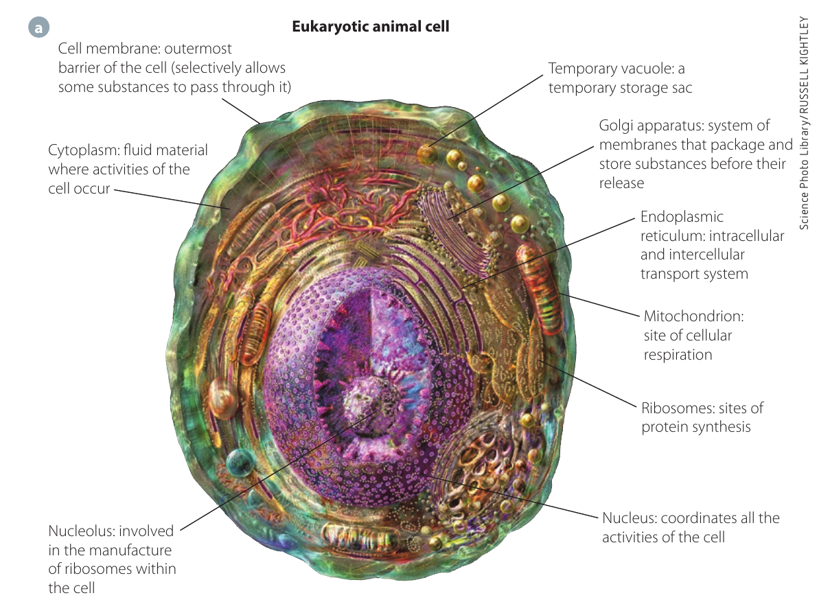
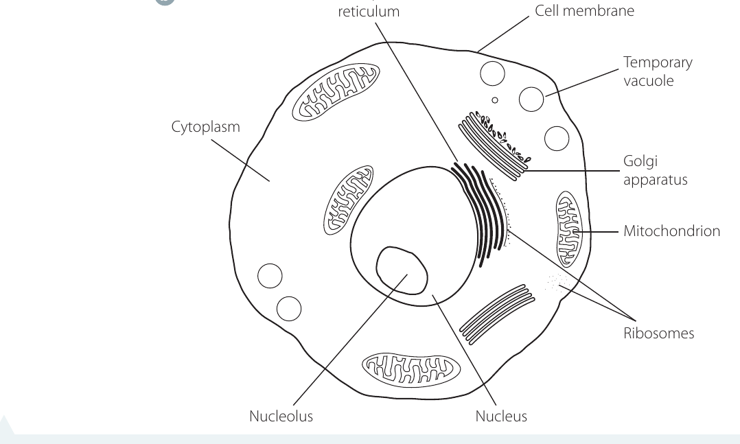
Cell membrane - the selective boundary
The cell membrane (also called the plasma membrane) forms the outer boundary of all cells. It separates the internal contents of the cell from the external environment. This membrane serves as a selective barrier, carefully controlling which substances can enter or leave the cell.
This selective property means the membrane is selectively permeable (sometimes termed semipermeable). Only certain molecules are permitted to pass through, whilst others are blocked. This selective permeability is crucial for maintaining the cell's internal environment and protecting it from harmful substances.
Both plant and animal cells possess a cell membrane. The membranes surrounding internal organelles also demonstrate this selective permeability, regulating the movement of substances between the cytoplasm and the organelle interior.
Protoplasm - the living content
Protoplasm refers to all the living material within a cell that is enclosed by the cell membrane. It is where essential life processes occur, including the production of cellular products and respiration.
The protoplasm consists of two main components:
- Nucleus - the control centre
- Cytoplasm - everything outside the nucleus
The cytoplasm contains a liquid called cytosol, which makes up approximately water. Dissolved within the cytosol are various chemical substances (such as chloride ions), along with suspended organelles and insoluble particles. This aqueous environment provides the medium in which all cellular chemical reactions take place.
Nucleus - the control and information centre
The nucleus (plural: nuclei) appears as a large, spherical or oval structure within the cytoplasm. It is colourless, transparent and has a slightly gel-like consistency compared to the surrounding cytoplasm. Most cells contain a single nucleus.
The nucleus stores all the genetic information required to control cellular activities. To effectively coordinate cell functions, the nucleus must communicate with the surrounding cytoplasm. A double nuclear membrane (nuclear envelope) surrounds the nucleus. This membrane is pierced by tiny nuclear pores that regulate the passage of substances between the nucleus and cytoplasm, enabling communication between these two regions.
Internal structure of the nucleus
The nucleoplasm (nuclear sap) is the liquid portion of the nucleus. Within this fluid is found chromatin material, composed of protein and nucleic acid. The nucleus stores DNA (deoxyribonucleic acid), a very large molecule that contains all the genetic information (the cellular 'blueprint') needed to control cell function. This DNA holds hereditary information that passes from one generation to the next.
Before a cell divides, the DNA must be copied so it can be transmitted to newly formed cells. During cell division, the chromatin condenses into short, thick rod-shaped structures called chromosomes. These structures take up stains easily during microscopy preparation, which is how they got their name (from Greek: chromo meaning coloured, and soma meaning body).
Chromosome Numbers Across Species
Each species has a characteristic chromosome number that remains constant across all individuals:
- Humans: chromosomes
- Platypus: chromosomes
- Lettuce: chromosomes
- Camels: chromosomes
The nucleolus is a dense, granular region visible within the nucleoplasm. It contains high concentrations of nucleic acid, primarily RNA (ribonucleic acid) with some DNA. The nucleolus is responsible for manufacturing ribosomes, which are essential cellular machinery for protein production.
Endoplasmic reticulum - transport and processing network
The outer nuclear membrane typically connects to an extensive network of flattened, interconnected membranes called the endoplasmic reticulum (ER). This network provides transport pathways between the nucleus and the cell's external environment, allowing intracellular transport (movement within the cell). The extensive folding of these membrane sheets dramatically increases the available surface area.
There are two types of ER:
Rough ER
Contains ribosomes attached to its surface, giving it a 'rough' appearance. Its functions include:
- Folding and processing proteins manufactured by the cell
- Synthesising lipids (fats)
Smooth ER
Lacks attached ribosomes. Its primary function is:
- Producing lipids, which are essential for membrane repair and construction
In plant cells, the ER can also transport substances between cells by passing through channels called cell pits in the cell wall, facilitating intercellular communication and material exchange.
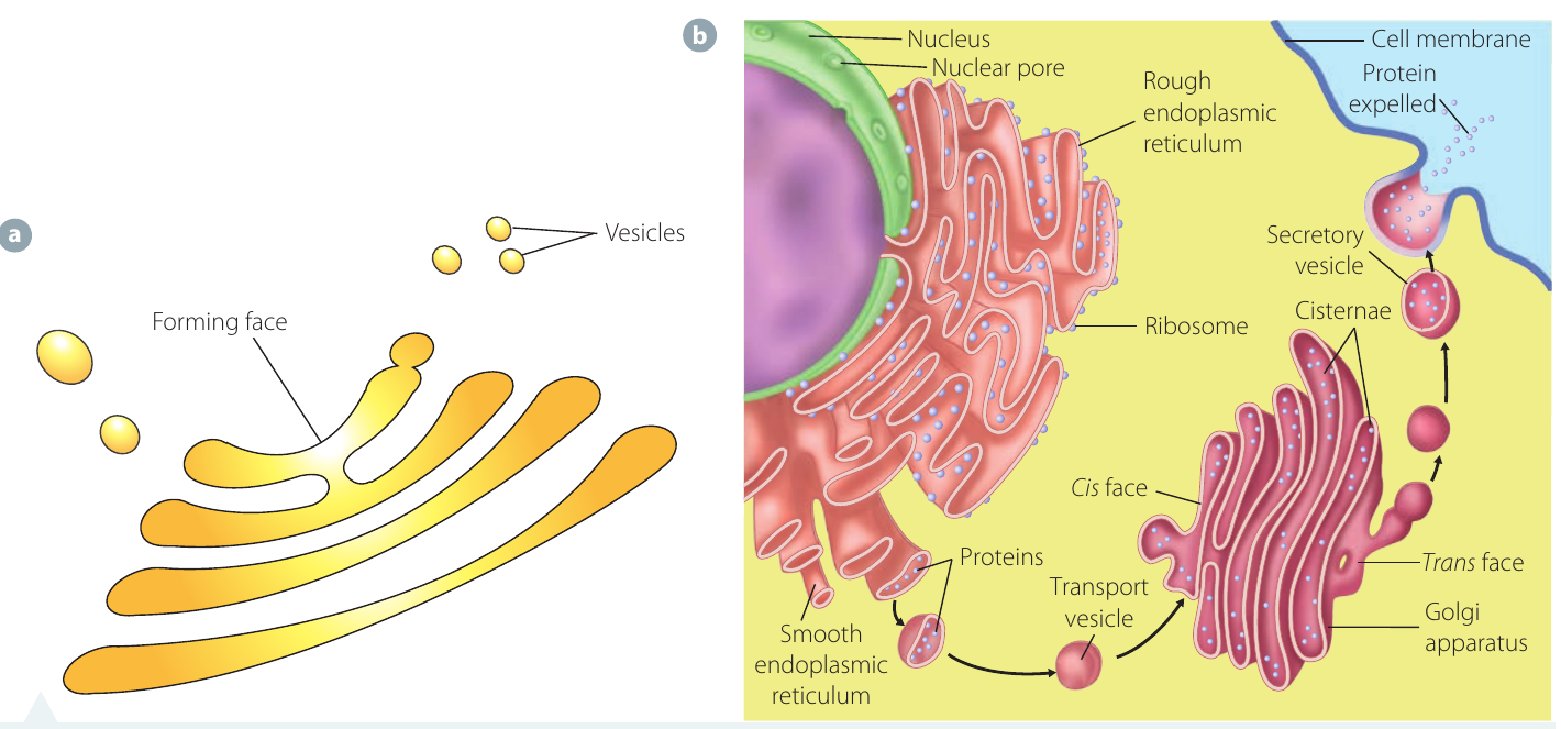
Ribosomes - protein synthesis machinery
Ribosomes are small organelles that appear as dense, rounded granules in electron micrographs. Their small size maximises their surface area relative to volume. Each ribosome is constructed from RNA and protein molecules.
Ribosomes serve as the cellular 'machinery' that reads the genetic instructions encoded in DNA and uses them to produce proteins. They join amino acids together in the correct sequence to form polypeptides, which are the building blocks of proteins.
Ribosomes can be found in two locations:
- Free-floating in the cytoplasm
- Attached to the surface of the ER
Newly synthesised proteins pass from the ribosomes into the ER, where they undergo folding into their functional three-dimensional shapes.
Golgi bodies - packaging and sorting centre
The Golgi body (also known as Golgi apparatus) consists of flat membranes arranged in stacks of to layers. Unlike the ER, the Golgi body does not have ribosomes attached to its surface. It is easily recognised by its characteristic curved shape on one surface, where small membrane-bound sacs called vesicles can be seen budding off. This surface is called the forming face, and the vesicles demonstrate the secretory function of the Golgi body.
The Golgi body performs three main functions:
- Processing cell products by adding proteins and carbohydrates
- Packaging cell products by wrapping them in membranes
- Sorting cell products by providing different membrane 'labels'
The features of these membrane packages determine the products' destinations - they may be transported to specific locations within the cell or secreted outside the cell entirely. This sophisticated sorting system ensures that each cellular product reaches its correct destination.
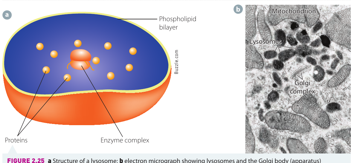
Lysosomes - digestion and recycling units
Over time, organelles within the cytoplasm wear out and reach their 'use-by date'. Rather than wasting the raw materials that comprise these organelles, cells have evolved an efficient recycling system. This task is performed by lysosomes (from 'lysis' meaning to break apart), which are special organelles found in animal cells.
Lysosomes are formed by the Golgi body and contain powerful digestive enzymes. These enzymes break down complex chemical compounds into simpler molecules. For example, proteins are split into individual amino acids, which can then be reused as building blocks for new compounds and organelles.
Programmed cell death
Sometimes lysosomes destroy an entire cell through a process called apoptosis or programmed cell death. This is a deliberate cellular action to eliminate old or damaged cells. The lysosome membrane ruptures, releasing digestive enzymes that break down all the cell's contents, killing the cell in a controlled manner.
Mitochondria - the cellular powerhouses
Mitochondria (singular: mitochondrion) are the 'powerhouses' of cells, generating energy through the process of cellular respiration. They are typically rod-shaped but can also be round, and they vary in both shape and size. Mitochondria are smaller than the nucleus and chloroplasts but larger than ribosomes.
The number of mitochondria in a cell correlates with the cell's energy requirements. Less active cells contain few mitochondria, whilst highly active cells may contain hundreds or even thousands.
Mitochondria in Active Liver Cells
Active liver cells, which perform numerous metabolic functions, contain between to mitochondria each. This high number reflects the enormous energy demands of liver tissue, which is responsible for processes such as detoxification, protein synthesis, and metabolism of nutrients.
Energy production
Just as machines require electrical energy to function, cells need energy to carry out their work. Mitochondria combine oxygen with sugars during cellular respiration to release energy in a form the cell can use - a chemical called ATP (adenosine triphosphate).
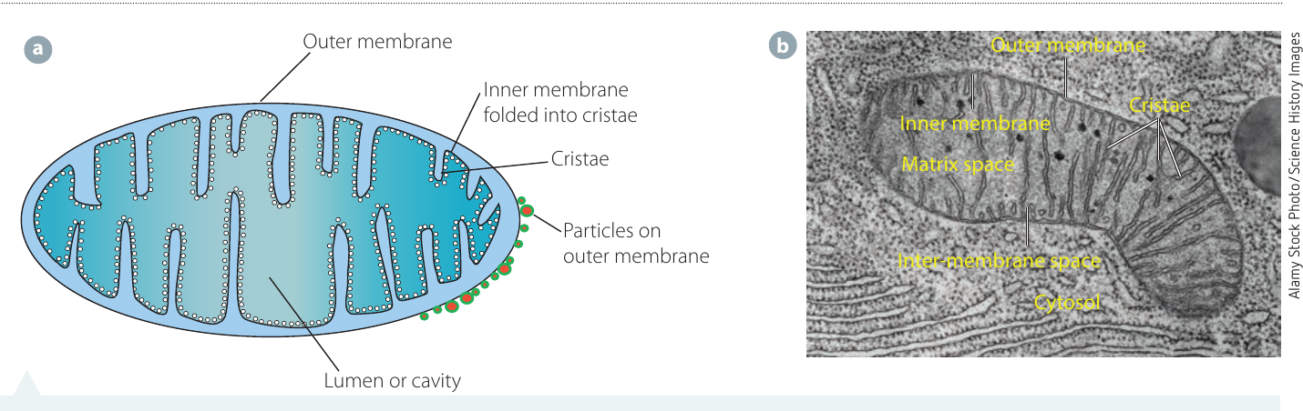
Structure of mitochondria
A double membrane surrounds each mitochondrion:
- Outer membrane - gives the mitochondrion its shape and allows small substances to pass in and out
- Inner membrane - folded into fine, finger-like projections called cristae, which increase the surface area for enzyme attachment
Groups of enzymes responsible for producing cellular energy appear as knob-like particles on the inside of the cristae. The central space is filled with fluid called the matrix, which contains mitochondrial DNA and enzymes. Interestingly, mitochondria can replicate themselves by pinching off and growing, particularly in very active cells or cells preparing to divide.
Vacuoles - storage and support structures
Vacuoles are large, permanent, fluid-filled compartments found in mature plant cells. Each vacuole consists of a watery solution called cell sap, enclosed by a single membrane called the tonoplast. The cell sap contains various dissolved substances including mineral salts, sugars and amino acids. It may also contain dissolved pigments that give cells their colour, such as the reds, pinks and purples visible in some flower petals.
Vacuoles perform two important functions:
Storage
Vacuoles store various materials needed by the plant cell.
Support
Vacuoles play a crucial role in providing structural support to plant cells. When filled with water, the vacuole pushes outwards against the cytoplasm, exerting pressure on the cell wall. This pressure keeps the cell wall firm. The outward pressure of the cell contents combined with the resistance of the cell wall makes the cell firm or turgid.
Small, temporary vesicles may occasionally appear in animal cells, but these do not contribute to cell support. Therefore, large permanent vacuoles that provide turgidity are considered an exclusive feature of plant cells.
Chloroplasts - photosynthesis factories
Chloroplasts are green organelles responsible for photosynthesis - the process of manufacturing sugar in plants using the energy of sunlight. Their green colour comes from a pigment called chlorophyll. Chloroplasts are not present in all plant cells; they are found only in the green tissues of plants capable of photosynthesis.
Under a light microscope, chloroplasts appear as green, disc-shaped structures smaller than the nucleus. An electron microscope is required to observe their detailed internal structure.
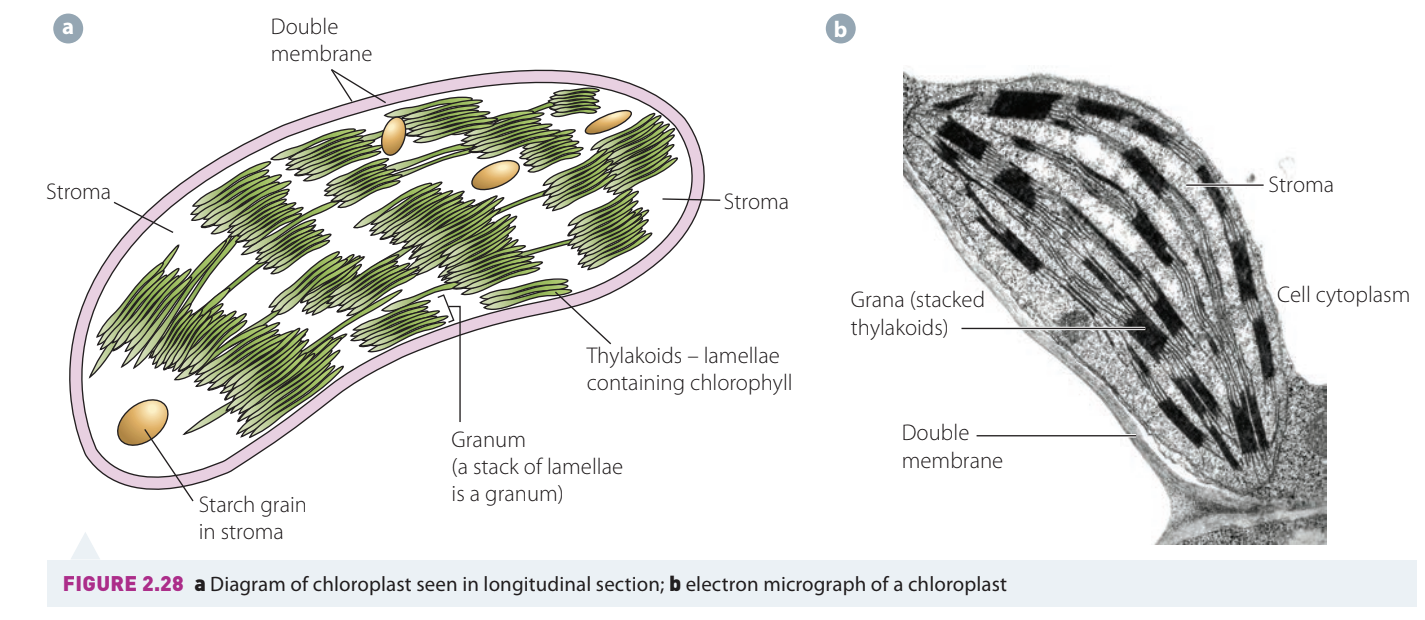
Structure of chloroplasts
Chloroplasts belong to a group of organelles called plastids, which have a biconvex shape. They are larger than mitochondria but, like mitochondria, contain their own DNA. The number of chloroplasts per cell varies.
A double membrane surrounds each chloroplast, allowing substances to pass between the cytoplasm and the chloroplast interior. Unlike mitochondria, the inner membrane of chloroplasts is not folded.
The liquid portion of the chloroplast is called the stroma. Within the stroma are stacks of membranes called thylakoids. Each stack of thylakoids is termed a granum (plural: grana), and chlorophyll molecules are located on these membranes.
Function in photosynthesis
The layered arrangement of membranes increases the surface area covered by chlorophyll, allowing large amounts of sunlight to be absorbed for photosynthesis. This captured light energy is then used to produce simple sugars.
All the enzymes required for photosynthesis are present in the stroma, and sugars produced during photosynthesis are stored in the stroma as starch grains.
Plant cell wall - structural support
The cellulose cell wall surrounding plant cells differs from the cell membrane inside it. Unlike the selectively permeable cell membrane, the cell wall is permeable to most molecules and does not regulate which substances enter the cell.
The cell wall's structure provides strength and support to plant cells. Strands of cellulose fibres have some elasticity and flexibility, allowing them to resist pressure. Some cell walls are thickened with additional chemicals that make them hard and woody (such as in tree trunks) or provide waterproofing (such as in cork or the waxy cuticle of leaves).
Centrioles - cell division structures
The centrosome is a dense, granular structure often found near the nucleus in animal cells. It consists of two centrioles, which play an important role during cell division. Centrioles form the spindle, a structure that holds chromosomes in position during cell division.
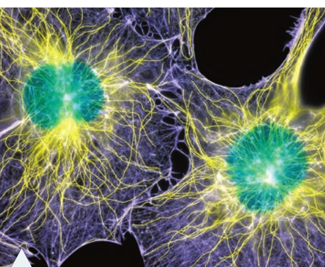
Cytoskeleton - the cellular framework
Organelles are not randomly scattered within cells. Their distribution is organised, and they are held in position by a network of tiny structures called the cytoskeleton. This network extends throughout the cytoplasm and consists of:
- Microtubules - hollow cylindrical structures
- Microfilaments - thin protein fibres
- Intermediate filaments - medium-sized fibres
The cytoskeleton serves multiple functions:
- Provides the framework for cell shape
- Enables cell movement
- Facilitates organelle movement within the cell
- Assists in cell division
Summary of organelle functions
| Organelle / Cell Structure | Function |
|---|---|
| Membranes | Selectively permeable boundaries, control the movement of substances into and out of the cell/organelle |
| Protoplasm | The living content of a cell that is surrounded by the cell membrane |
| Nucleus | The control and information centre |
| Endoplasmic reticulum | Transport and processing of proteins and lipids |
| Golgi bodies | Packaging and sorting the products |
| Ribosomes | Protein synthesis |
| Lysosomes | Digestion and cell destruction |
| Mitochondria | Cellular respiration – production and storage of energy (ATP) |
| Vacuoles | Storage and support |
| Chloroplasts | Photosynthesis |
| Plant cell wall | Shape and support |
| Centrioles | Spindle production in cell division |
| Cytoskeleton | Cell shape, organelle placement and movement and cell division |
Remember!
Key Points to Remember:
-
Organelles are specialised membrane-bound structures that each perform specific functions to ensure efficient cell operation.
-
Surface area maximisation is a common feature of many organelles, achieved through folding (mitochondria, ER) or stacking (Golgi body, chloroplasts) of membranes.
-
Plant and animal cells share most organelles but differ in three key structures: plant cells uniquely possess cell walls, chloroplasts and large permanent vacuoles, whilst animal cells contain centrioles and lysosomes.
-
The nucleus is the control centre containing DNA, which stores all genetic information needed to direct cellular activities and is passed on during cell division.
-
Energy production occurs in mitochondria through cellular respiration, producing ATP that powers all cellular processes - more active cells contain more mitochondria.