Cell Membranes (HSC SSCE Biology): Revision Notes
Cell Membranes
Introduction to cell membranes
Cell membranes are essential structures that separate the internal environment of cells from their external surroundings. Even with the powerful magnification of electron microscopes, we cannot directly observe the detailed molecular structure of these membranes. This limitation means scientists have had to develop theoretical models to understand how cell membranes are organised.
Because the detailed structure of cell membranes cannot be seen directly, scientists must develop and test models based on experimental evidence. These models are then refined or replaced as new evidence becomes available.
After many years of research and testing different ideas, the scientific community developed the fluid mosaic model of cell membranes. This model was proposed in 1972 by two scientists, Singer and Nicolson, and remains our accepted understanding today. The model has been validated because it successfully explains the behaviour of membranes, matches their estimated surface area, aligns with chemical analysis results, and accounts for observations from electron microscopy studies. Most importantly, it explains nearly all the functions we observe in cell membranes.
The fluid mosaic model
Overview of the model
Cell membranes have a crucial role: they regulate what enters and exits the cell. They are described as selectively permeable, which means they allow only certain molecules or ions to pass through whilst blocking others. This selective barrier enables cells to maintain stable internal conditions that differ from their external environment.
The fluid mosaic model describes the membrane structure using a vivid metaphor: a 'lipid sea' with 'many and various proteins floating on and in it'. The term 'fluid' refers to the ability of the membrane to flow and change shape like a two-dimensional liquid. The term 'mosaic' describes the pattern created by various protein molecules embedded throughout the membrane structure.
Understanding the Model's Name
The name 'fluid mosaic model' comes from two key features:
- Fluid: The phospholipids and proteins can move laterally within the membrane, giving it liquid-like properties
- Mosaic: The scattered pattern of different protein molecules resembles tiles in a mosaic artwork
The foundation of this model is a lipid bilayer - a double layer of lipid molecules. Within this lipid layer, specialised protein molecules are positioned in various patterns, much like tiles in a mosaic artwork. Some proteins can move sideways through the membrane, whilst others remain fixed in specific locations. Both the phospholipids and proteins work together to control the movement of substances between the inside and outside of the cell.
Lipid component
Phospholipid structure
The 'fluid' portion of the cell membrane consists of two layers of phospholipids, forming what we call a phospholipid bilayer. Understanding the structure of individual phospholipid molecules helps us understand why they arrange themselves this way.
Each phospholipid molecule has two distinct parts:
- A head containing a phosphate group
- Two tails made of fatty acid chains
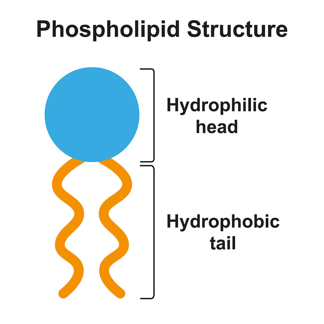
The phosphate group in the head makes it hydrophilic (meaning 'water loving'). This region can absorb water or dissolve in water. In contrast, the fatty acid tails are hydrophobic (meaning 'water hating'). These tails avoid water and cannot dissolve in it.
Phospholipid bilayer arrangement
When phospholipid molecules come together to form a membrane, they automatically arrange themselves in a specific way due to their chemical properties. The hydrophilic heads position themselves facing outward - toward the watery cytoplasm on one side and toward the watery external environment on the other side. Meanwhile, the hydrophobic tails point inward, facing each other and away from any water.
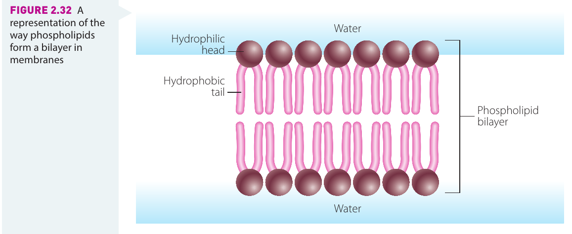
The Bilayer Arrangement Rule: "Heads OUT, Tails IN"
The phospholipid bilayer always arranges itself with:
- Hydrophilic (water-loving) heads facing the aqueous environments on both sides
- Hydrophobic (water-hating) tails tucked away in the middle, shielded from water
This arrangement is not a choice - it happens automatically due to the chemical properties of phospholipids when they are in a watery environment.
This double-layered arrangement is called a bilayer. The structure is not rigid or fixed; instead, the phospholipid molecules can move around within their layer, giving the membrane its 'fluid' quality. This fluidity is essential for many membrane functions.
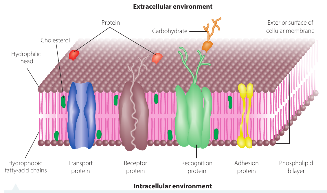
Cholesterol and membrane flexibility
Within the phospholipid bilayer of animal cells, molecules of cholesterol are scattered throughout the structure. Cholesterol increases the membrane's flexibility, allowing it to bend and move without breaking. Plant cells use a different type of lipid called phytosterol to achieve the same effect.
The lipid components give all cellular membranes important properties. Membranes can be flexible, allowing cells to change shape and grow. They can also repair themselves if damaged. This self-repair ability is crucial during processes like cell division, when membranes must break apart and then reassemble around the newly formed daughter cells.
The self-repair capability of cell membranes is possible because the phospholipid bilayer is held together by weak forces rather than strong chemical bonds. If the membrane is punctured or torn, phospholipids naturally move to fill the gap, restoring the bilayer structure.
This lipid bilayer structure forms the foundation not only of the cell's outer membrane but also of all the membranes surrounding organelles inside the cell.
Membrane proteins
Distribution and structure of proteins
Protein molecules are scattered throughout the lipid bilayer. Some proteins extend all the way through the membrane from one side to the other, creating channels that allow certain materials to cross. Other proteins are only partially embedded in the membrane, sitting within just one layer.
Scientists describe these proteins as 'floating' in the lipid bilayer 'like icebergs in a lipid sea', which creates the mosaic pattern that gives the model its name. Some proteins are anchored in fixed positions, whilst others can move freely within the membrane.
Types of membrane proteins and their functions
Different types of proteins perform specialised functions within the cell membrane:
Transport proteins act as passageways or channels that allow specific substances to move across the membrane. Some create temporary or permanent pores (openings), whilst others form active carrier systems that help transport materials. These proteins ensure that necessary substances can enter the cell and waste products can exit, even though they might not be able to pass through the lipid bilayer on their own.
Without transport proteins, many essential molecules (such as glucose and ions) would be unable to cross the hydrophobic core of the lipid bilayer. These proteins provide specific pathways for substances that cannot pass through the phospholipid layer directly.
Receptor proteins enable cellular communication. Different types of cells have different receptor proteins on their surfaces. These receptors respond only to specific chemical signals, such as hormones. When a hormone binds to its matching receptor, it triggers a specific response in the cell, giving each cell type its specialised functions.
How Receptor Proteins Work
Consider insulin, a hormone that regulates blood sugar:
- Insulin molecules circulate in the bloodstream
- Only cells with matching insulin receptors can detect and respond to insulin
- When insulin binds to its receptor, it triggers the cell to take up glucose from the blood
- Cells without insulin receptors cannot respond to insulin, even when it's present
This specificity allows different cell types to respond differently to the same chemical environment.
Recognition proteins, also called glycoproteins, consist of a protein molecule with a carbohydrate molecule attached to it. These proteins serve as identification markers on the cell surface. They display unique patterns called antigens or marker molecules that identify which organism or tissue the cell belongs to. The immune system uses these markers to distinguish between the body's own cells (marked as 'self') and foreign particles like bacteria or viruses (marked as 'non-self'). This recognition system ensures that immune cells attack only foreign invaders whilst leaving the body's own cells unharmed.
Recognition Proteins and the Immune System
Glycoproteins act as identity cards for cells. Each person has unique marker molecules on their cell surfaces, which is why:
- Organ transplants must be carefully matched between donor and recipient
- Incompatible blood transfusions can trigger immune reactions
- The immune system can identify and destroy foreign cells like bacteria
Without these markers, the immune system couldn't distinguish between 'self' and 'non-self', making it impossible to fight infections effectively.
Adhesion proteins link neighbouring cells together. In multicellular organisms, these proteins help maintain the three-dimensional structure by holding cells in their proper positions relative to one another. This cell-to-cell connection is essential for forming tissues and organs.
Together, these various proteins enable cell-to-cell interaction, cellular communication, and the controlled exchange of substances between the cell and its external environment.
Key Points to Remember:
-
Cell membranes are selectively permeable, controlling which substances can enter or leave the cell to maintain stable internal conditions.
-
The fluid mosaic model describes the cell membrane as a flexible phospholipid bilayer with embedded proteins, like a 'lipid sea' with 'floating proteins' creating a mosaic pattern.
-
Phospholipids have hydrophilic (water-loving) heads and hydrophobic (water-hating) tails that naturally arrange into a bilayer with heads facing outward toward water and tails facing inward away from water.
-
Cholesterol (in animals) and phytosterol (in plants) are dispersed throughout the membrane to increase flexibility and enable self-repair.
-
Membrane proteins include:
- Transport proteins - allowing substances to cross
- Receptor proteins - receiving chemical signals
- Recognition proteins (glycoproteins) - identifying the cell to the immune system
- Adhesion proteins - linking cells together