Sizes of Cells (HSC SSCE Biology): Revision Notes
Sizes of Cells
Introduction to the microscopic world
Modern biology has expanded our ability to observe increasingly smaller structures in living organisms. We've moved beyond measuring objects in centimetres and millimetres. Today, scientists routinely work with measurements in micrometres (also called microns) and even nanometres. We now live in what biologists call the "nano age" - an era where we can study life at the molecular and atomic level.
Understanding these tiny measurements is essential for studying cells, bacteria, viruses, and the molecules that make up living things. Each unit of measurement allows us to explore a different level of biological organization.
Understanding metric units for cell measurement
In biology, we use the metric system to measure objects of vastly different sizes. The table below shows how metres relate to smaller units of measurement:
Here are the key relationships you need to know:
| Unit | Symbol | Relationship to metre | Reciprocal |
|---|---|---|---|
| Metre | m | m | - |
| Centimetre | cm | m cm | cm m |
| Millimetre | mm | m mm | mm m |
| Micrometre (micron) | µm | m µm | µm m |
| Nanometre | nm | m nm | nm m |
Converting between units
When converting between units, remember these useful relationships:
- cm mm
- mm µm
- µm nm
Each step down the scale represents a 1000-fold decrease in size (except cm to mm, which is only 10 times smaller).
Key conversions to memorize:
- mm µm
- µm nm
These two conversions are fundamental for working with cell measurements in biology. Each step represents multiplying or dividing by 1000.
Worked Example: Unit Conversions
Converting cm to mm:
Converting mm to µm:
Converting nm to µm:
What can we see at different scales?
Different tools allow us to observe structures at different size ranges. The diagram below illustrates what can be seen by the naked eye, light microscopes, and electron microscopes:
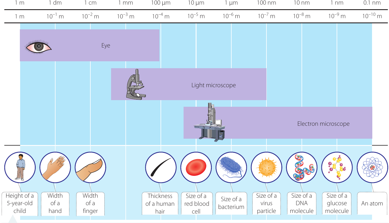
Naked eye viewing range
The human eye can see objects down to approximately mm (or µm). This includes:
- Large organisms and body parts (metres to centimetres)
- The thickness of a human hair (approximately µm)
Light microscope range
Light microscopes use visible light to magnify specimens. They can resolve objects down to approximately nm (or µm). This allows us to see:
- Most eukaryotic cells ( µm)
- Red blood cells ( µm diameter)
- Bacteria ( µm)
- Large cellular organelles
Electron microscope range
Electron microscopes use beams of electrons instead of light, achieving much higher resolution. They can visualize structures as small as nm. This includes:
- Viruses ( nm)
- Large molecules like DNA
- Individual proteins
- Even atoms ( nm)
Why do we need different types of microscopes?
The resolution limit of each tool determines what it can see. Light has a limited wavelength, which restricts light microscopes to objects larger than about nm. Electron beams have much shorter wavelengths, allowing electron microscopes to resolve much smaller structures down to the atomic level.
Comparing sizes of biological structures
Biological structures span an enormous range of sizes - from whole organisms that we can see easily, down to individual atoms that are invisible even under the most powerful microscopes.
The scale below shows the relative sizes of different objects, from macroscopic to nanoscopic:
Macroscopic level (visible to naked eye):
- Height of a 5-year-old child: approximately m
- Width of a hand: approximately dm ( m)
- Width of a finger: approximately cm ( m)
- Thickness of human hair: approximately µm ( m)
Microscopic level (requires light microscope):
- Red blood cell: µm diameter ( m)
- Bacterium: µm ( m)
Nanoscopic level (requires electron microscope):
- Virus particle: nm ( to m)
- DNA molecule: approximately nm diameter ( m)
- Glucose molecule: approximately nm ( m)
- Atom: approximately nm ( m)
Drawing scaled diagrams of cells
When studying cells under a microscope, it's important to create accurate drawings that represent their true proportions. Scaled diagrams help you maintain this accuracy.
What is a scale bar?
A scale bar is a reference line included in a diagram that shows the relationship between the size of your drawing and the actual size of the specimen. Without a scale bar, there's no way to know how large the real object is.
Scale bars typically represent a convenient whole number of micrometres (such as µm, µm, or µm) and are usually cm in length on your diagram. The key is to choose a simple, memorable number that makes it easy for others to interpret your drawing.
Creating a scale bar: step-by-step process
To draw a cell to scale with an appropriate scale bar, follow these steps:
Step 1: Determine the actual size of the object you want to draw (for example, from measurements taken using a microscope).
Step 2: Decide on a suitable size for your drawing (typically cm for a cell diagram).
Step 3: Calculate the scale using this formula:
Scale Calculation Formula:
This formula is essential for all scaled biological drawings. Make sure you understand how to apply it!
Step 4: The result should be a simple whole number. This number tells you what distance your scale bar represents.
Step 5: Draw your diagram to the chosen size, then add a scale bar at the bottom showing the calculated measurement.
Worked Example: Drawing a Human Cheek Cell to Scale
Given information:
- Actual diameter of cell: µm
- Actual diameter of nucleus: µm
- Proposed diameter of drawing: cm
Step 1: Calculate the scale
This means that cm on our drawing represents µm in reality.
Step 2: Calculate the nucleus size in the drawing
The nucleus is µm in real life. Using our scale:
Step 3: Create the drawing
- Draw a roughly circular shape with a diameter of cm to represent the cell membrane (use a single solid line)
- Draw the nucleus as a circle with diameter cm ( mm) inside the cell
- Add a scale bar at the bottom: draw a line cm long and label it " µm"
The completed diagram would look like this:
Key points for biological drawings:
- Use a single, solid line for outlines (no sketchy or double lines)
- Draw in proportion based on your scale
- Label important structures clearly
- Always include a scale bar
- Keep drawings simple and clear
Size ranges of different cells and organisms
Different types of cells and organisms vary enormously in size. Understanding these size differences helps you identify what type of structure you're looking at and which microscopy technique would be needed to observe it.
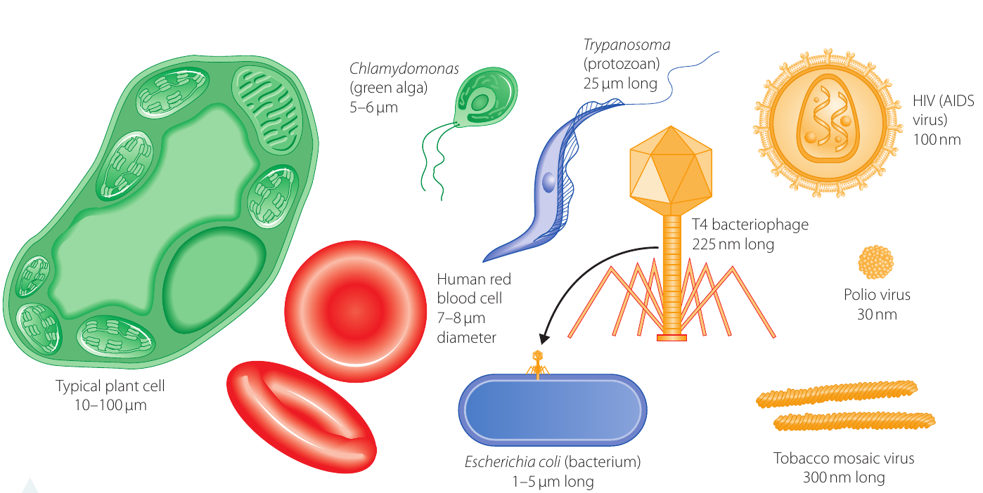
Eukaryotic cells
Plant cells are typically the largest cells:
- Size range: µm
- Visible under light microscope
- Contain chloroplasts, cell wall, and large vacuoles
Animal cells are generally smaller than plant cells:
- Human red blood cells: µm diameter
- Human cheek cells: approximately µm diameter
- Most animal cells: µm
- Visible under light microscope
Prokaryotic cells
Bacteria are much smaller than eukaryotic cells:
- Escherichia coli (E. coli): µm long
- Most bacteria: µm
- Visible under light microscope, but only just
- No membrane-bound nucleus or organelles
Protists and algae
Protozoans vary in size:
- Trypanosoma (causes sleeping sickness): approximately µm long
- Can be similar in size to eukaryotic cells
Green algae:
- Chlamydomonas: µm
- Visible under light microscope
Viruses
Viruses are much smaller than cells and require electron microscopy:
- Tobacco mosaic virus: nm long
- T4 bacteriophage: nm long
- HIV (AIDS virus): nm diameter
- Polio virus: nm diameter
- All measured in nanometres, not micrometres
- Cannot be seen with a light microscope
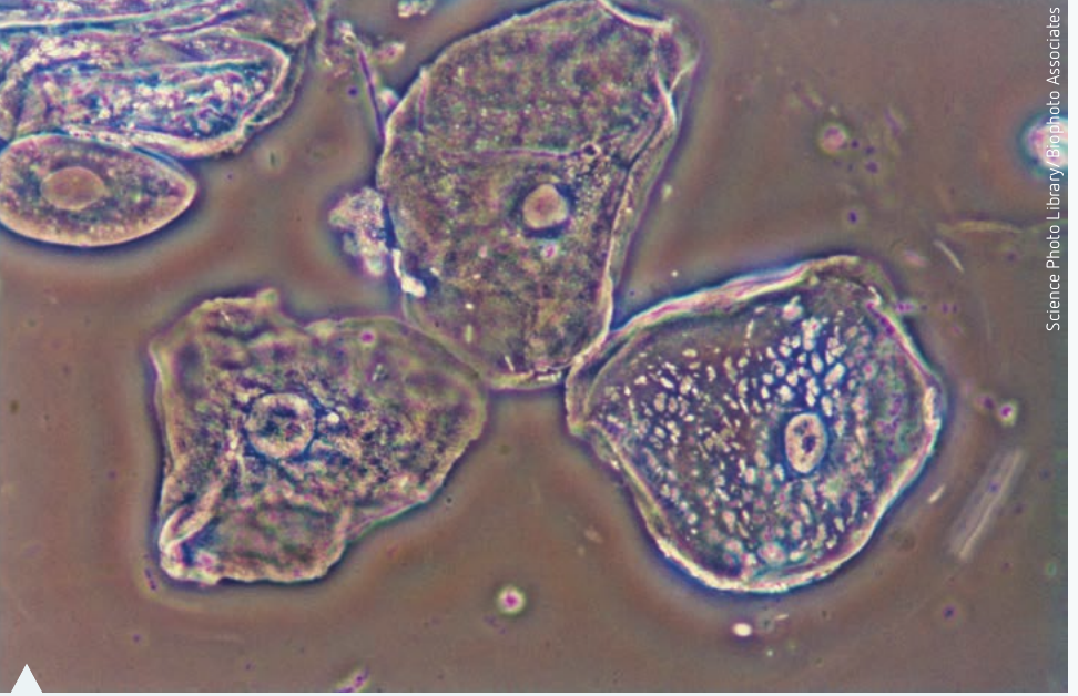
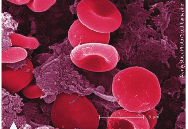
The image below shows red blood cells under an electron microscope with a scale bar for reference. Notice how the scale bar allows you to determine the actual size of these cells.
Comparing prokaryotes and eukaryotes
Understanding the Key Differences:
Prokaryotes (bacteria like E. coli):
- No nucleus
- No membrane-bound organelles
- Generally µm in size
- Simpler structure
Eukaryotes (plant cells, animal cells, protists):
- Have a membrane-bound nucleus
- Contain membrane-bound organelles
- Generally µm in size
- More complex structure
Remember: Plant cells are typically larger than animal cells, which are larger than bacteria!
Key Points to Remember:
-
The metric system uses powers of 10: Each unit is related to the metre by factors of , , , and so on. This makes conversions straightforward.
-
Different tools reveal different size ranges: The naked eye can see down to about µm, light microscopes can resolve structures down to about nm, and electron microscopes can visualize objects as small as nm.
-
Cells vary enormously in size: Plant cells ( µm) are typically larger than animal cells ( µm), which are larger than bacteria ( µm). Viruses ( nm) are even smaller and require electron microscopy to see.
-
Scale bars are essential: When drawing cells, always include a scale bar calculated using the formula: Scale = Actual length ÷ Length of drawing. This allows others to understand the true size of the specimen.
-
Key conversions to memorize: mm µm and µm nm. These conversions are fundamental for working with cell measurements in biology.