Mammalian Digestive System (HSC SSCE Biology): Revision Notes
Mammalian Digestive System
Introduction to heterotrophs and digestion
Heterotrophs are organisms that must obtain all their nutrients by consuming food from external sources. Unlike autotrophs that can make their own food, heterotrophs rely on ingesting complex foodstuffs to supply energy and the raw materials needed to build organic compounds. These complex foods are broken down by the digestive system into simpler molecules that can be absorbed into the bloodstream and used throughout the body.
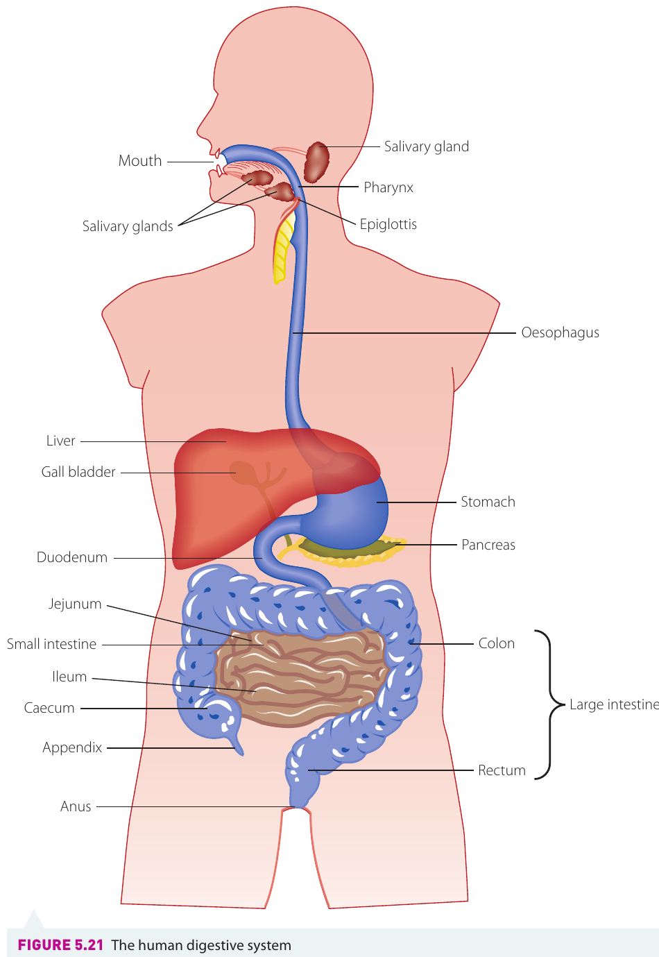
The human digestive system is a complex series of organs working together to process the food we eat. It includes the mouth, oesophagus, stomach, small intestine, large intestine, and several accessory organs such as the liver, pancreas, and gall bladder.
The key difference between heterotrophs and autotrophs lies in their nutritional strategy. Autotrophs (like plants) can manufacture their own food through photosynthesis, whilst heterotrophs (like animals) must consume pre-existing organic matter from their environment.
The process of digestion
Digestion is the process of breaking down large and complex food particles into much smaller and simpler molecules. This breakdown occurs through two complementary mechanisms: mechanical digestion and chemical digestion. The ultimate goal of digestion is to reduce food particles to a size small enough to pass through the intestinal walls and enter the bloodstream.
Mechanical digestion
Mechanical digestion involves the physical breakdown of food into smaller pieces. This process begins in the mouth, where different types of teeth work together to cut, tear, chew, and grind food. The tongue helps manipulate food during chewing, ensuring thorough breakdown. Later, the churning movements of the stomach continue this mechanical process.
The purpose of mechanical digestion is twofold. First, it breaks food into smaller pieces that are easier to swallow and move through the digestive tract. Second, and more importantly, it increases the surface area of food particles, making them more accessible to digestive enzymes during chemical digestion.
Why is surface area so important?
Think of a large cube of sugar versus the same amount of sugar granules. The granules dissolve much faster because more of the sugar is exposed to the water at once. The same principle applies to digestion - smaller pieces with greater surface area allow enzymes to work more efficiently.
Chemical digestion
Chemical digestion uses specialised proteins called digestive enzymes to chemically break down large, complex molecules into their simpler building blocks. Each enzyme targets specific types of molecules:
- Carbohydrates are broken down into simple sugars such as glucose
- Proteins are broken down into amino acids
- Lipids (fats) are broken down into glycerol and fatty acids
- Nucleic acids are broken down into nucleotides
These simpler molecules can then be absorbed through the intestinal wall and transported to cells throughout the body where they are needed.
Pathway through the digestive system
Food follows a specific path through the digestive system, with different organs performing specialised functions at each stage.
Mouth
Digestion begins the moment food enters the mouth. Here, mechanical digestion starts as the teeth break food into smaller pieces through cutting, tearing, and grinding actions. This increases the surface area of the food, making it more accessible to enzymes.
At the same time, chemical digestion begins with the release of salivary amylase, an enzyme produced by the salivary glands. This enzyme mixes with the food during chewing and begins breaking down starch (a complex carbohydrate) into maltose (a simpler sugar). The tongue plays an important role by manipulating the food during chewing and mixing it with saliva.
Once the food has been thoroughly chewed and mixed with saliva, the tongue shapes it into a ball-like mass called a bolus. This bolus is then swallowed and moves into the oesophagus.
Oesophagus
The oesophagus is a muscular tube that connects the mouth to the stomach. When the bolus is swallowed, a flap of tissue called the epiglottis covers the entrance to the trachea (windpipe). This prevents food from entering the respiratory system and ensures it travels down the correct pathway.
The epiglottis plays a critical safety role. When it fails to close properly during swallowing, food or liquid can enter the trachea, causing choking. This is why we're told not to talk or laugh whilst eating - these actions can interfere with the epiglottis closing properly.
The bolus doesn't simply fall down the oesophagus due to gravity. Instead, coordinated muscular contractions called peristalsis push the bolus along the tube. These wave-like contractions of the circular muscles surrounding the oesophagus move the food towards the stomach. During this journey, the chemical digestion of starch by salivary amylase continues.
The stomach
The stomach is a J-shaped, muscular organ that plays a crucial role in both mechanical and chemical digestion. At both the entrance and exit of the stomach are narrow openings controlled by circular muscles called sphincters.
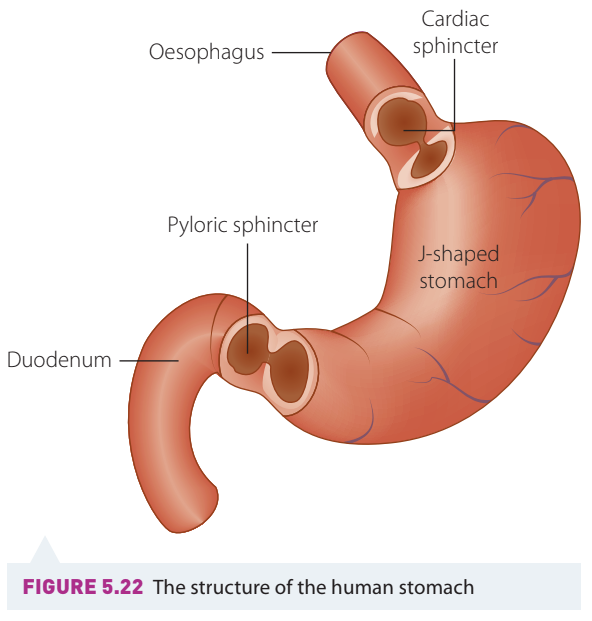
The cardiac sphincter controls entry into the stomach from the oesophagus, whilst the pyloric sphincter controls exit into the small intestine. These sphincters regulate the flow of material into and out of the stomach.
Once food enters the stomach, the muscular walls contract and relax in a churning motion. This continues the mechanical digestion begun in the mouth. The bolus breaks apart and mixes with gastric juices secreted by the stomach wall, forming a semi-liquid mixture called chyme.
Gastric juices contain several important components:
- Water - helps dissolve and mix substances
- Hydrochloric acid - creates an acidic environment with a pH of to
- Pepsinogen - an inactive enzyme precursor
- Pepsin - the active enzyme form
The acidic environment serves multiple purposes. It kills many bacteria present in food, denatures proteins making them easier to digest, and converts pepsinogen into its active form, pepsin. A protective layer of mucus lines the stomach wall, preventing the acid from damaging the stomach tissue itself.
Why produce pepsinogen instead of pepsin directly?
Pepsinogen is an inactive form of the enzyme. If the stomach cells produced active pepsin directly, the enzyme would start digesting the cells themselves! By producing the inactive form and only activating it in the stomach cavity, the cells protect themselves from self-digestion. The protective mucus layer provides additional defence.
The enzyme pepsin begins the chemical breakdown of proteins. It breaks the long chains of amino acids in proteins into shorter segments called peptides. Pepsin also breaks down nucleic acids (DNA and RNA) present in food into their component nucleotides.
The chyme typically remains in the stomach for approximately six hours before gradually moving into the small intestine.
The small intestine
The small intestine is the longest part of the digestive tract, measuring approximately metres in length in adults. Despite its name referring to its narrow diameter, this highly folded organ contains three distinct regions: the duodenum (the first section), the jejunum (the middle section), and the ileum (the final section).
The three regions of the small intestine each have distinct roles:
- Duodenum - where most digestion is completed
- Jejunum - where most absorption occurs
- Ileum - where final absorption takes place
A simple way to remember the order: Don't Just Ignore = Duodenum, Jejunum, Ileum
Chyme enters the small intestine gradually through the pyloric sphincter. As it enters the duodenum, it triggers the release of hormones that stimulate the pancreas to secrete pancreatic juices into this region.
Pancreatic juices contain a mixture of important substances:
- Bicarbonate ions - neutralise the acidic chyme from the stomach, creating a more suitable pH for intestinal enzymes
- Amylase - continues breaking down carbohydrates
- Trypsin - continues breaking down proteins
- Lipase - breaks down lipids (fats)
When lipids are present in the chyme, another substance called bile is released into the duodenum. Bile is produced by the liver and stored in the gall bladder. Unlike digestive enzymes, bile doesn't chemically break down fats. Instead, it acts like a detergent, breaking large fat globules into smaller droplets through a process called emulsification. This increases the surface area of fats, making them more accessible to the enzyme lipase, which then chemically breaks the lipids into fatty acids and glycerol molecules.
Most digestion is completed in the duodenum. From here, the digested food moves into the jejunum, where most absorption occurs.
Absorption in the small intestine
The jejunum is specially adapted for absorption. Its inner wall is lined with millions of tiny, finger-like projections called villi (singular: villus). These structures dramatically increase the surface area available for absorption, making the process much more efficient.
Each villus has several features that enhance absorption:
- Single-cell-thick walls - allow molecules to pass through easily
- Moist surface - facilitates diffusion
- Rich blood supply - a network of tiny capillaries wraps around each villus
- A lacteal - a vessel of the lymphatic system runs through the centre
Different products of digestion are absorbed into different transport systems:
-
Glucose and amino acids are absorbed into the blood capillaries
-
Fatty acids and glycerol are absorbed into the lacteal (lymph system)
-
Water is also absorbed here
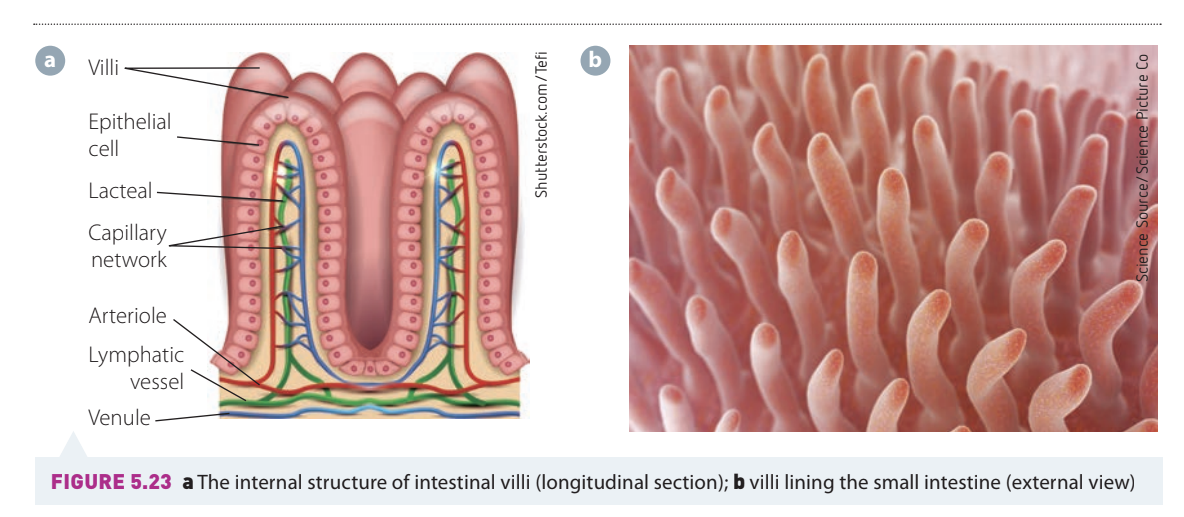
The massive surface area provided by villi is crucial for efficient absorption. If the small intestine were just a smooth tube, it would need to be many times longer to absorb the same amount of nutrients. The villi allow for maximum absorption in a compact space.
The products move through the villi walls by diffusion (moving from high to low concentration) or active transport (using energy to move against a concentration gradient). From the blood capillaries and lymph vessels, these nutrients are distributed throughout the body to wherever they are needed.
The liver - an accessory gland
Although not part of the digestive tract itself, the liver plays a vital role in processing digested food. Once nutrients are absorbed into the bloodstream from the small intestine, they travel directly to the liver.
The liver acts as the central metabolic hub of the body. It regulates the levels of sugars, glycogen (stored carbohydrate), and proteins in the blood, ensuring they remain balanced. The liver also performs an important detoxification function, removing harmful substances from the blood before it circulates to the rest of the body.
The large intestine
After all digestible nutrients have been absorbed in the small intestine, the remaining material moves into the large intestine. This leftover material consists primarily of water, mineral salts, and dietary fibre that the body cannot digest.
The large intestine has two main sections: the colon and the rectum. In the colon, water and some mineral salts are reabsorbed back into the bloodstream. This reabsorption concentrates the remaining waste material into a more solid form.
Beneficial bacteria in the colon
The colon houses billions of beneficial bacteria that live in a symbiotic relationship with our bodies. These bacteria feed on undigested material and produce vitamins A and K as byproducts, which are then absorbed into our bloodstream. This mutually beneficial relationship helps us obtain essential vitamins whilst providing the bacteria with nutrients and a habitat.
The remaining waste material, now called faeces, is moved into the rectum through peristalsis. The faeces are stored in the rectum until they are eliminated from the body through the anus in a process called egestion.
The fate of digestive products
Once absorbed into the bloodstream, the products of digestion are transported throughout the body where they serve various purposes. Cells can reassemble these simple molecules into more complex structures needed by the body.
Structural uses include:
- Lipids and proteins become part of cell membranes
- Proteins form muscle fibres and connective tissue
- Proteins are used to make enzymes and hormones
Energy storage includes:
- Fatty tissue stores lipids beneath the skin and around organs
- Glycogen (a form of "animal starch") is stored in the liver and muscles
- Unlike carbohydrates and fats, proteins cannot be stored and must be used or broken down
Why can't proteins be stored?
Unlike carbohydrates (stored as glycogen) and fats (stored in adipose tissue), the body has no mechanism for storing excess proteins. Any amino acids not immediately needed for building proteins or other molecules are broken down, with the nitrogen-containing parts converted to urea and excreted. This is why a consistent dietary protein intake is important.
This efficient system ensures that the nutrients we obtain from food are used effectively throughout the body for growth, repair, and energy production.
Remember!
Key Points to Remember:
-
Digestion involves both mechanical and chemical processes. Mechanical digestion physically breaks food into smaller pieces (teeth, stomach churning), whilst chemical digestion uses enzymes to break large molecules into smaller ones.
-
Different organs have specialised roles. The mouth begins digestion, the stomach continues it with acid and pepsin, the small intestine completes most digestion and absorption, and the large intestine absorbs water and forms waste.
-
Enzymes are specific to their substrates. Amylase breaks down carbohydrates to sugars, pepsin and trypsin break down proteins to amino acids, and lipase (with help from bile) breaks down lipids to fatty acids and glycerol.
-
The small intestine is the main site of absorption. Villi provide a large surface area, and nutrients are absorbed into either blood capillaries (glucose, amino acids) or lacteals (fatty acids, glycerol).
-
The digestive system is a continuous pathway. Food travels from mouth → oesophagus → stomach → small intestine (duodenum, jejunum, ileum) → large intestine (colon, rectum) → anus, with each section performing specific functions.