Apoptosis (VCE SSCE Biology): Revision Notes
Apoptosis
Introduction to apoptosis
Have you ever wondered why most people don't have webbed fingers or toes? During foetal development, our fingers and toes are initially fused together by skin. As we grow, specific cells between our digits undergo a controlled death process, separating our fingers and toes. When this process doesn't work properly, babies can be born with webbed digits, a condition called syndactyly that affects approximately one in every 2,000-3,000 births. This controlled cell death is called apoptosis.
Apoptosis is the controlled death of cells in the body. Also known as programmed cell death, it is a natural and essential process that plays a vital role in our development and daily bodily functions.
The scale of apoptosis
Our bodies contain approximately 30-40 trillion cells. Remarkably, scientists estimate that around 300 million cells die every minute through apoptosis and are typically replaced by healthy cells. This constant turnover helps maintain tissue health and removes cells that are damaged, malfunctioning, or simply no longer needed.
When cells begin to malfunction, sustain damage, or become unnecessary, they receive signals that trigger apoptosis, leading to their eventual death. This process is highly regulated and occurs without causing inflammation or damage to surrounding tissues.
Pathways of apoptosis
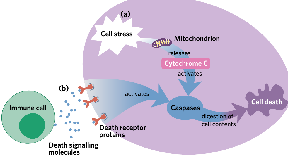
There are two distinct pathways that can initiate apoptosis: the mitochondrial pathway and the death receptor pathway. Both pathways ultimately lead to the activation of caspase enzymes, which are catalysts that cleave specific intracellular proteins during apoptosis. Once caspases are activated, the two pathways follow nearly identical steps.
Mitochondrial pathway (intrinsic pathway)
The mitochondrial pathway is the pathway of apoptosis which is initiated by the detection of internal cellular damage. Also known as the intrinsic pathway, this route responds to problems within the cell itself.
When internal cellular components (particularly DNA) become damaged, mitochondria detect this damage and respond by releasing cytochrome c (a protein embedded in the inner mitochondrial membrane) into the cytosol. The released cytochrome c binds with other cytosolic proteins to form a complex structure called an apoptosome. This apoptosome then activates caspase enzymes, initiating the apoptosis process.
Death receptor pathway (extrinsic pathway)
The death receptor pathway is the pathway of apoptosis which is initiated by the reception of extracellular death signalling molecules. Also known as the extrinsic pathway, this route responds to external signals.
Death signalling molecules can be released by immune cells and are recognised by death receptor proteins located on the cell surface. When these signalling molecules bind to death receptor proteins, they directly activate caspase enzymes, initiating apoptosis.
Memory Aid: Intrinsic vs Extrinsic
To remember which pathway is which, note that mitochondria are found inside cells. Therefore, the mitochondrial pathway is the internal (intrinsic) pathway of apoptosis, triggered by internal signals. This means the death receptor pathway must be the external (extrinsic) pathway of apoptosis, triggered by signals from outside the cell.
Apoptosis vs necrosis
It's important to distinguish apoptosis from another form of cell death called necrosis. Necrosis is the unregulated death of cells initiated by significant damage. Unlike apoptosis, necrosis causes cells to swell, burst, and release their contents into the surrounding environment.
Key Difference: Necrosis leads to inflammation and damage in nearby cells and tissues, making it a harmful process rather than a controlled, protective one like apoptosis.
Stages of apoptosis
Following caspase activation and the initiation of apoptosis, the process continues through four distinct stages:
The Four Stages of Apoptosis
Stage 1: Activation of caspases The mitochondria detect internal DNA damage and release cytochrome c, or death receptors on the cell surface receive external death signals. Either way, caspase enzymes become activated.
Stage 2: Digestion of cell contents Activated caspases cleave (cut) intracellular proteins throughout the cell. This protein breakdown leads to the digestion and dismantling of cellular organelles.
Stage 3: Cell shrinkage As intracellular material is broken down, both the cell and its nucleus shrink and condense. The cell becomes smaller and more compact.
Stage 4: Membrane blebbing and formation of apoptotic bodies As the cytoskeleton is digested, the cell loses its structural integrity. The plasma membrane begins to warp and bulge outward in a process called blebbing (the bulging of the plasma membrane to form apoptotic bodies). These bulges eventually pinch off from the cell, forming membrane-enclosed vesicles called apoptotic bodies (vesicles containing cell contents that are released from a dying cell during apoptosis and engulfed by phagocytes).
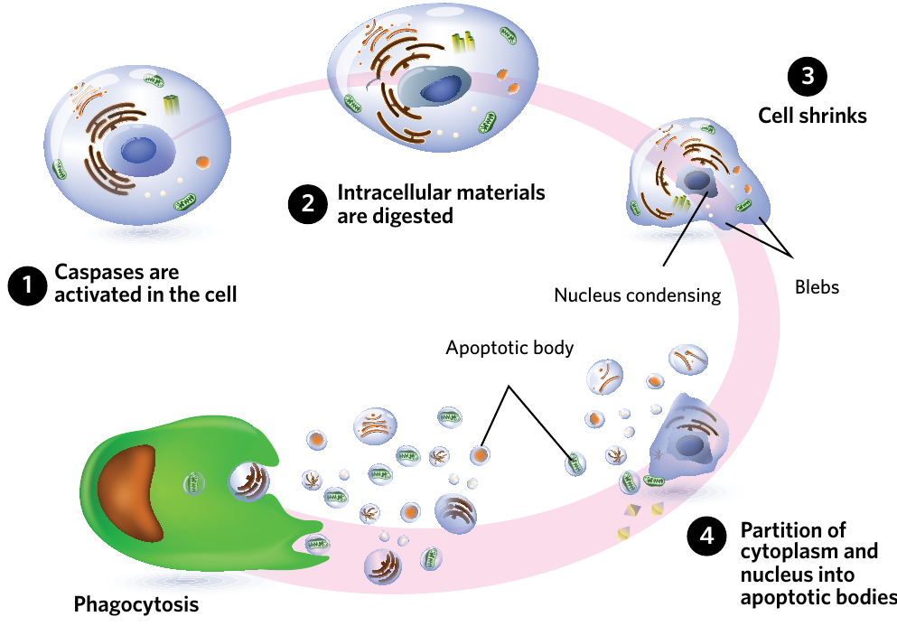
Cleanup Process:
After apoptosis is complete, phagocytes (cells of the immune system responsible for engulfing and destroying harmful microorganisms and foreign material) engulf and digest the free-floating apoptotic bodies through phagocytosis (endocytosis of solid material or food particles). This cleanup process ensures that the remains of dead cells don't cause inflammation or harm to surrounding tissues.
When apoptosis goes wrong
Proper functioning of apoptosis is vital for healthy organism development and maintenance. However, when apoptosis malfunctions, serious diseases can result.
Decreased apoptosis: cancer development
When apoptosis fails to occur at the appropriate rate, damaged or abnormal cells can continue to replicate, potentially leading to cancer.
Cell cycle checkpoints
Recall that the eukaryotic cell cycle is regulated by checkpoints that monitor cell health:
- G1 checkpoint – inspects for DNA damage before the cell commits to DNA replication
- G2 checkpoint – confirms that DNA has correctly replicated during S phase
- Metaphase checkpoint – confirms that spindle fibres have correctly attached to chromosome centromeres
When errors are detected at any checkpoint, the cell should either repair the damage or undergo apoptosis. However, apoptosis failure isn't always due to checkpoint errors. For example, cells may stop expressing functional death receptor proteins, preventing death signalling molecules from initiating apoptosis. When the rate of apoptosis decreases too much, abnormal cells survive and continue dividing.
Tumour formation
When apoptosis rates are insufficient, cell growth can increase exponentially, resulting in the formation of a tumour (a mass of abnormal cells). Tumours are classified into two categories:
Benign tumours are relatively slow-growing masses of cells that are generally enclosed within a capsule. This capsule prevents the abnormal cells from separating and invading other parts of the body. While benign tumours can cause problems by pressing on nearby structures, they don't spread throughout the body.
Malignant tumours develop when cells from benign tumours mutate further and gain the ability to invade nearby tissues and/or enter the bloodstream or lymphatic system. Once in circulation, these cells can travel to distant parts of the body and establish new tumours. Malignant tumours are also known as cancerous cells (abnormal cells with the ability to invade nearby tissue and migrate to other parts of the body).
Cancer Defined:
Cancer is a disease caused by the uncontrolled replication of cells with the ability to migrate to other parts of the body. The key difference between benign and malignant tumours is that only malignant tumours can undergo metastasis (the migration of tumour cells from the primary tumour site to distant parts of the body).
Characteristics of cancer cells
| Characteristic | Description |
|---|---|
| Self-sufficiency | Typically, cells require chemical growth signals to initiate cell replication. However, in tumour cells, they can replicate without these signals by either producing their own chemical signals, or by permanently activating cell growth and replication pathways. |
| Antigrowth deactivation | There are many different mechanisms present in cells to prevent cell replication when it is not needed. In tumour cells, these mechanisms can be disabled, thereby allowing cell replication to initiate. |
| Increased survival | Due to mutations in the regulation of the cell cycle, apoptosis no longer functions correctly in tumour cells. Tumour cells are also capable of replicative immortality, which theoretically allows them to divide forever, enhancing their survival. However, in practice, due to limitations such as the inability for blood vessels to form in the centre of tumours and provide the necessary nutrients, tumour cells can still die. |
| Blood supply formation | Tumour cells can form new blood vessels when growing to maintain adequate nutrient and oxygen supply. |
| Tissue invasion and metastasis | When benign tumour cells become malignant/cancerous they are capable of invading nearby layers of tissue and migrating to other parts of the body away from the primary tumour site, typically via the bloodstream or lymphatic system (metastasis). |
The table above shows five hallmark characteristics of cancer cells. Note that the final characteristic—tissue invasion and metastasis—is unique to malignant cancer cells and doesn't occur in benign tumours.
Increased apoptosis: neurological disorders
While decreased apoptosis can lead to cancer, increased apoptosis also causes serious health problems. When apoptosis occurs at an abnormally high rate, healthy cells undergo programmed cell death unnecessarily.
Many neurological disorders (diseases affecting the nervous system) are linked to increased rates of apoptosis in brain cells, particularly neurones. As these cells die, the total number of neurones and neurological connections in the brain decreases. This leads to common symptoms including difficulty with movement, mood changes, and significant decreases in cognitive ability.
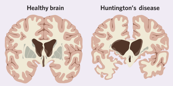
Huntington's Disease Example:
Huntington's disease is one example of a neurological disorder where excessive apoptosis causes large tissue gaps in the brain. The comparison above shows a healthy brain versus a brain affected by Huntington's disease, where you can see the dramatic loss of brain tissue due to excessive cell death.
Remember!
Key Takeaways:
-
Apoptosis is programmed cell death – a natural, controlled process where approximately 300 million cells die every minute in our bodies and are replaced by healthy cells.
-
Two pathways initiate apoptosis – the mitochondrial pathway (intrinsic) responds to internal cellular damage, while the death receptor pathway (extrinsic) responds to external death signals. Both activate caspase enzymes.
-
Apoptosis follows four stages – caspase activation, digestion of cell contents, cell shrinkage, and membrane blebbing to form apoptotic bodies, which are then cleaned up by phagocytes.
-
Decreased apoptosis can cause cancer – when apoptosis fails, damaged cells continue dividing and can form tumours. Malignant tumours (cancer) can invade other tissues and spread through metastasis, unlike benign tumours which remain localised.
-
Increased apoptosis causes neurological disorders – excessive programmed cell death in the brain leads to loss of neurones and brain tissue, causing symptoms like movement difficulties and cognitive decline, as seen in Huntington's disease.