The Eukaryotic Cell Cycle (VCE SSCE Biology): Revision Notes
The Eukaryotic Cell Cycle
Introduction to the cell cycle
The eukaryotic cell cycle is a continuous, highly organised process that allows cells to grow and produce new cells. This cycle is essential for growth, development, tissue repair, and reproduction in all eukaryotic organisms. Unlike prokaryotic cells which divide through binary fission, eukaryotic cells follow a more complex pathway involving multiple carefully regulated stages.
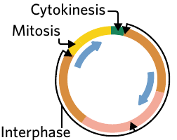
The cell cycle consists of three major stages that occur in sequence: interphase, mitosis, and cytokinesis. Each stage plays a crucial role in ensuring that daughter cells receive the correct genetic information and cellular components.
The cycle is continuous, meaning that once cells complete cytokinesis, they can immediately enter interphase again to prepare for another round of division. However, not all cells continuously divide - some exit the cycle and enter a resting state when division is not needed.
The three main stages of the cell cycle
Interphase
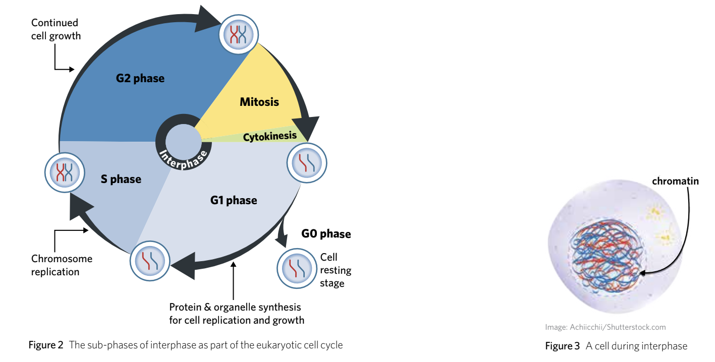
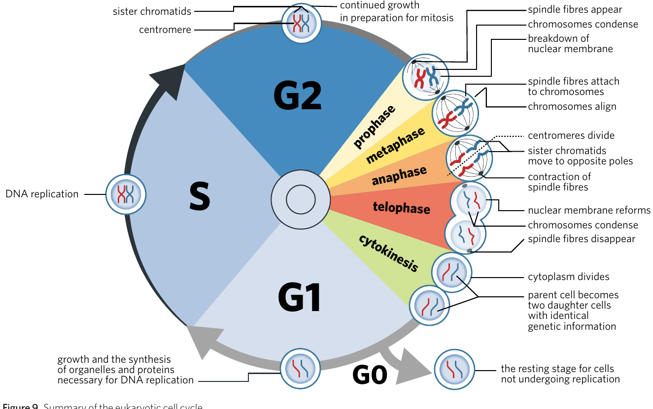
Interphase is the first stage of the eukaryotic cell cycle which involves cellular growth and duplication of chromosomes. It is composed of three phases: G1, S, and G2.
This is the longest stage of the cell cycle, typically lasting much longer than mitosis and cytokinesis combined. During interphase, cells carry out their normal functions while simultaneously preparing for potential division.
Mitosis
Mitosis is the second stage of the eukaryotic cell cycle, which involves the complete separation of sister chromatids and nuclei.
This stage ensures that genetic material is accurately divided between the two forming cells. Mitosis itself consists of four distinct sub-stages that we will explore in detail later in this note.
Cytokinesis
Cytokinesis is the division of the cytoplasm and formation of two daughter cells.
This final stage physically separates one cell into two complete, independent cells. Each daughter cell receives approximately half of the original cell's cytoplasm and organelles.
Interphase in detail
Interphase is when cells spend most of their time. During this period, the cell grows larger, replicates its DNA, and produces the proteins and organelles necessary for division. The DNA during interphase exists as loosely packed chromatin - chromosomes (DNA and proteins) that have been unwound and loosely packed during interphase - rather than as the tightly coiled chromosomes visible during mitosis.
Understanding chromatin vs chromosomes:
During interphase, DNA exists as chromatin - a loosely packed, thread-like form that allows the cell to access genes for normal functions. Only during mitosis does chromatin condense into the tightly coiled chromosomes that are visible under a microscope. This condensation is essential for the orderly separation of genetic material.
Interphase consists of three main sub-phases (G1, S, and G2), with an additional optional phase (G0) that cells can enter if they do not need to divide.
The G1 phase
The G1 (Gap 1) phase is the first growth period of interphase. During this phase, the cell actively grows and prepares for DNA replication by:
- Increasing the volume of its cytosol (the fluid component of cytoplasm)
- Synthesising proteins that will be needed for DNA replication
- Replicating its organelles such as mitochondria and ribosomes
This phase ensures the cell is large enough and has sufficient resources before committing to DNA replication. At the end of G1, the cell reaches a critical decision point: it can either proceed to the S phase to continue the cycle, or exit the cycle and enter the G0 phase.
The G0 phase
The G0 (Gap 0) phase is a resting stage where cells that are not required to replicate remain. Cells in G0 can be categorised as either quiescent or terminally differentiated.
Quiescent cells are dormant cells which can re-enter the cell cycle when needed. For example, liver cells are generally quiescent. When the liver is damaged or partially removed, these cells can re-enter the cell cycle to regenerate the lost tissue.
Terminally differentiated cells have fully specialised and no longer replicate. These cells remain in G0 indefinitely and cannot return to the cell cycle.
Most nerve cells (neurons) in the brain and spinal cord are terminally differentiated, which is why nervous system injuries are often permanent and why protective equipment like helmets is so important. Unlike quiescent cells, these neurons cannot re-enter the cell cycle to repair damage.
The S phase
The S (Synthesis) phase is when DNA replication occurs. This is arguably the most critical phase of the cell cycle because the cell must accurately duplicate its entire genome.
During the S phase, each chromosome - a structure composed of DNA tightly wrapped around histone proteins that carries the genetic information (genes) of a cell - is replicated to produce two identical copies. These copies are called sister chromatids - the two identical halves of a replicated chromosome.
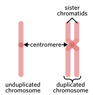
The sister chromatids remain joined together at a region called the centromere - the structure which holds sister chromatids together. While the sister chromatids are connected, they are still considered a single chromosome. Only when they separate during mitosis does each chromatid (one half of a double-stranded chromosome) become an individual chromosome.
Understanding diploid cells and chromosome numbers
In humans, most of our cells are somatic cells - any cell that is not a reproductive cell (such as sperm and egg cells). Somatic cells contain two sets of chromosomes, one inherited from each parent. Cells with two sets of chromosomes are described as diploid - cells or organisms that have two sets of chromosomes ().
Humans have 23 pairs of chromosomes, giving us a diploid number of (23 pairs × 2 = 46 total chromosomes). After the S phase, each human somatic cell still contains 46 chromosomes, but each chromosome now consists of two sister chromatids joined at the centromere. This means the DNA content has doubled, even though the chromosome count remains at 46.
Worked Example: Calculating Diploid Number
The diploid number can be calculated using the formula:
For humans with 23 chromosome pairs:
This means human somatic cells contain 46 total chromosomes.
Other examples:
- Sheep: (27 pairs)
- Potatoes: (24 pairs)
Note: A higher chromosome number does not necessarily indicate a more complex organism.
The G2 phase
The G2 (Gap 2) phase is the final stage of interphase. During this phase, the cell continues to grow and makes final preparations for mitosis. The activities in G2 are similar to those in G1, including:
- Increasing the volume of the cytosol
- Synthesising proteins, specifically those needed for the mitotic process
By the end of G2, the cell has completed all necessary preparations and is ready to enter mitosis.
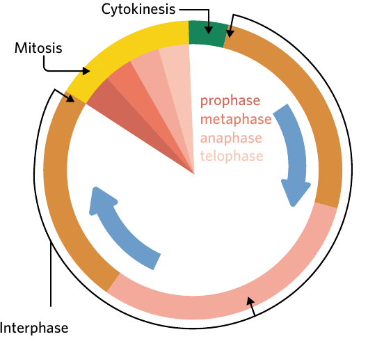
Mitosis
Mitosis is the stage where the replicated chromosomes are separated and two new nuclei form. This ensures that each daughter cell receives an identical and complete set of genetic information. Mitosis consists of four distinct sub-stages: prophase, metaphase, anaphase, and telophase.
Helpful Mnemonic for Remembering the Stages:
To help remember these stages in order, you can use this helpful mnemonic:
Isaac Please Make Another Two Cells
- I = Interphase
- P = Prophase
- M = Metaphase
- A = Anaphase
- T = Telophase
- C = Cytokinesis
This mnemonic helps you remember not just the mitosis stages, but the entire sequence including interphase and cytokinesis!
Let's examine each stage of mitosis in detail.
Prophase
Prophase marks the beginning of mitosis and involves several important changes in the cell:
Chromosome condensation occurs - the shortening and thickening of chromosomes as DNA is tightly wrapped around histone proteins. This condensation makes the chromosomes visible under a microscope, whereas during interphase they existed as loosely packed chromatin threads.
Centrioles - cylindrical structures composed of protein which form the spindle fibres during mitosis and meiosis - migrate to opposite ends (poles) of the cell. As they move, spindle fibres - structures which aid in the movement of chromosomes to either pole of the cell during mitosis and meiosis - begin to form between them.
The nuclear membrane breaks down and disintegrates, allowing the spindle fibres to access the chromosomes. The nucleolus (the dense region inside the nucleus) also disappears during this stage.
Metaphase
During metaphase, the spindle fibres fully develop and attach to the centromere of each chromosome. These spindle fibres guide the chromosomes towards the centre of the cell.
The chromosomes line up along the equator - the centre line between opposite ends of the cell that the chromosomes line up on during metaphase. This central line is also called the metaphase plate.
The alignment of chromosomes at the equator is crucial for ensuring that each daughter cell receives one copy of each chromosome. If chromosomes are not properly aligned, the cell will not proceed to the next stage - this is monitored by the metaphase checkpoint.
Anaphase
Anaphase is characterised by the separation of sister chromatids. The spindle fibres contract, which causes the centromere holding the sister chromatids together to split. Once separated, each chromatid is now considered an individual chromosome.
The spindle fibres then pull the separated chromatids to opposite poles of the cell. This ensures that each end of the cell receives one copy of each chromosome. By the end of anaphase, both poles of the cell have a complete set of chromosomes.
In animal cells, a cleavage furrow - an indentation of the plasma membrane during cytokinesis - begins to form during late anaphase, preparing for cell division.
Telophase
Telophase is essentially the reverse of prophase. The chromosomes arrive at opposite poles of the cell and begin to decondense, becoming less tightly packed. New nuclear membranes form around each set of chromosomes, creating two distinct nuclei within the same cell. The spindle fibres break down and disappear.
At this point, the cell has successfully created two genetically identical nuclei, each containing the same genetic information as the original cell. The cell is now ready for cytokinesis.
Cytokinesis
After mitosis creates two nuclei, cytokinesis divides the cytoplasm to produce two separate daughter cells - the formation of a new cell following cell replication. The mechanism of cytokinesis differs between animal and plant cells due to structural differences, particularly the presence of a rigid cell wall in plant cells.
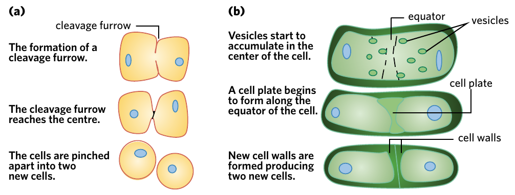
Cytokinesis in animal cells
In animal cells, cytokinesis occurs through the formation and contraction of a cleavage furrow. This furrow is an indentation that forms in the plasma membrane during anaphase and telophase.
The cleavage furrow deepens as proteins inside the cell contract, much like a drawstring being pulled. The furrow continues to deepen until it reaches the centre of the cell, at which point the plasma membrane pinches completely in two. This separates the original cell into two independent daughter cells.
Cytokinesis in plant cells
Plant cells cannot use a cleavage furrow because they have a rigid cell wall that cannot be pinched inward. Instead, plant cells form a cell plate - a component involved in the formation of a cell wall.
Small membrane-bound sacs called vesicles, containing cell wall materials, accumulate at the equator of the cell. These vesicles fuse together to form the cell plate. The cell plate grows outward from the centre until it reaches and fuses with the existing cell wall. As the cell plate develops, new plasma membranes and cell walls form on either side, effectively separating the cell into two daughter cells.
Key Difference: Animal vs Plant Cytokinesis
- Animal cells: Use a cleavage furrow that pinches inward like a drawstring
- Plant cells: Form a cell plate that grows outward from the centre
This difference exists because plant cells have a rigid cell wall that cannot be pinched, requiring the construction of a new wall from the inside out.
Cell cycle regulation
The cell cycle is carefully regulated by checkpoints that monitor the cell for errors and ensure conditions are suitable for division. There are three major checkpoints: the G1 checkpoint, the G2 checkpoint, and the metaphase checkpoint. At each checkpoint, the cell assesses whether it should proceed to the next stage, pause for repairs, or undergo programmed cell death if damage is irreparable.
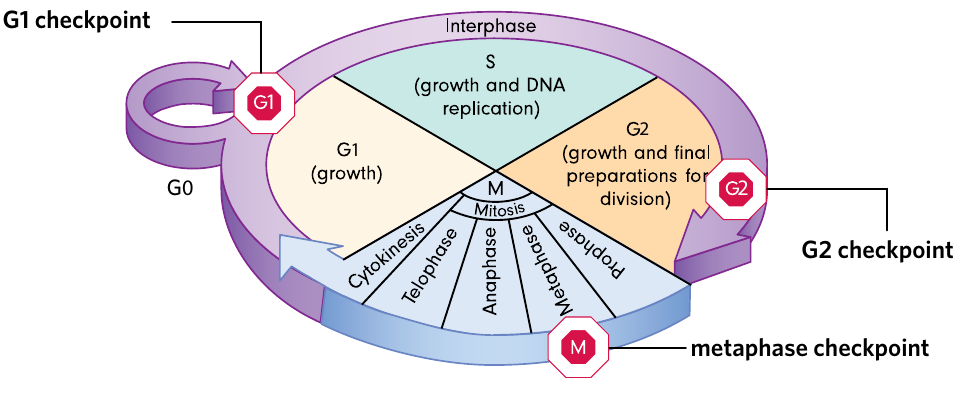
The G1 checkpoint
The G1 checkpoint occurs at the end of the G1 phase, before the cell commits to DNA replication. This checkpoint is sometimes called the restriction point. The cell verifies several conditions:
- Has the cell grown to an adequate size?
- Has the cell synthesised enough proteins for DNA replication?
- Is the DNA damaged from previous rounds of division or from environmental factors?
- Are there sufficient nutrients and oxygen available (favourable conditions for mitosis)?
If all conditions are met, the cell proceeds to the S phase. If conditions are unfavourable, the cell may enter the G0 phase and wait until conditions improve.
If DNA damage is severe and irreparable, the cell may undergo programmed cell death. This mechanism prevents damaged cells from dividing and potentially becoming cancerous. It's better for the organism to lose one cell than to allow a damaged cell to produce many faulty daughter cells.
The G2 checkpoint
The G2 checkpoint occurs at the end of the G2 phase, after DNA replication but before mitosis begins. This checkpoint ensures:
- Has DNA replication occurred properly during the S phase?
- Is there any DNA damage that needs repair?
- Does the cell have enough resources and energy for mitosis?
This checkpoint is the final quality control before the cell commits to the energy-intensive process of mitosis. If problems are detected, the cycle pauses for repairs. Cells with irreparable damage undergo programmed cell death.
The metaphase checkpoint
The metaphase checkpoint, also called the spindle checkpoint, occurs during metaphase of mitosis. This checkpoint monitors:
- Have all chromosomes correctly attached to spindle fibres?
- Are all chromosomes properly aligned at the equator of the cell?
The cell will not proceed to anaphase until all chromosomes are correctly positioned and attached. This is crucial because premature separation could result in daughter cells receiving incorrect numbers of chromosomes. Once all chromosomes are properly aligned and attached, the cell proceeds to anaphase.
Summary
The eukaryotic cell cycle is a sophisticated and highly regulated process that enables cells to grow and reproduce. The cycle consists of three major stages:
Interphase is the longest stage, during which cells grow, replicate their DNA, and prepare for division. It includes the G1 phase (growth and organelle replication), the S phase (DNA replication creating sister chromatids), the G2 phase (final preparation), and the optional G0 phase (resting state for cells not dividing).
Mitosis separates the replicated chromosomes into two identical nuclei through four sub-stages. In prophase, chromosomes condense and become visible, centrioles move to opposite poles, and the nuclear membrane breaks down. In metaphase, chromosomes align at the cell's equator. In anaphase, sister chromatids separate and move to opposite poles. In telophase, nuclear membranes reform around each set of chromosomes.
Cytokinesis divides the cytoplasm and creates two daughter cells. Animal cells use a cleavage furrow that pinches the cell in two, while plant cells form a cell plate that grows outward to divide the cell.
The cell cycle is monitored by three checkpoints (G1, G2, and metaphase) that ensure the cell is ready to proceed and that no errors have occurred. Cells with irreparable damage undergo programmed cell death rather than producing faulty daughter cells.
Key Points to Remember:
- The eukaryotic cell cycle consists of three main stages: interphase (growth and DNA replication), mitosis (nuclear division), and cytokinesis (cytoplasmic division).
- Interphase has three phases: G1 (growth), S (DNA synthesis creating sister chromatids), and G2 (preparation for mitosis), plus the optional G0 resting phase.
- Mitosis involves four stages that can be remembered using the mnemonic "Isaac Please Make Another Two Cells": Interphase, Prophase, Metaphase, Anaphase, Telophase, Cytokinesis.
- Animal cells divide by forming a cleavage furrow that pinches inward, while plant cells form a cell plate that grows outward because of their rigid cell wall.
- Three checkpoints (G1, G2, and metaphase) monitor the cell cycle to ensure proper growth, accurate DNA replication, and correct chromosome alignment before division proceeds.