The Hindbrain, Midbrain, Forebrain, and Cerebral Cortex (VCE SSCE Psychology): Revision Notes
The Hindbrain, Midbrain, Forebrain, and Cerebral Cortex
Introduction to brain regions
The human brain is divided into four major regions, each with specialized structures and functions. These regions are the hindbrain, midbrain, forebrain, and cerebral cortex. Understanding these divisions helps us grasp how different areas of the brain contribute to behaviour and mental processes.
Optional: Basics of the nervous system
This section reviews foundational knowledge about neurons and the nervous system organization.
The role of neurons
Neurons are specialized cells that form the nervous system. The human body contains approximately 100 billion neurons, which link together to create neural pathways that transmit information throughout the body. These cells serve as the functional units of the nervous system, each with specialized roles such as receiving information, transmitting signals between neurons, and sending messages to cells and muscles.
Structure of a neuron
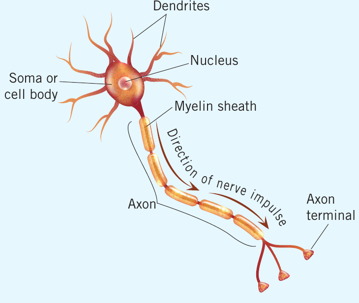
A typical neuron consists of several key components:
Dendrites are branching projections extending from the cell body that receive information from other neurons. Some dendrites contain additional projections called dendritic spines, which play a role in learning and memory by allowing thousands of connections between neurons.
Soma (cell body) serves as the coordinating centre of the neuron. It receives information from dendrites and passes it down the axon. The soma contains the nucleus with the neuron's genetic material.
Axon is a fibre that transmits signals, allowing the neuron to send information to multiple other cells. Electrical messages (nerve impulses) travel along the axon in one direction only.
Myelin is a fatty substance covering most axons. It acts as insulation, preventing signal leakage and speeding up information transmission through the nervous system.
Axon terminals are structures at the axon's end that store and release neurotransmitters—chemical messengers that carry information across synapses to the next neuron.
Synapse is the gap between neurons where neurotransmitters travel from the axon terminal of one neuron to the dendrite or cell body of the next.
The one-directional flow of information through neurons—from dendrites through the soma and axon to axon terminals—ensures orderly and efficient signal transmission throughout the nervous system. This directional property is crucial for coordinated bodily functions and responses.
Organization of the nervous system
The nervous system has two main divisions: the central nervous system (CNS) and peripheral nervous system (PNS).
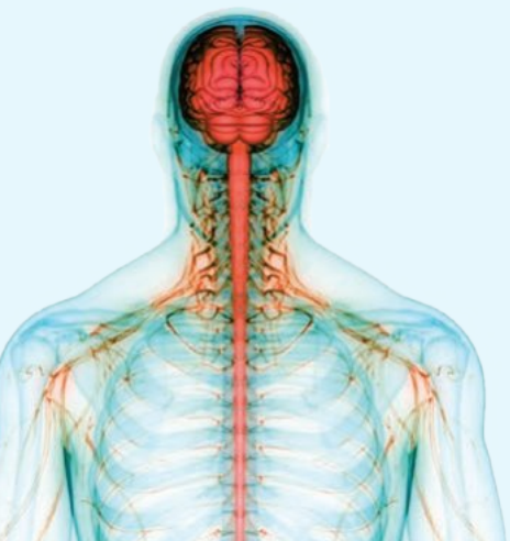
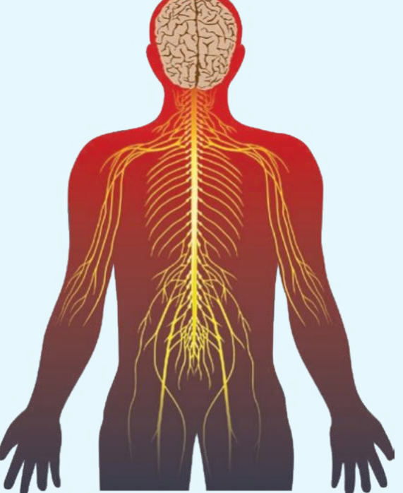
The CNS comprises the brain and spinal cord. Protected by the skull and vertebral column, the CNS receives information from the PNS, processes it, and activates appropriate body responses.
The spinal cord extends from the brainstem down to the lumbar region. It carries sensory information upward to the brain and motor information downward from the brain to organs, glands, and muscles.
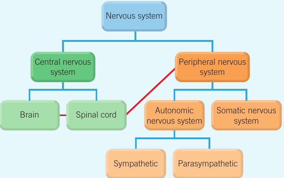
The PNS consists of all nerves outside the CNS. It relays information from sense organs, muscles, and glands to the CNS and delivers instructions back to these structures. The PNS has two subdivisions:
Somatic nervous system carries sensory information to the CNS and transmits motor instructions from the CNS to skeletal muscles for voluntary movements.
Autonomic nervous system controls internal organs and glands unconsciously. It has two subdivisions:
- Sympathetic nervous system: Activates the fight-or-flight-or-freeze response during dangerous or stressful situations
- Parasympathetic nervous system: Activates the rest-and-digest response after stress has passed
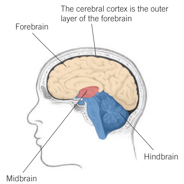
The hindbrain
The hindbrain is located in the lower rear part of the brain and comprises three main structures: the cerebellum, medulla oblongata, and pons. This region supports vital bodily processes including breathing and sleep.
The hindbrain supports vital processes essential for survival, including breathing and heart rate regulation. Because these functions are critical to life, damage to hindbrain structures—particularly the medulla oblongata—can be life-threatening.
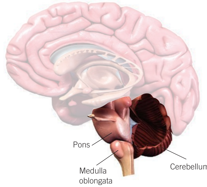
Cerebellum
The cerebellum receives commands from the cerebral cortex and coordinates their execution. It plays a role in coordinating voluntary movements, maintaining balance and posture, and controlling movements associated with speech and vision.
When we make movements involving multiple muscle groups—such as picking up a cup—the cerebellum ensures smooth, coordinated action. It activates the required muscles and controls the degree of their engagement, often without conscious thought.
The cerebellum also contributes to learning and memory by storing motor control information. This enables us to develop skills like playing an instrument or riding a bicycle.
Mnemonic for hindbrain structures: "Can Monkeys Play?" helps you remember the three main structures: Cerebellum, Medulla oblongata, and Pons.
Damage to the cerebellum can result in balance problems, difficulties detecting visual motion, and loss of muscle coordination.
Medulla oblongata
The medulla oblongata sits at the brain's base where the brainstem connects to the spinal cord. It contains control centres for many autonomic functions including heart rate, breathing, salivating, blood pressure, swallowing, vomiting, and sneezing.
Given the vital nature of these functions, damage to this area can cause death or severe health problems.
Pons
The pons is located above the medulla oblongata and below the midbrain. It acts as a bridge connecting the cerebellum and cerebral cortex. Additionally, the pons is involved in sleep, arousal, facial expressions, and hearing.

The midbrain
The midbrain sits at the topmost part of the brainstem in the brain's centre, connecting upper and lower brain areas. It is involved in auditory and visual processing, motor control, pain inhibition, and reward-based learning patterns. The midbrain contains two important structures: the substantia nigra and reticular formation.
Substantia nigra
The substantia nigra represents one of the brain's largest collections of dopamine-producing neurons. Dopamine is a neurotransmitter involved in movement and coordination.
Damage to nerve cells in the substantia nigra is linked to Parkinson's disease, which features tremors and movement difficulties. This connection highlights the critical role of dopamine in coordinating smooth, controlled movements.
Reticular formation and reticular activating system
The reticular formation is a complex arrangement of neuron clusters in the midbrain, connected to a network running from the hindbrain to the forebrain. Its neurons play a role in maintaining arousal, consciousness, and motor control.
Within the reticular formation lies the reticular activating system (RAS). When stimulated, the RAS causes alertness and awakening. It has ascending and descending pathways extending into various brain areas and the spinal cord. The neuron groupings comprising the RAS are responsible for attention, arousal, muscle control, and the ability to focus.
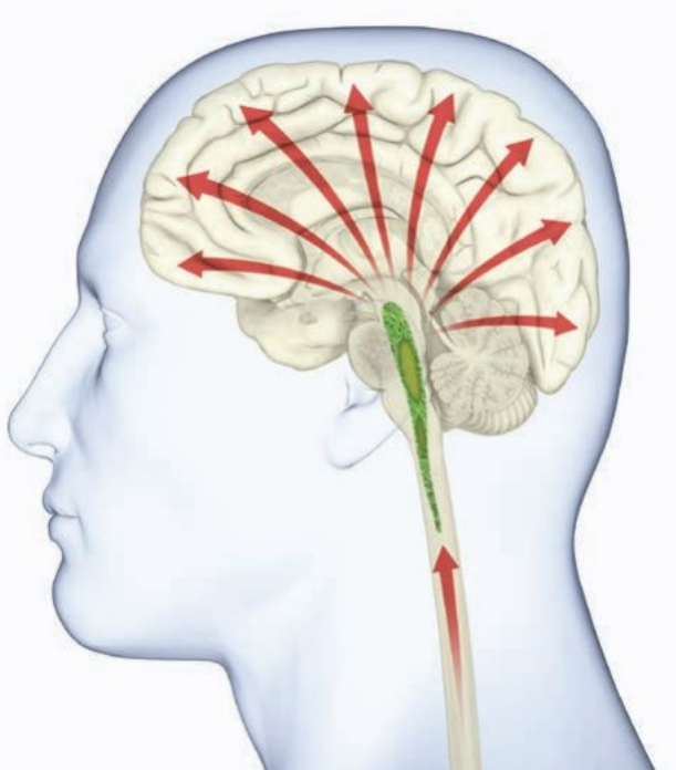
One key RAS function is filtering unnecessary information so only important stimuli get through. For example, the RAS explains why you might learn a new word and then start noticing it everywhere—your RAS creates a filter for what you focus on, presenting you with relevant information from incoming sensory data. This selective attention mechanism helps prevent sensory overload.

The forebrain
The forebrain is the brain's largest region. Its neurons connect with the midbrain and hindbrain, playing an important role in coordinating brain activity. The forebrain comprises the entire cerebrum, thalamus, hypothalamus, pineal gland, and limbic system. It is involved in a wide range of bodily functions, as well as learning, memory, thinking, and perception.
Hypothalamus
As its name suggests ("hypo" meaning "under"), the hypothalamus sits directly beneath the thalamus. About the size of an almond, it connects the hormonal and nervous systems via the pituitary gland. The hypothalamus regulates hormone release to maintain homeostasis—the body's stable internal environment—through controlling body temperature, hunger, thirst, and sleep.
Different body systems send signals to the brain when they encounter environmental changes. The brain alerts the hypothalamus to any unbalanced factors requiring attention. The hypothalamus responds by releasing hormones or signalling hormone release into the bloodstream to restore balance.
The hypothalamus forms part of the limbic system—a set of interconnected brain structures that play a role in how we experience emotions like sadness and anger. The limbic system also contributes to behaviour control and long-term memory formation.
Hypothalamus damage can lead to issues controlling body temperature, persistent hunger after eating (potentially developing into an eating disorder), sleep problems, and changes in sex drive.
Thalamus
The thalamus has two halves located side-by-side, each in a separate brain hemisphere, near the brain's centre within the forebrain. Its most important function is relaying information to relevant cerebral cortex sections for additional processing. Specifically, sensory information (except smell) comes via the thalamus, where processing and relay occur.
The thalamus also plays a major role in regulating arousal through connections with the reticular formation and RAS. Damage to this area may cause an individual to enter a coma.
Additionally, the thalamus contributes to attention, helping us focus on important information by filtering the vast amount of sensory information we encounter.
The thalamus acts as a sensory relay station—nearly all sensory information (except smell) passes through the thalamus before reaching the cerebral cortex. This makes it essential for processing and directing sensory input to appropriate brain regions.
Issues arising from thalamus damage depend on which part is affected. Examples include numbness, hypersensitivity, visual field loss, and decreased taste.
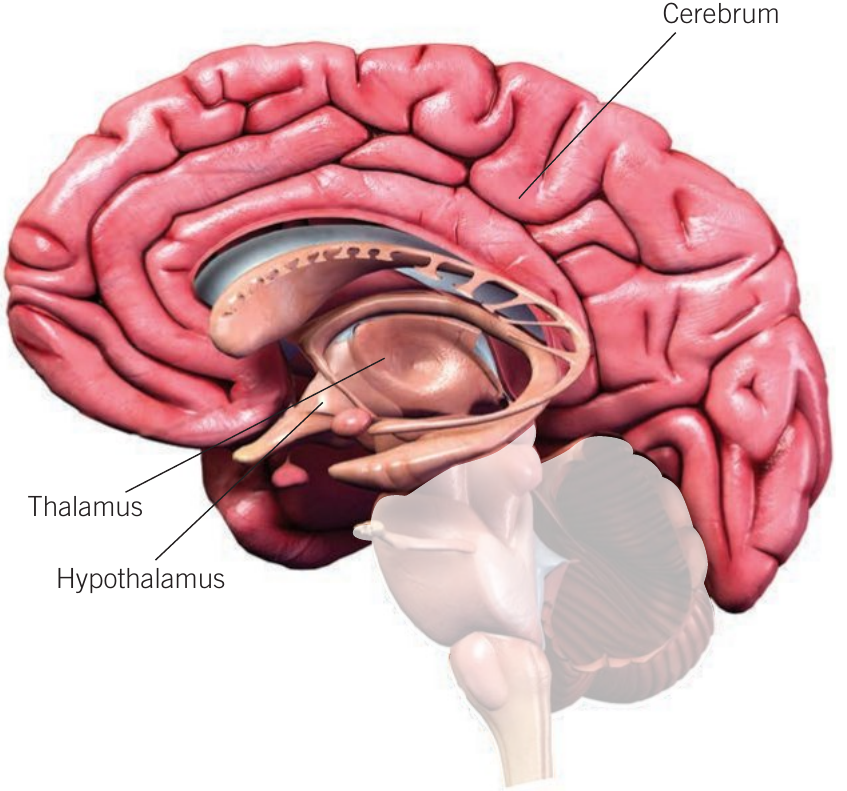
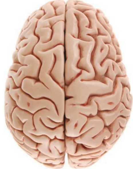
Cerebrum
The cerebrum is the forebrain's largest part and the brain's uppermost section. It contains two cerebral hemispheres that are separate but partially joined by the corpus callosum, which allows information exchange between hemispheres. The cerebrum directs conscious motor activities and receives and processes various sensory information. The outer layer of the cerebral hemispheres is the cerebral cortex.
The cerebral cortex
The cerebral cortex is the thin layer of neurons covering the cerebrum's outer region. It accounts for approximately half the brain's weight and consists primarily of grey matter. The cerebral cortex surface is extensively folded, providing room for additional neurons.
Specific areas of the cerebral cortex are dedicated to specific functions—this is called localization of function. For example, the visual cortex receives and processes information from the eyes. However, most cerebral cortex areas perform a wide array of functions.
The cerebral cortex forms extensive connections with subcortical areas below it. Therefore, it plays a role in various brain functions including processing complex sensory information, initiating voluntary movements, language, symbolic thinking, and emotion regulation.
Three Functional Areas of the Cerebral Cortex:
To understand this large brain portion, it is often simplified into three areas:
- Sensory areas: Receive and process sensory information
- Motor areas: Initiate voluntary movement
- Association areas: Integrate information from multiple brain regions, facilitating complex cognitive processes such as language, creativity, and decision-making
Cerebral hemispheres
The two cerebral hemispheres extend from the brain's front to back. Although they look alike and perform many identical functions, they are not the same. They are commonly referred to as the left and right brain hemispheres.
Each hemisphere controls motor and sensory functions on the body's opposite side. For example, the left hemisphere receives sensory information from the body's right side and controls movement on that side. The opposite is true for the right hemisphere. The positions of these sensory and motor areas are similar for both hemispheres.
Each hemisphere has areas in which it is dominant. When one hemisphere has a specialized function not possessed (or controlled to a lesser extent) by the other, this is called hemispheric specialisation. However, whilst hemispheric specialisation occurs, both hemispheres play at least some role in all functions, acting in a coordinated manner.
Mnemonics for hemispheric specialisation:
- Left hemisphere: "Language Lives Left" (left hemisphere is typically dominant for language)
- Right hemisphere: "Right is for Recognition" (right hemisphere specializes in spatial and visual recognition)
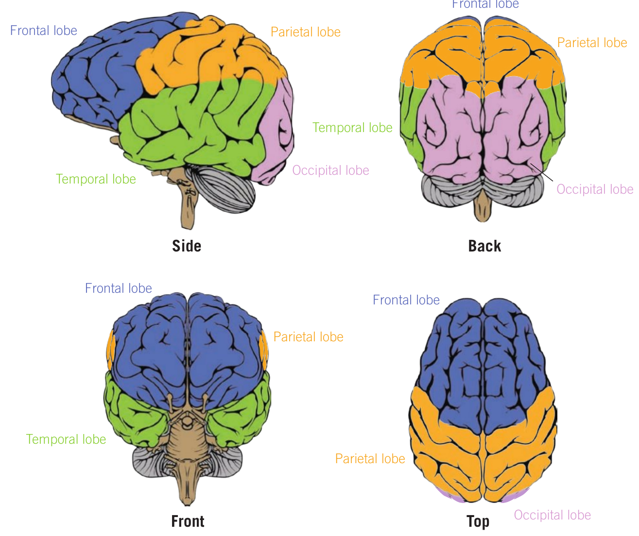
Cortical lobes of the cerebral cortex
Both cerebral hemispheres comprise four cortical lobes: the frontal, parietal, temporal, and occipital lobes. Each is made up of motor, sensory, and association areas.
Mnemonic for cortical lobes: "Four Types of Pizza Ordered" helps you remember: Frontal, Temporal, Parietal, Occipital.
Frontal lobe
The frontal lobe is located in the upper forward half of both left and right cerebral hemispheres. This brain area is important for planning, sequencing, and executing voluntary motor activity. Three areas within the frontal lobe play a role in these functions: the prefrontal cortex, premotor cortex, and primary motor cortex.
The prefrontal cortex is an important association area involved in planning the required motor sequence to carry out voluntary movement. It also contributes to various other brain functions including reasoning, problem-solving, emotional regulation, attention, symbolic thinking, and initiating and inhibiting behaviours.
Once the prefrontal cortex plans a motor sequence, it passes this to the premotor cortex, which sits just behind the prefrontal cortex in the frontal lobe. The premotor cortex prepares the movement sequence and sends this information to the frontal lobe's rear portion—the primary motor cortex.
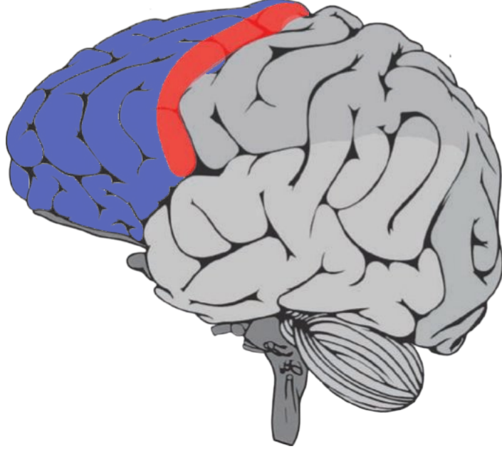
The primary motor cortex sends neural messages to skeletal muscles, initiating and executing voluntary movements. The left primary motor cortex controls voluntary movements on the body's right side, whilst the right controls movements on the left side.
Different primary motor cortex regions are responsible for movements associated with different body areas. Body areas requiring precision movements and over which we have substantial control (such as fingers and thumbs) are devoted greater motor cortex area. Less space is allotted to areas requiring coarse or uncomplicated movements or areas over which we have less control (such as back or thigh muscles).
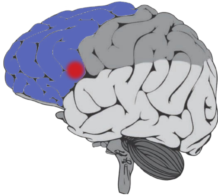
Broca's area is another frontal lobe part, located in the left hemisphere next to the primary motor cortex. This brain area contributes to clear and fluent speech by coordinating the movements of muscles involved in this process. Broca's area also sends and receives messages from other brain areas involved in language, such as those assisting with understanding word meanings, contributing to overall speech production.
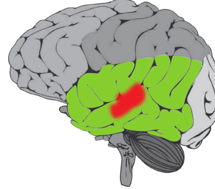
Temporal lobe
Located below the frontal and parietal lobes, the temporal lobe sits just above the ear. It plays a role in receiving and processing sounds from the ears, as well as in memory, emotional responses to sensory information, and some visual perception (such as recognizing faces and identifying objects).
The primary auditory cortex, found in the temporal lobe, assists in identifying and responding to sound. Different parts of the cortex respond to different sound types, such as high or low pitch.
Wernicke's area is a specialized area of the left temporal lobe that plays a role in understanding the sounds involved in speech, helping us comprehend the words we hear. Wernicke's area also contributes to speech production and has connections to Broca's area in the frontal lobe.
Key Difference Between Speech Areas:
| Broca's area | Wernicke's area |
|---|---|
| Responsible for contributing to clear and fluent speech through coordinating the movements of the muscles involved in this process | Involved in understanding speech through processing the meaning of sounds |
Both areas work together for complete language function, but damage to each produces different effects on language abilities.
Parietal lobe
The parietal lobe is found behind the frontal lobe but does not extend all the way to the brain's back. This lobe comprises areas involved in spatial awareness (judging our body's position in space), spatial reasoning, attention, and receiving and processing somatosensory information.
The parietal lobe contains the primary somatosensory cortex, located just behind the primary motor cortex and at the front of the parietal lobe in both left and right hemispheres.
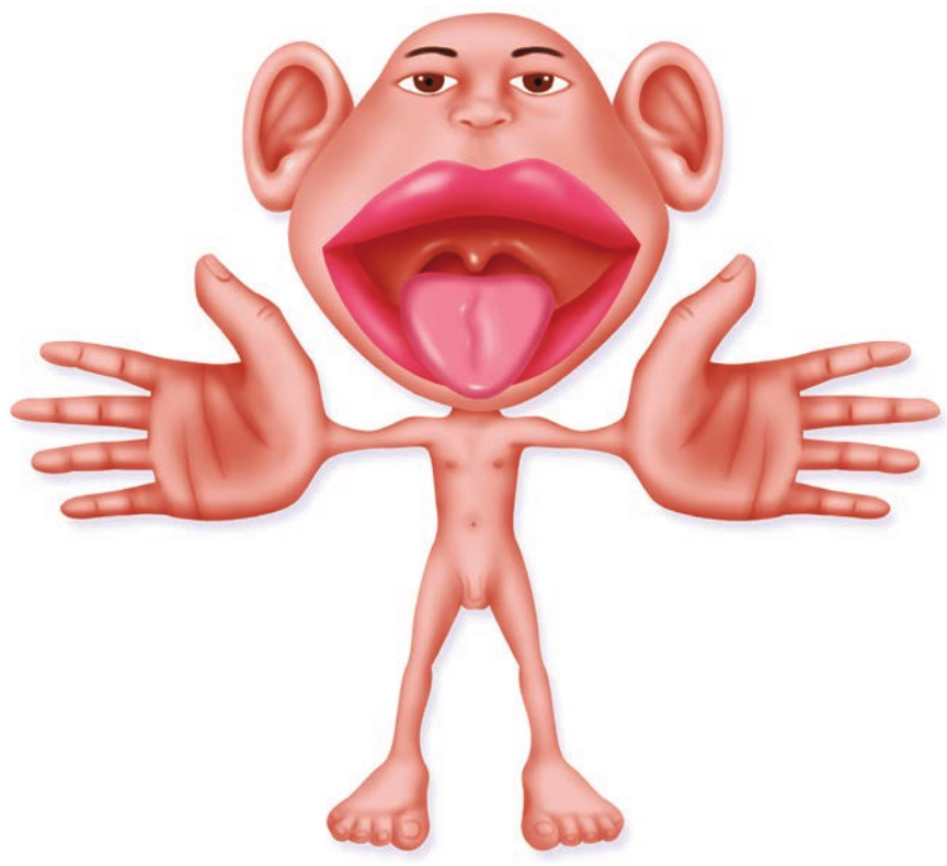
The primary somatosensory cortex receives and processes sensory information from various body areas including fingers, hands, arms, feet, legs, face, lips, tongue, and genitals. Similar to the primary motor cortex, the left primary somatosensory cortex receives and processes sensory information from the body's right side and vice versa.
Body areas that are more sensitive than others are devoted more area in the primary somatosensory cortex. For example, the fingers are allotted greater area than the knee. This explains why we have such fine tactile discrimination in our fingertips compared to less sensitive body regions. The below image shows a somatosensory homunculus – a human body modified so that body regions are given sizes in proportion to the corresponding area they have in the primary somatosensory cortex.
Occipital lobe
The occipital lobe plays a role in vision. Located at the rear of each cerebral hemisphere, this brain area is divided into multiple areas associated with vision, including the primary visual cortex.
The primary visual cortex receives and processes information from visual sensory receptors in both eyes' retinas. The left half of each eye sends information to the visual cortex in the left occipital lobe, whilst the right half of each eye sends information to the visual cortex in the right occipital lobe. In this way, each hemisphere receives and processes half of the visual information.
Key Functions of the Four Cortical Lobes:
| Frontal lobe | Temporal lobe | Parietal lobe | Occipital lobe |
|---|---|---|---|
| Important in the planning, sequencing and executing of voluntary motor activity | Plays a role in receiving and processing sounds from the ears, as well as in memory and emotional responses to sensory information | Involved in spatial awareness (judging our body's position in space), spatial reasoning, attention and receiving and processing somatosensory information | Plays a role in vision |
Remember!
Key Points to Remember:
- The brain is divided into four major regions: hindbrain, midbrain, forebrain, and cerebral cortex
- The hindbrain (cerebellum, medulla oblongata, pons) supports vital processes including breathing, balance, and coordination
- The midbrain contains the substantia nigra and reticular formation, playing a role in arousal, consciousness, and motor control
- The forebrain includes the hypothalamus (homeostasis), thalamus (sensory relay), and cerebrum (conscious motor activity)
- The cerebral cortex comprises four lobes: frontal (motor planning and execution), temporal (auditory processing), parietal (somatosensory processing), and occipital (visual processing)
- Hemispheric specialisation means each hemisphere has dominant functions, though both work together in all processes
- Broca's area (frontal lobe) controls speech production while Wernicke's area (temporal lobe) controls speech comprehension