Using an Optical Microscope (AQA A-Level Biology): Revision Notes
Using an Optical Microscope
Introduction to optical microscopes
The optical microscope is an essential tool for biologists because many biological structures are too small to observe with the naked eye. Tissues, cells and organelles require magnification to be studied in detail.
Optical microscopes are fundamental instruments in biology, enabling scientists to explore the microscopic world that would otherwise remain invisible to human observation.
Optical microscopes work by directing light through a thin specimen on a glass slide. The light passes through several lenses to create a magnified image that can be viewed through an eyepiece. Different objective lenses can be rotated into position to provide varying levels of magnification.
Components of an optical microscope
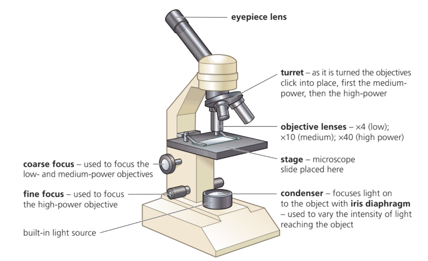
Main structural parts
- Eyepiece lens - where you look through to observe the specimen
- Turret - rotating mechanism that holds the objective lenses and allows you to switch between magnification powers
- Objective lenses - typically ×4 (low power), ×10 (medium power), and ×40 (high power)
- Stage - platform where the microscope slide is placed
- Condenser - focuses light onto the specimen using an iris diaphragm to control light intensity
- Built-in light source - provides illumination from below the specimen
The three standard objective lens magnifications (×4, ×10, ×40) provide progressively higher detail resolution. Always remember that higher magnification requires more precise focusing and better specimen preparation.
Focusing mechanisms
- Coarse focus - used for initial focusing with low and medium-power objectives
- Fine focus - used for precise focusing, particularly with high-power objectives
Preparing specimens for observation
Creating thin specimens
Specimens must be transparent or very thin to allow light to pass through effectively. Different materials require different preparation methods:
- Plant tissue (epidermis from leaves) - naturally thin enough or can be peeled off
- Animal tissue - must be sectioned using a sharp blade to create thin slices
- Living organisms (water fleas) - naturally transparent and can be observed alive
Using solutions to prevent damage
Isotonic solutions have the same water potential as cell cytoplasm and prevent osmotic damage to cells. For most animal cells, a drop of sugar or salt solution works well. Plant cells may require water, but thin layers dry quickly under the microscope lamp heat, so a drop of water beneath the cover slip prevents dehydration.
Proper specimen preparation is critical for successful microscopy. Poor preparation can result in damaged cells, unclear images, or complete failure to observe the intended structures.
Using stains to enhance visibility
Some cellular structures are naturally coloured and easy to observe, such as chloroplasts in plant cells or red blood cells. However, many structures are transparent and require staining to become visible.
Chromosomes can be observed during cell division by adding acetic-orcein stain, which colours DNA and makes chromosomes visible in different stages of mitosis. The stain works by binding to genetic material and creating contrast against the cytoplasm.
Always use small volumes of stain - excess stain obscures detail
Making measurements with graticules
A graticule is a transparent disc with an engraved scale that fits into the eyepiece and acts like a ruler overlaid on the specimen image. However, graticules have no fixed units and must be calibrated using a stage micrometre.
Graticules are useless without proper calibration. Never attempt to make measurements with an uncalibrated graticule as your results will be meaningless.
Calibration process
The stage micrometre has a precise scale engraved in micrometres. By viewing both the graticule and stage micrometre together, you can determine how many micrometres each graticule division represents. This calibration value remains constant for each objective lens, allowing accurate measurements of cellular structures.
Summary
Key Points to Remember:
- Optical microscopes use light and lenses to magnify small biological structures for detailed observation
- Always begin observations with low power (×4) before progressing to higher magnifications
- Specimens must be thin and transparent - use appropriate solutions to prevent cell damage
- Stains help visualise transparent structures like chromosomes during cell division
- Graticules require calibration with stage micrometres to make accurate measurements