Radiography Test (Leaving Cert Engineering): Revision Notes
Radiography Test
What is radiography testing?
Radiography testing is a non-destructive testing method that uses X-rays to detect internal flaws and defects in materials and components. This technique allows engineers to examine the inside of materials without causing any damage to the test piece.
The method works on the principle that X-rays can penetrate through materials but are absorbed differently depending on the material's density and thickness. This creates a visual image that reveals hidden defects inside the component being tested.
The effectiveness of radiography testing relies on the differential absorption of X-rays by different materials. Dense materials absorb more radiation, while voids and cracks allow more X-rays to pass through, creating contrast in the final image.
How radiography testing works
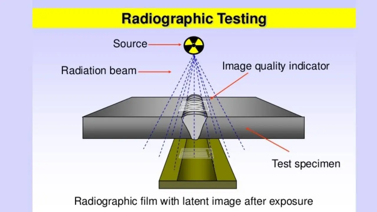
The radiography testing process involves several key steps that work together to create a clear image of internal defects:
Radiation source creation: A radiation source produces X-rays through the rapid movement of electrons in a focused direction. This creates a controlled beam of electromagnetic radiation that can penetrate materials.
Penetration and absorption: The X-ray beam passes through the test component. Different materials and thicknesses absorb varying amounts of radiation. Areas with flaws, cracks, or voids allow more X-rays to pass through, while dense, solid material blocks more radiation.
The contrast in absorption is what makes radiography testing so effective. Defects essentially create "shadows" on the photographic film, making internal flaws visible to trained inspectors.
Image formation: A photographic film is placed on the opposite side of the component from the X-ray source. When X-rays reach the film, they create an image based on how much radiation passed through each area of the material.
Image interpretation: The resulting image shows dark areas where X-rays passed through easily (indicating flaws or less dense material) and brighter areas where dense material resisted the X-rays. Internal defects appear as tone changes on the film, making them easily identifiable.
Applications and advantages
Radiography testing proves particularly effective for detecting internal flaws in various engineering components. The method can identify cracks, voids, inclusions, and other defects that would be impossible to see from the surface.
Key advantages:
- Detects internal defects without damaging the component
- Creates permanent photographic records of test results
- Highly effective at revealing hidden flaws
- Suitable for various materials and component shapes
The permanent record aspect is particularly valuable for quality assurance and regulatory compliance, as the photographic evidence can be stored and reviewed later if needed.
Limitations and considerations
Despite its effectiveness, radiography testing comes with significant drawbacks that must be carefully considered:
Cost factors: The equipment and setup required for radiography testing make it extremely expensive to implement. This high cost often limits its use to critical components where safety is paramount.
Radiation Safety Warning
The method carries serious radiation risks to operators and nearby personnel. Strict safety protocols, protective equipment, and trained technicians are essential to prevent harmful exposure. Never attempt radiography testing without proper certification and safety measures.
Operational requirements: The testing process requires specialised facilities, proper ventilation, and controlled access areas to ensure radiation safety.
Critical Safety Requirements
- Specialised radiation-shielded facilities
- Trained and certified operators only
- Personal protective equipment (PPE)
- Radiation monitoring equipment
- Controlled access areas with proper signage
Key Points to Remember:
- Radiography testing uses X-rays to detect internal flaws without damaging the test component
- The method creates images on photographic film that show density differences in materials
- Dark areas on the film indicate flaws or voids, while bright areas show dense, solid material
- The technique is highly effective but comes with high costs and significant radiation safety risks
- Proper training and safety protocols are essential due to the hazardous nature of X-ray radiation