Enzymes (HSC SSCE Biology): Revision Notes
Enzymes
Introduction to enzymes
Enzymes are specialised protein molecules that play a crucial role in controlling and regulating all the chemical reactions that occur within living cells. Without these remarkable molecules, cellular reactions would proceed so slowly that life as we know it would not be possible. Think of enzymes as the cell's workforce—they make sure that essential chemical processes happen quickly and efficiently.
Every single chemical reaction in your body—from digesting food to replicating DNA—depends on enzymes. Without them, these reactions would take thousands of years to complete, making life impossible.
Enzymes as biological catalysts
Enzymes function as biological catalysts. A catalyst is a substance that speeds up a chemical reaction without being permanently changed or consumed in the process. This means that after an enzyme helps a reaction occur, it remains intact and can be used again and again to catalyse the same reaction multiple times. This reusability is one of the key features that makes enzymes so efficient and valuable to cells.
Metabolism and enzyme diversity
Metabolism refers to the complete set of all chemical reactions taking place within a living organism. In a single cell, over different chemical reactions can occur simultaneously. Remarkably, each of these reactions is controlled by its own specific enzyme. This means that living organisms contain as many different types of enzymes as there are types of chemical reactions—an incredible diversity of molecules working together to sustain life.
Understanding activation energy
For a chemical reaction to occur, the reactant molecules must collide with sufficient energy to break their existing chemical bonds. The minimum amount of energy required to initiate a chemical reaction is called the activation energy. Without enough activation energy, reactant molecules simply cannot be transformed into products, even if they collide.
Understanding Activation Energy Through a Simple Reaction
Consider a simple chemical reaction:
where and are reactants and and are products.
Enzymes work by lowering the activation energy needed for a reaction to proceed. They achieve this by binding to the reactant molecules (called substrates) and holding them in a specific orientation that makes the reaction much more likely to occur. By reducing the energy barrier, enzymes dramatically increase the rate at which cellular reactions take place.
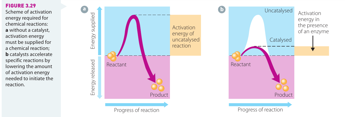
The diagram above illustrates how enzymes reduce activation energy. In an uncatalysed reaction (left), a large amount of activation energy must be supplied before the reaction can proceed. When an enzyme is present (right), the activation energy requirement is significantly reduced, allowing the reaction to occur more readily.
Properties of enzymes
Protein structure and the active site
Enzymes are composed of protein molecules that are often highly folded into complex three-dimensional shapes. This folding creates a specific region on the enzyme's surface called the active site. The active site has a particular chemical shape and structure that is crucial to the enzyme's function.
The active site is where the reactant molecules, known as substrates, temporarily attach to the enzyme. When a substrate binds to the active site, a substrate-enzyme complex is formed. This complex provides the ideal environment for the chemical reaction to take place. Once the reaction is complete and products are formed, they are released from the active site. The enzyme itself remains unchanged and is immediately available to catalyse another reaction with new substrate molecules.
Enzyme specificity
One of the most important properties of enzymes is their specificity. Each enzyme is designed to catalyse only one particular type of chemical reaction. This specificity exists because the shape of the active site will only accommodate substrates that match that particular shape.
The Lock-and-Key Analogy
Think of enzyme specificity like a lock and key—only the correct key (substrate) will fit into a specific lock (enzyme's active site). This means that a single enzyme cannot catalyse multiple different reactions; it is highly specialized for one specific job.
Models of enzyme activity
Scientists have developed models to explain how substrates bind to the active site of an enzyme. These models help us understand and predict enzyme behaviour.
The lock-and-key model
The original model proposed to explain enzyme-substrate interaction was the lock-and-key model. This model suggested that the active site of an enzyme is rigid and has a fixed shape. The substrate molecule is thought to have a complementary shape that fits exactly into the active site, much like a key fitting precisely into a lock.
According to this model:
- The substrate approaches the enzyme
- The substrate fits exactly into the rigid active site
- The substrate-enzyme complex forms
- The reaction is rapidly catalysed
- Products are released and the enzyme returns to its original state
The induced-fit model
Further research and testing of predictions led scientists to develop an improved model called the induced-fit model. This is the currently accepted model of enzyme action. Unlike the lock-and-key model, the induced-fit model recognises that proteins are not rigid structures—they are flexible and dynamic.
The Currently Accepted Model
The induced-fit model is the modern, scientifically accepted explanation of how enzymes work. Unlike the older lock-and-key model, it recognizes that enzymes are flexible molecules that can change shape to better accommodate their substrates.
The induced-fit model proposes that when a substrate binds to the active site, it induces (causes) the enzyme to change its shape slightly. This conformational change allows the enzyme to fit more tightly around the substrate, creating an even better fit than would be possible with a rigid structure. This flexibility enhances the enzyme's catalytic efficiency.
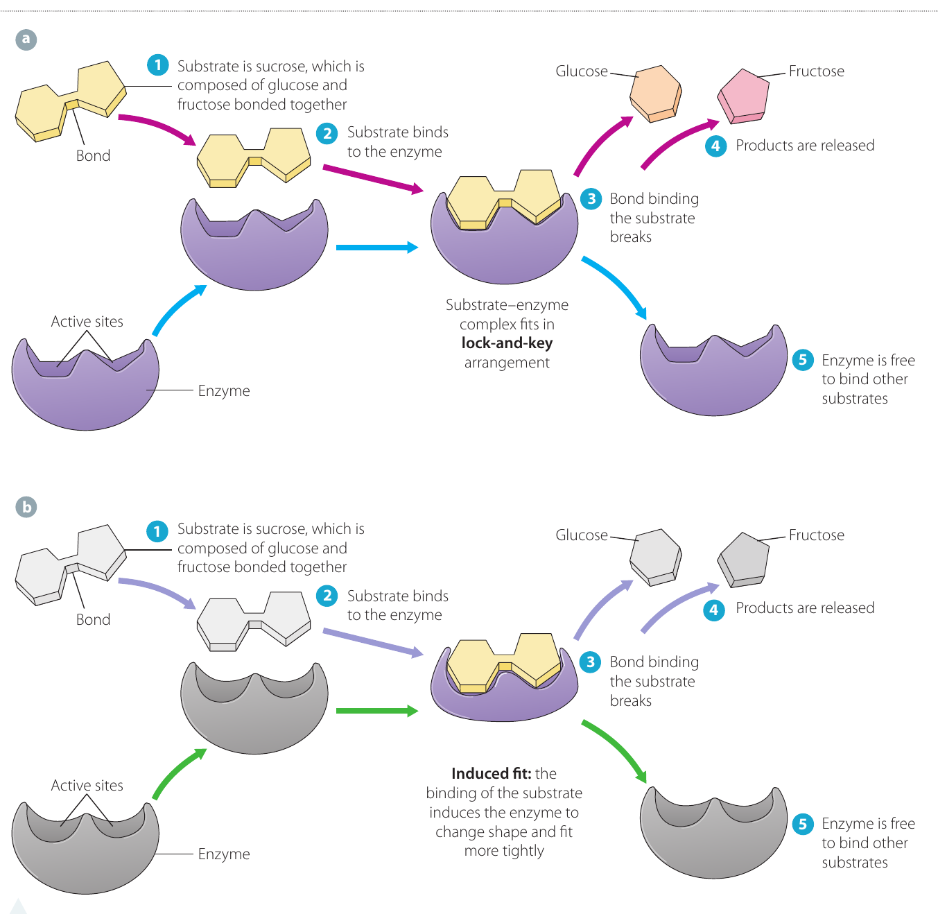
The diagram above compares both models. In the lock-and-key model (panel a), the enzyme maintains its shape throughout the reaction. In the induced-fit model (panel b), notice how the enzyme changes shape when the substrate binds, allowing for a tighter, more effective fit. Both diagrams show the breakdown of sucrose (composed of glucose and fructose bonded together) into its component parts.
Factors affecting enzyme activity
Enzymes require specific conditions to function at their optimal efficiency. Changes to these conditions can significantly affect how well an enzyme works, or whether it works at all.
Temperature sensitivity
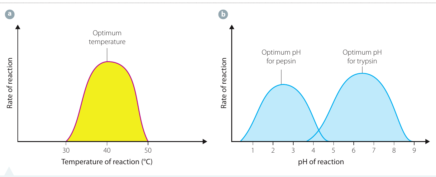
Enzymes are highly sensitive to temperature changes. Each enzyme functions best at a particular temperature, known as its optimum temperature. For most enzymes in living organisms, this optimum temperature is around . However, some organisms that inhabit extreme environments have enzymes with much higher or lower optimal temperatures. In humans, the optimum temperature for most enzymes is approximately —normal body temperature.
How temperature affects enzyme activity
As temperature increases, the rate of enzyme-catalysed reactions also increases. This occurs because higher temperatures provide more kinetic energy to molecules, causing them to move faster and collide more frequently. The reaction rate continues to increase until the optimum temperature is reached, at which point enzyme activity is at its maximum level.
However, above the optimum temperature, enzyme activity begins to decline rapidly. At temperatures between approximately , enzyme activity stops completely. This happens because excessive heat energy causes the protein structure to bend, flex, and eventually break down. The shape of the active site changes so dramatically that it can no longer accommodate its substrate. When an enzyme loses its structure and function due to excessive heat, we say it has been denatured.
Denaturation is Irreversible
Denaturation due to heat is irreversible—the enzyme cannot regain its function even if the temperature is lowered. This is why high fevers can be dangerous; they can permanently damage essential enzymes in the body. However, changes caused by cold temperatures are often reversible, and the enzyme can usually resume its function when temperature returns to normal.
Excessive cold also affects enzyme activity by slowing down molecular motion and changing the enzyme's shape. However, unlike heat denaturation, changes caused by cold temperatures are often reversible. When the temperature returns to normal, the enzyme can usually resume its function.
pH sensitivity
The pH scale measures how acidic or alkaline a substance is. Acidic solutions have pH values below , neutral solutions have a pH of exactly , and alkaline (or basic) solutions have pH values above .
How pH affects enzyme activity
Each enzyme has its own narrow range of pH values within which it functions most efficiently. This is called the enzyme's optimum pH. Outside this optimal range, the enzyme's activity decreases. Just as with temperature, extremes of pH alter the three-dimensional shape of the enzyme molecule, affecting the shape of the active site and reducing or eliminating the enzyme's ability to bind substrates effectively.
Extreme pH values, whether very acidic or very alkaline, cause enzymes to denature. This denaturation is similar to that caused by high temperatures and is usually irreversible.
pH specificity examples
Most enzymes that function inside cells work best at or near neutral pH (around ). However, enzymes in the digestive system often function in environments with quite different pH values:
Examples of pH Adaptation in Digestive Enzymes
- Pepsin is a protein-digesting enzyme found in gastric juice in the stomach. It functions optimally in the highly acidic environment of the stomach, with an optimum pH around .
- Trypsin is another protein-digesting enzyme, but it works in the small intestine where conditions are more alkaline. Its optimum pH is around .
These examples show how enzymes are perfectly adapted to work in their specific environments within the body.
The graph in the image above shows these different pH optima for pepsin and trypsin, illustrating how different enzymes are adapted to function in different environments within the body.
Substrate concentration and enzyme activity
The concentration of substrate molecules available also significantly affects the rate of enzyme-catalysed reactions.
The relationship between substrate concentration and reaction rate
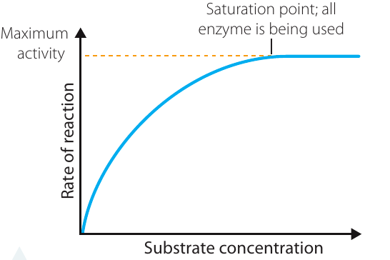
When substrate concentration is low, there are plenty of free enzyme molecules available to catalyse reactions. As substrate concentration increases, more enzyme molecules become occupied with substrate molecules, and the reaction rate increases proportionally. This is because there are more substrate molecules available to bind with the enzyme active sites.
However, this increase in reaction rate cannot continue indefinitely. Eventually, all available enzyme molecules are occupied—they are all actively catalysing reactions. At this point, the enzyme is working at its maximum turnover rate. This point is called the saturation point. At saturation, enzymes are working as fast as they possibly can.
Beyond the saturation point
If substrate concentration continues to increase beyond the saturation point, the reaction rate remains constant and does not increase further. Why? Because all enzyme molecules are already occupied and working at maximum capacity. Any additional substrate molecules must simply wait until an enzyme becomes available after completing its current reaction.
Understanding the Saturation Point
The only way to increase the reaction rate beyond the saturation point would be to add more enzyme molecules to the system, thereby increasing the enzyme concentration rather than the substrate concentration. This is a crucial concept in understanding enzyme kinetics and is often tested in examinations.
Investigation 3.3: A practical investigation to model the action of enzymes in cells
This practical investigation allows you to observe enzyme activity in action and explore how substrate concentration affects enzyme function. The investigation uses catalase, a real enzyme found in all living organisms, to model enzyme behaviour in cells.
Background: Catalase and hydrogen peroxide
Catalase is an enzyme present in virtually all living organisms. Its vital role is to catalyse the breakdown of hydrogen peroxide (), a toxic by-product of cellular respiration. If hydrogen peroxide is allowed to accumulate in cells, it becomes toxic and can cause cell death. Catalase protects cells by breaking down hydrogen peroxide into two harmless substances: water and oxygen gas.
The Catalase Reaction
The chemical equation for this reaction is:
This equation shows that two molecules of hydrogen peroxide are broken down by catalase to produce two molecules of water and one molecule of oxygen gas. The gas bubbles produced make it easy to measure enzyme activity.
Purpose of the investigation
This investigation serves two purposes:
- To model the action of enzymes in cells, demonstrating the substrate-enzyme binding action and the reusability of enzymes
- To investigate the effect of substrate concentration on enzyme activity
The investigation uses hydrogen peroxide as the substrate and catalase (found naturally in potato tissue) as the enzyme. Because oxygen gas is one of the products of this reaction, we can measure enzyme activity by observing and measuring the height of oxygen bubbles produced.
Hypothesis
Based on our understanding of enzyme kinetics and the saturation point concept:
The activity of the enzyme will increase as the substrate concentration increases until it reaches saturation point, after which the activity of the enzyme will remain constant.
Materials
- test tubes (same size)
- Test tube rack
- Hydrogen peroxide (6%)
- 10 mL measuring cylinders
- Cork borer or blender
- 30 cm rule
- Marking pen
- Potato
- Labels
- Stopwatch
Risk assessment
Before beginning any practical investigation, it's essential to identify hazards and plan how to manage them safely.
Safety First
Always complete a risk assessment before starting practical work. Identify potential hazards and plan how to manage them to ensure safe working conditions.
| What is the hazard? | What risk does the hazard pose? | How can you safely manage this risk? |
|---|---|---|
| Hydrogen peroxide | Toxic if ingested; eye and skin irritant | Wear safety goggles and disposable gloves. Do not ingest. |
| Potato | Can be a skin irritant | Wear disposable gloves. |
Method
Step 1: Label the seven test tubes with the numbers 1 to 7.
Step 2: Measure the quantities of hydrogen peroxide and water specified in the table below using measuring cylinders. Place these measured volumes into the appropriately numbered test tube.
| Test tube no. | Volume (mL) | Volume (mL) |
|---|---|---|
| 1 | 10 | 0 |
| 2 | 10 | 0 |
| 3 | 8 | 2 |
| 4 | 6 | 4 |
| 5 | 4 | 6 |
| 6 | 2 | 8 |
| 7 | 0 | 10 |
This dilution series creates different concentrations of hydrogen peroxide (substrate), allowing us to investigate the effect of substrate concentration on enzyme activity. Test tube 1 serves as a control (no enzyme), test tube 2 has the highest substrate concentration, and test tube 7 is a control with no substrate.
Step 3: Using the marker pen, mark the level of liquid in each test tube. This baseline mark will help you measure bubble height later.
Step 4: Using a cork borer, obtain six cylinders of potato of the same diameter.
Step 5: Cut the potato cylinders so that they are all of equal length. This ensures that each test tube receives the same amount of enzyme (catalase).
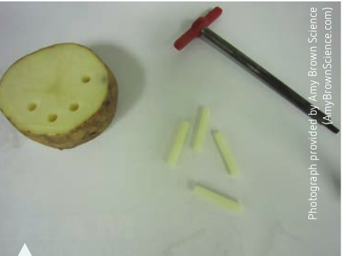
Alternative method: Instead of using potato cylinders, you can purée cubes of potato with a small amount of water in a blender. You would then measure a few millilitres of this puréed potato into each test tube. This method may provide more consistent results as the enzyme is more evenly distributed.
Step 6: Place a cylinder of potato into each of the test tubes numbered 2–7. Do not add potato to test tube 1—this will serve as a control. Allow the reaction to proceed for approximately 5 minutes.
Step 7: At the completion of this time, place another mark on each test tube to indicate the maximum height that the bubbles reached. The bubbles are composed of oxygen gas produced by the enzyme-catalysed breakdown of hydrogen peroxide.
Step 8: Measure the difference between the level of the liquid and the height that the bubbles reached in each test tube. This measurement represents the activity of the enzyme—more bubbles indicate greater enzyme activity. Record your results in a table like the one shown below.
Step 9: Compare your results with those of other groups in the class to assess the reliability of the investigation.
Results
Record your measurements in this table:
| Test tube no. | Height of bubbles (cm) (Activity of enzyme) |
|---|---|
| 1 | |
| 2 | |
| 3 | |
| 4 | |
| 5 | |
| 6 | |
| 7 |
Graphing: Create a line graph of your results. Remember that the independent variable (substrate concentration, represented by test tube number) goes on the x-axis, and the dependent variable (enzyme activity, measured as bubble height) goes on the y-axis.
Discussion questions
Working through these questions will help you understand what your results mean and how well your investigation modelled enzyme activity:
- Identify:
- a) The enzyme (catalase in the potato)
- b) The substrate in this model of enzyme activity (hydrogen peroxide)
- Describe how test tubes 1 and 2 model the action of enzymes in cells. (Test tube 1 contains substrate but no enzyme, showing that the reaction does not occur without the enzyme. Test tube 2 contains both substrate and enzyme, demonstrating that the enzyme is necessary for the reaction to proceed.)
- Was this a valid model? Justify your answer. Consider whether the model accurately represented the key features of enzyme activity: substrate-enzyme binding, reusability of enzymes, and the effect of substrate concentration.
- Identify the following variables:
- a) Independent variable (the factor you deliberately changed: substrate concentration/hydrogen peroxide concentration)
- b) Dependent variable (the factor you measured: enzyme activity/bubble height)
- c) Controlled variables (factors kept constant: amount of enzyme, temperature, pH, time allowed for reaction, size of test tubes, etc.)
- Why was test tube 1 with no potato added used? This is the control, allowing you to confirm that bubbles only form when the enzyme is present.
- Discuss the validity of the experimental design. Consider whether the method appropriately tested the hypothesis and modelled enzyme behaviour in cells.
- Describe the trend obtained on your graph. You should observe increasing enzyme activity as substrate concentration increases, potentially levelling off at higher concentrations if the saturation point was reached.
- Is the hypothesis supported by these results? Evaluate whether your data matches the prediction that enzyme activity increases with substrate concentration until reaching a saturation point.
- Compare your results with other groups in the class and comment on the reliability of the investigation. If results were similar across different groups, this suggests high reliability. Differences might indicate experimental errors or variations in technique.
Conclusion
Write a conclusion that relates both aims of the investigation to your results. Your conclusion should:
- State whether the investigation successfully modelled enzyme action in cells
- Explain what your results showed about the effect of substrate concentration on enzyme activity
- Reference specific data from your results
- State whether the hypothesis was supported or not supported by the evidence
Remember!
Key Points to Remember:
-
Enzymes are biological catalysts made of protein that speed up cellular reactions by lowering activation energy. They remain unchanged after the reaction and can be reused many times.
-
The active site is the specific region on an enzyme where substrates bind. Enzyme specificity exists because the active site's shape only accommodates matching substrate shapes.
-
The induced-fit model is the currently accepted explanation of enzyme action. It describes how enzymes change shape slightly when substrates bind, creating a tighter, more effective fit than the older lock-and-key model suggested.
-
Temperature and pH both significantly affect enzyme activity. Each enzyme has an optimum temperature and optimum pH at which it works best. Extreme temperatures or pH values cause enzymes to denature (lose their structure and function) irreversibly.
-
Substrate concentration affects reaction rate up to the saturation point, where all enzyme molecules are occupied and working at maximum capacity. Beyond saturation, adding more substrate does not increase the reaction rate.