Cell Replication (HSC SSCE Biology): Revision Notes
Cell Division – Mitosis and Meiosis
Introduction to cell division
Cell division is essential for life. It allows organisms to grow, repair damaged tissues, and reproduce. Understanding how cells divide helps us appreciate how genetic information is passed from one generation of cells to the next.
There are two main types of cell division:
- Mitosis – produces two identical daughter cells for growth and repair
- Meiosis – produces four non-identical gametes (sex cells) for sexual reproduction
Why do cells need to divide?
Cells divide to ensure that genetic information is accurately transmitted from parent cells to daughter cells. During division, each daughter cell must receive an exact copy of the genetic instructions contained in DNA. This is crucial for maintaining the stability and proper functioning of organisms.
Mitosis
What is mitosis?
Mitosis is the type of nuclear division that produces two genetically identical daughter cells. Each daughter cell receives the same number and type of chromosomes as the parent cell.
In unicellular organisms, mitosis is used for asexual reproduction – one organism becomes two. In multicellular organisms, mitosis is essential for growth and tissue repair.
Role and importance of mitosis
Mitosis plays four critical roles:
Growth of multicellular organisms
- Enables development from a single fertilised egg (zygote) into an embryo
- Supports continued growth from infant to adult
- Relies on mitotic division followed by cell enlargement and specialisation
Repair and replacement
- Fixes damaged tissues
- Replaces worn-out cells
- Examples include skin cells constantly being replaced, and healing of wounds
Your body replaces millions of cells every day through mitosis. For instance, your outer skin layer is completely renewed approximately every 2-4 weeks, and red blood cells are replaced roughly every 120 days.
Asexual reproduction
- Allows organisms to reproduce without gametes
- Used in plant cuttings and cloning
- One parent produces genetically identical offspring
Genetic stability
- Ensures precise and equal distribution of chromosomes to each daughter nucleus
- All resulting cells have identical genetic information
- Maintains consistency across all body cells
Embryonic stem cells and differentiation
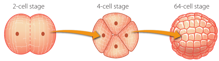
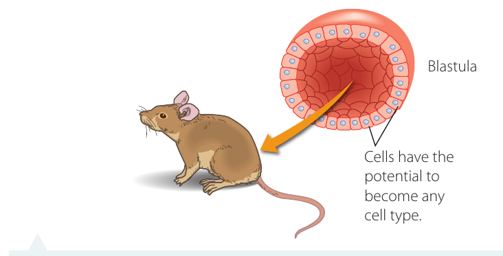
Most multicellular organisms start as a single fertilised egg cell called a zygote. This cell divides repeatedly to form an embryo.
Embryonic stem cells are special cells in early embryos that are pluripotent – they have the potential to develop into any type of tissue in the body. These cells can divide repeatedly and then specialise (differentiate) to form specific tissues like muscle, nerve, or blood cells.
The difference between pluripotent and multipotent stem cells is significant:
- Pluripotent (embryonic stem cells) = can become ANY cell type in the body
- Multipotent (adult stem cells) = can only become SPECIFIC cell types
In mature organisms, not all cells continue to divide:
- Adult stem cells exist in specific locations (e.g., bone marrow, skin base layer)
- These are multipotent – they can only form specific types of cells
- For example, bone marrow stem cells can make different blood cells, but not brain cells
- Some cells, like nerve cells, may last a lifetime and rarely divide
- These non-dividing cells are said to be in the G₀ phase
In plants, tissue capable of division is called meristem and is found at growing tips of stems and roots.
The cell cycle
Cell division occurs in a repeating sequence called the cell cycle. Mitosis is only one part of this cycle, typically taking about 1–2 hours. A complete cell cycle in actively dividing cells takes approximately 18–22 hours.
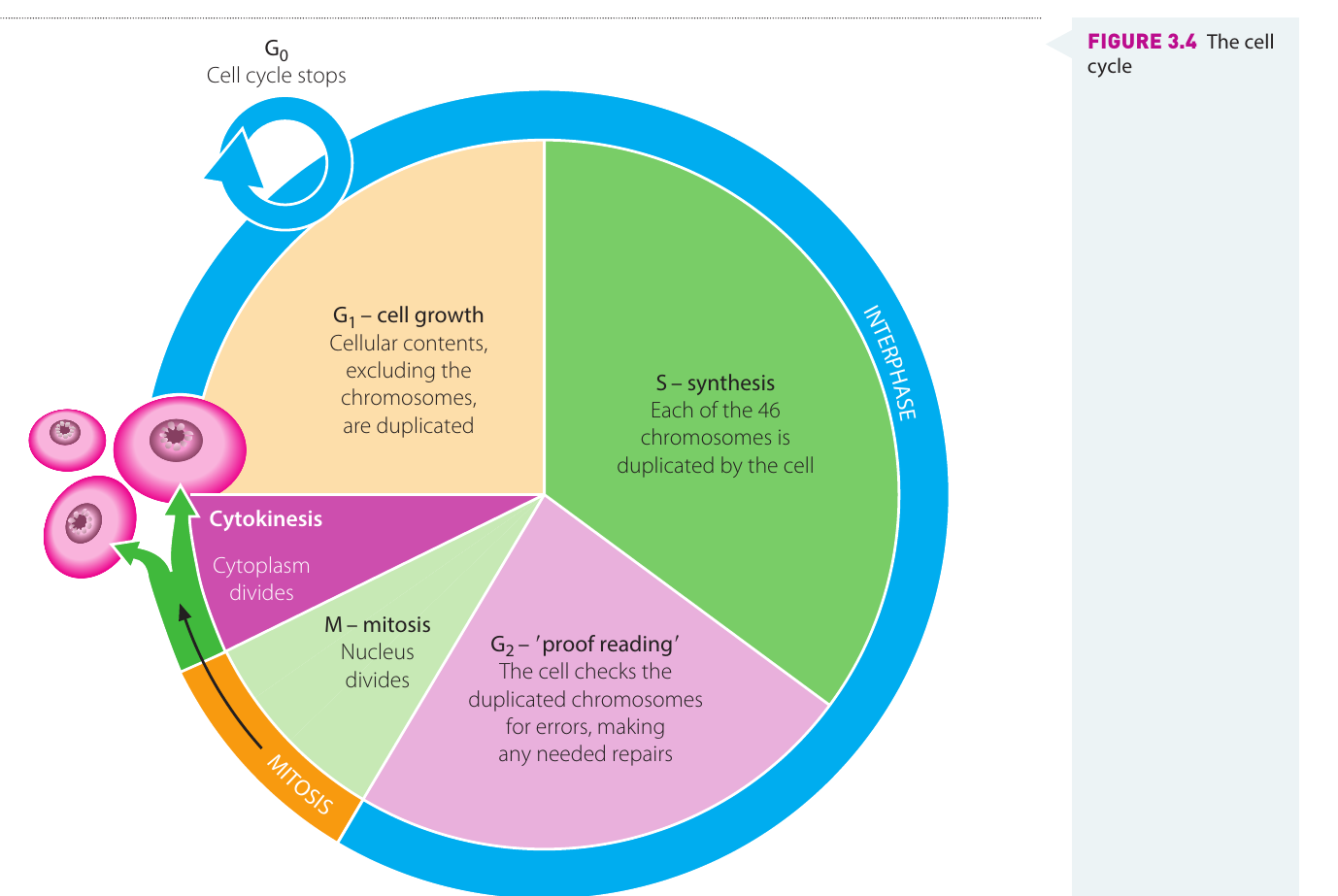
The cell cycle has four main phases:
G₁ phase (Gap 1) – Cell growth
- Cell enlargement occurs
- Cellular contents (except chromosomes) are duplicated
- Metabolic changes prepare the cell for division
- Cell reaches a commitment point and enters the next phase
S phase (Synthesis) – DNA replication
- DNA replicates (copies itself)
- Each chromosome is duplicated
- At the end, the cell has two identical copies of each chromosome
G₂ phase (Gap 2) – Quality checking
- Enzymes check duplicated chromosomes for errors
- Any mistakes are corrected
- Cytoplasmic materials accumulate ready for division
M phase (Mitosis and cytokinesis)
- Nucleus divides (mitosis)
- Cytoplasm divides (cytokinesis)
- Results in two separate daughter cells
Following cytokinesis, each daughter cell enters G₁ again if it continues to divide. During G₁, cells increase in size by taking in nutrients – these come from digestion in animals and photosynthesis in plants.
Cells that no longer need to divide enter the G₀ phase and only re-enter the cell cycle under special circumstances.
Understanding chromosome structure
Before learning about mitosis stages, it's important to understand key terms:
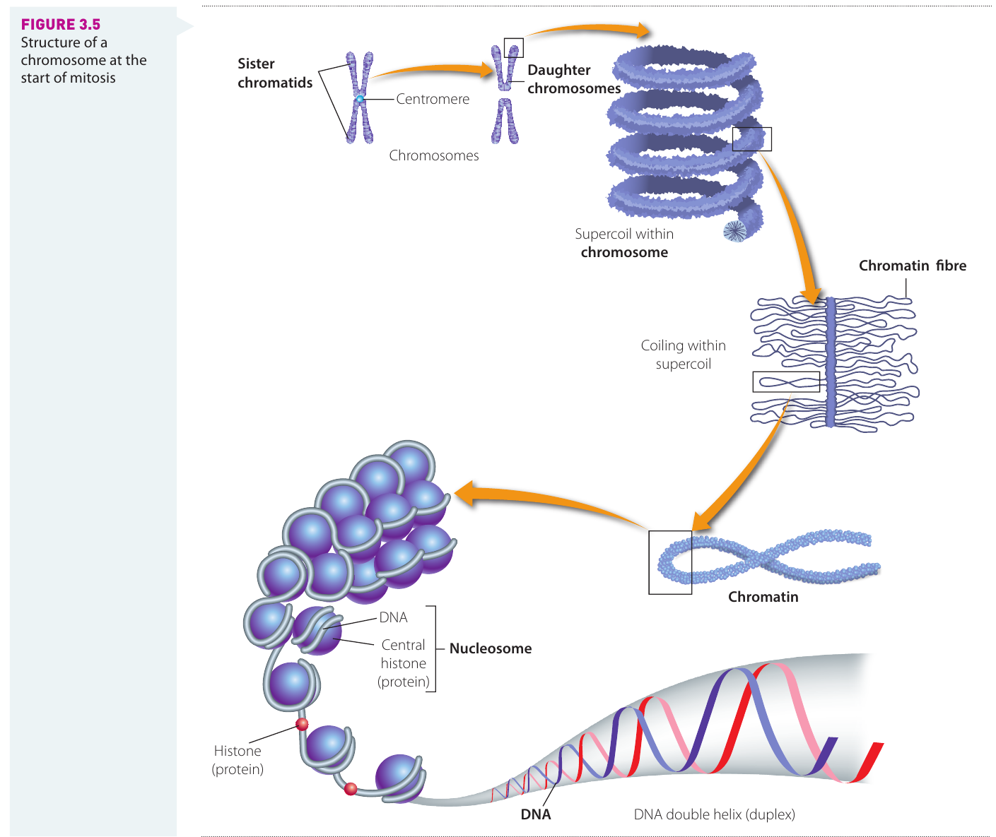
- Chromatin – DNA wound around proteins in a non-dividing cell; appears diffuse and spread out
- Chromosome – condensed, visible structure containing DNA that appears during cell division
- Gene – a section of DNA that codes for a specific inherited characteristic (e.g., eye colour, height)
- Sister chromatids – two identical copies of a chromosome joined at the centromere
- Centromere – the structure that holds sister chromatids together
- Daughter chromosomes – separated chromatids after they move to opposite poles during cell division
Chromosome Numbers in Different Organisms
Each organism has a fixed number of chromosomes:
- Humans: chromosomes
- Platypus: chromosomes
- Lettuce: chromosomes
These numbers remain constant for each species, ensuring genetic stability across generations.
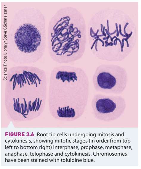
Stages of mitosis
Although mitosis is continuous, we describe it in four main stages: prophase, metaphase, anaphase, and telophase (remember: PMAT).
Interphase (before mitosis begins)
- Occurs during the S phase of the cell cycle
- DNA replicates, making identical copies
- DNA appears as diffuse chromatin, not yet visible as individual chromosomes
- In early prophase, DNA begins to separate into chromosomes
Prophase – Chromosomes become visible
- Chromatin material shortens and thickens by coiling
- DNA separates into visible chromosomes (under a light microscope)
- Each chromosome contains two identical copies of DNA called sister chromatids
- Sister chromatids are joined by a single centromere
- Nuclear membrane breaks down
- Spindle fibres begin to form across the cell
Metaphase – Chromosomes line up
- Chromosomes line up across the centre (equator) of the cell
- Each chromosome is attached to spindle fibres by its centromere
- Each chromosome still consists of two sister chromatids
Anaphase – Chromosomes separate
- Proteins in the centromere are cleaved (broken)
- Sister chromatids separate from each other
- Each chromatid is now called a daughter chromosome
- Spindle fibres contract
- Daughter chromosomes are pulled to opposite poles (ends) of the cell
- Movement is assisted by the centromere
Telophase – Two nuclei form
- Daughter chromosomes gather at opposite poles
- Spindle fibres break down
- Nuclear membrane reforms around each set of chromosomes
- Nucleolus reappears in each nucleus
- Nuclear division (mitosis) is now complete
- Result: two nuclei with identical chromosomes to each other and the parent cell
Remember PMAT for the stages of mitosis:
- Prophase – chromosomes become visible
- Metaphase – chromosomes line up at the equator
- Anaphase – sister chromatids separate and move apart
- Telophase – two nuclei form
Cytokinesis
Cytokinesis is the division of the cytoplasm. It begins while the nucleus is completing telophase. This process separates the two newly formed daughter nuclei, ensuring each cell has only one nucleus.
Cytokinesis differs between animal and plant cells:
Animal cells
- The cytoplasm constricts in the centre between the two nuclei
- Cell membrane pinches inward (like squeezing a balloon in the middle)
- Eventually the membrane meets and separates into two cells
Plant cells
- A cell plate forms during telophase
- Thickenings appear on spindle fibres at the equator
- These join to form a cell plate made of pectin compounds
- Cellulose is deposited on either side
- This forms a new cell wall separating the two daughter nuclei
Key Difference: Animal cells pinch inward to divide, while plant cells build a new wall down the middle. This difference exists because plant cells have rigid cell walls that cannot be pinched inward like flexible animal cell membranes.
The outcome of mitosis and cytokinesis is two daughter cells with chromosomes identical to each other and to the original parent cell.
Daughter cells then enlarge during G₁ until they reach the same size as the original adult cell. The nucleus controls all cell activities. If the cytoplasm becomes too large relative to the nucleus, the nucleus cannot effectively control cell functioning, which may trigger another round of cell division.
Telomeres and ageing
As people age, some dividing cells reach a point where they can no longer divide. Scientists have discovered this relates to changes at the ends of chromosomes.
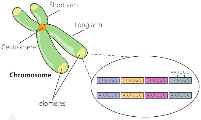
A telomere is a DNA-protein region at each end of a chromosome. Telomeres have several functions:
- Prevent chromosomes from sticking to each other
- Protect the genome from degradation
- Prevent unnecessary recombination and repair
- Help extend the life of a cell
Children have longer telomeres than older people. With each cell division, a small amount of DNA is lost from the telomeres, making them shorter. Once telomeres reach a certain length, the cell stops dividing, leading to cell ageing (senescence) and/or death.
There is variation in:
- The initial length of telomeres people are born with
- The rate at which telomeres shorten
- How lifestyle choices influence telomere shortening
This area has become important in research into ageing and disease. Scientists are investigating whether protecting or lengthening telomeres could slow ageing or prevent age-related diseases.
Meiosis
What is meiosis?
Meiosis is the type of cell division that occurs in the sexual reproductive organs of plants and animals. It produces gametes (sex cells) with half the number of chromosomes of the parent cell.
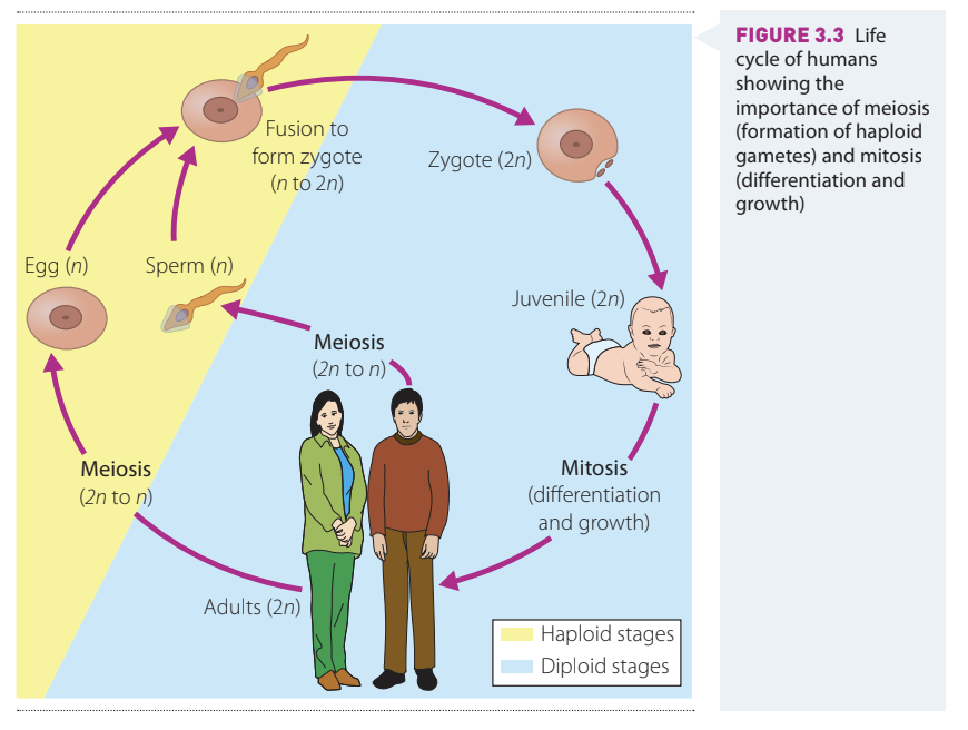
Role and importance of meiosis
During sexual reproduction, two parents contribute genetic material to offspring. To prevent the chromosome number from doubling in each generation, a special mechanism is needed.
Meiosis is a reduction division that ensures:
- Each parent contributes only half their chromosomes to offspring
- The chromosome number of each species is maintained (not doubled)
- Genetic variation is introduced into the population
Key features of meiosis
Before meiosis begins, DNA replicates. The cell then undergoes two successive divisions:
Meiosis I
- The diploid cell (with paired chromosomes) divides into two haploid cells
- Chromosome number is halved
Meiosis II
- The two cells each divide again
- Results in four haploid daughter cells (called a tetrad)
Each daughter cell (gamete) has half the original number of chromosomes that the parent cell had.
The resulting gametes are:
- In animals: egg cells (in ovaries) and sperm cells (in testes)
- In seed-producing plants: pollen grains (in anthers) and egg cells (in ovules)
Gametes are often called "vehicles of inheritance" because they carry genes from one generation to the next.
Meiosis also introduces genetic variation into a species through processes that will be covered in more detail in Chapter 5.
Why DNA needs to replicate exactly
An organism's genetic code contains instructions for:
- Every biochemical process that allows it to function
- All physical features and body size
- How genes are expressed (read and processed)
- How biological information is translated from DNA to proteins
When a cell divides, it is vital that DNA passed to each daughter cell is an exact copy. This ensures:
In unicellular organisms
- Genetic stability of the species is maintained
- Offspring are identical to the parent
In multicellular organisms
- All body cells contain the same genetic code
- Cells can function in a controlled and coordinated way
- Proteins are produced correctly
- Body structures develop properly
Along chromosomes are sequences of base pairs forming genes. Genes code for proteins, which:
- Make up a large proportion of cell structure (bones, hair, skin, muscles, connective tissue)
- Regulate cell functioning as enzymes
- Control every biochemical reaction (respiration, photosynthesis)
A change in DNA, called a mutation, may:
- Change the protein produced
- Have a harmful effect on the cell by interfering with structure or function
- Sometimes have no effect
- Occasionally be beneficial
During mitosis, DNA is replicated precisely and copies of all genes are divided equally between resulting cells. The cells are therefore clones of each other.
Replication of DNA outside the nucleus
Although each cell has one nucleus, it contains many organelles in the cytoplasm, such as:
- Mitochondria – cells requiring large amounts of energy have millions of these
- Chloroplasts – photosynthesising plant cells have many of these
A small amount of DNA is located in these organelles in the cytoplasm. This non-nuclear DNA or extrachromosomal DNA carries genes important to cell metabolism.
During cytokinesis, when the cytoplasm divides, organelles are distributed between the two daughter cells. To maintain adequate numbers, mitochondria and chloroplasts must replicate themselves.
These organelles:
- Contain their own small amounts of DNA
- Replicate independently of the nucleus
- Restore their numbers by the time daughter cells have grown to full size
Mitochondrial DNA (mtDNA) is:
- Much shorter than nuclear DNA
- Arranged in a single circle
- Inherited only from the mother
In human gamete production:
- All mitochondria inherited by offspring are in the cytoplasm of the egg cell
- Sperm cells have very little cytoplasm (mostly nucleus and tail)
- Children inherit all their mtDNA from their mother
- No mtDNA is inherited from the father
Therefore, studies of mtDNA reflect maternal inheritance over many generations.
Key Points to Remember:
- Mitosis produces two genetically identical daughter cells for growth, repair, and asexual reproduction
- Meiosis produces four non-identical haploid gametes for sexual reproduction and introduces genetic variation
- The cell cycle has four phases: G₁ (growth), S (DNA synthesis), G₂ (checking), and M (mitosis and cytokinesis)
- The stages of mitosis are: Interphase → Prophase → Metaphase → Anaphase → Telophase (remember PMAT)
- Cytokinesis differs between animal cells (pinching) and plant cells (cell plate formation)
- Telomeres shorten with each cell division and are linked to ageing
- Exact DNA replication is essential for genetic stability and proper cell functioning
- Mitochondrial DNA (mtDNA) is inherited only from the mother and replicates independently of the nucleus