DNA and Polypeptide Synthesis (HSC SSCE Biology): Revision Notes
Proteins in Living Things
Introduction to proteins
Proteins are the most abundant organic molecules found in cells. They perform two critical roles in living organisms. First, they create the fundamental structure of cells. Second, they carry out all cellular work by controlling chemical reactions that occur within cells.
The shape of each protein is essential to how it functions. Proteins bind with other molecules to perform their roles, so both their physical shape and chemical properties (including electrical charge and how they interact with water) determine their specific functions.
Chemical structure of proteins
Elements in proteins
Proteins are composed of five chemical elements:
- Carbon (C)
- Hydrogen (H)
- Oxygen (O)
- Nitrogen (N)
- Sulfur (S) - sometimes present
These elements combine to form amino acids, which are the building blocks of all proteins.
Amino acids and polypeptides
There are approximately different amino acids. These can be arranged in chains containing up to amino acids. Each amino acid contains:
- An amino group ()
- An acid group ()
- A variable 'R' group (hydrocarbon chain) that differs between amino acids
The diagram below shows the structure of two amino acids - proline and glycine:
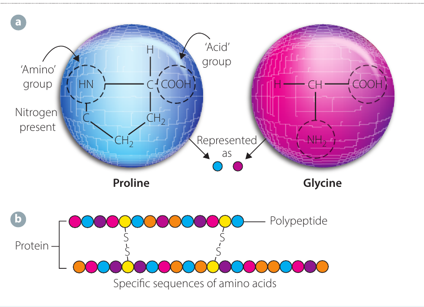
Amino acids in a chain are connected by peptide bonds - chemical forces of attraction that hold the sequence together. A chain of amino acids linked by peptide bonds is called a polypeptide.
Important distinction:
- If a single chain of amino acids is longer than - amino acids and folded in a specific manner, it is called a protein
- If the chain is shorter than - amino acids and combines with other chains to form a functional protein, it is called a polypeptide
One or more polypeptides can twist together into a particular shape, creating the overall structure of a protein. The sequence and arrangement of amino acids determines the type of protein, similar to how letters of the alphabet combine to form words and sentences.
Physical structure of proteins
Proteins have a hierarchy of organisation described at four levels: primary, secondary, tertiary and quaternary structure.
Primary structure
The primary structure is the linear sequence of amino acids in a polypeptide chain. This is the most basic level of protein structure - simply the order in which amino acids are arranged.
The primary structure determines all higher levels of protein folding because the sequence determines which amino acids are available to form bonds with each other.
Secondary structure
The secondary structure refers to the three-dimensional arrangement of the polypeptide chain. This structure forms when the amino acid chain becomes linked by hydrogen bonds.
There are two main types of secondary structure:
- Alpha helix: The polypeptide twists into a spiral shape. This is typical of fibrous proteins
- Pleated sheet: The polypeptide folds into a sheet-like structure held together by hydrogen bonds
Tertiary structure
The tertiary structure is seen in more complex proteins, particularly globular proteins. Further folding occurs when forces of attraction (such as disulfide bonds) form between different parts of the secondary structure. This causes the polypeptide to fold into a complex three-dimensional shape.
The tertiary structure is crucial because it determines the protein's ability to bind with other molecules and therefore determines how effectively it functions.
Quaternary structure
Quaternary structure occurs when a protein is made up of two or more polypeptide chains that link together to create an even more complex three-dimensional structure.
Key point: Proteins made from a single polypeptide chain have primary, secondary and tertiary structure. Proteins made from multiple polypeptide chains also have quaternary structure.
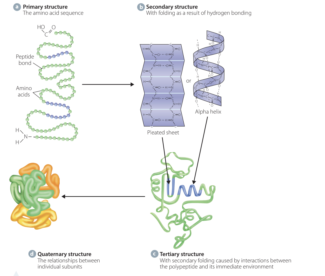
A single protein molecule may contain more than one type of structure. For example, spider silk contains pleated sheets joined by less ordered alpha helices.
Impact of amino acid changes
The primary structure (amino acid sequence) determines the secondary and tertiary structure of the protein. If an incorrect amino acid is inserted into a polypeptide chain, this may change the bonding properties of an essential part of the protein. This can alter the secondary, tertiary and quaternary structures, potentially changing or preventing the protein from functioning.
Example - Sickle Cell Anaemia:
In this inherited blood disease, a single amino acid change occurs in the protein haemoglobin. The amino acid glutamate is replaced by valine. This small change distorts the shape of the haemoglobin protein (changing it to long fibres) and affects its ability to transport oxygen. This is not lethal unless inherited from both parents, but demonstrates how even a single amino acid substitution can significantly impact protein function.
Occasionally, an amino acid change may improve protein function. Such changes are the source of random variation in populations that natural selection can act upon.
Types of proteins
Proteins can be classified into three main structural types based on their shape and composition.
Fibrous proteins
Fibrous proteins are long, stringy proteins that are insoluble in water. They form structural components of cells and tissues. Together with water, they create the basic structure of protoplasm (the cytoskeleton).
Examples:
- Collagen: A long, stringy protein that is coiled and very strong. Commonly found in skin, ligaments and tendons
- Elastin: Found in skin, ligaments and tendons alongside collagen
- Keratin: Found in hair and nails
The diagram below compares collagen (a fibrous protein) with haemoglobin (a globular protein):
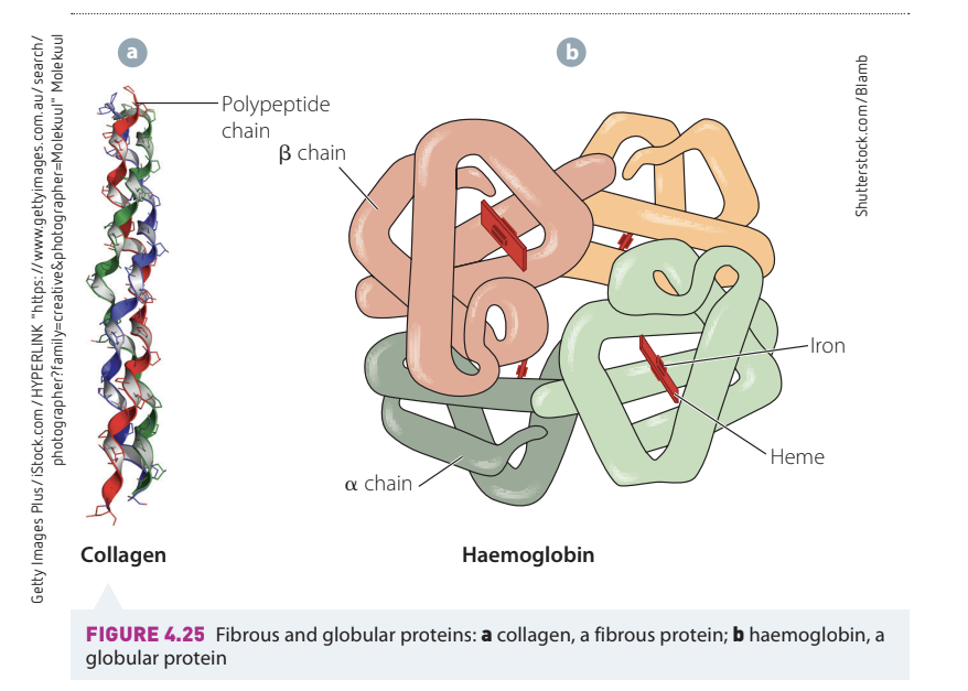
Globular proteins
Globular proteins are usually spherical in shape, compact, and soluble in water. They typically perform functional roles rather than structural roles.
Examples:
- Haemoglobin: A transport protein in blood that carries oxygen
- Immunoglobulins (antibodies): Defence proteins that fight pathogens
- Hormones: Chemical messengers
- Enzymes: Catalysts that speed up biochemical reactions
Conjugated proteins
Some proteins are linked to a non-protein component called a cofactor. When the cofactor is tightly bound, it is termed a prosthetic group and may be organic or an inorganic metallic ion.
Example: The blood pigment haemoglobin contains inorganic iron as its prosthetic group, which is essential for binding oxygen.
If a cofactor is loosely bound to an enzyme, it is known as a coenzyme (often an organic molecule such as a vitamin).
Functions of proteins
The biological properties of proteins depend on their interactions with other molecules. The tertiary structure and three-dimensional shape of a protein determines its ability to bind tightly and specifically with other molecules, and therefore determines its ability to function effectively.
Proteins are reusable, and reactions between them and their binding molecules (called ligands) are reversible.
Structural proteins - support and movement
Structural proteins provide support and enable movement in cells and organisms.
Support
Structural proteins are often fibrous and found in connective tissues such as:
- Skin, cartilage and bone
- Tendons and ligaments
- Shells in invertebrates
- Hair, nails and hooves in vertebrates
Tubulin (a protein in microtubules) forms the cytoskeleton, which maintains cell shape.
Movement
Several proteins enable movement:
Microtubules: These allow movement in cells. For example:
- Cilia and flagella move as microtubules slide along each other
- Spindle fibres contract by a similar mechanism during cell division
Contractile proteins: In muscle, the protein actin slides along another protein called myosin, allowing the muscle to contract. This is shown in the diagram below:
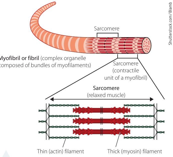
Actin is also found in microfilaments in cells. These are responsible for:
- Contraction of the cytoplasm, allowing the cell membrane to pinch off during cytokinesis in animal cells
- The crawling movement of protists such as amoeba
Enzymes - control of biochemical reactions
Enzymes are protein molecules involved in all biochemical aspects of cellular metabolism. They catalyse (speed up) reactions such as:
- Chemical respiration pathways
- Digestion
- DNA replication, repair and transcription
The shape of an enzyme's active site determines its binding specificity and therefore its ability to function. Enzymes are particularly important in gene functioning - they replicate, repair and transcribe DNA to make new proteins.
Proteins for cell communication, cell signalling and biological recognition
Cells communicate using chemical signals. Many of these signals involve proteins.
Membrane transport proteins
Some proteins embedded in cell membranes form channels to transport substances between cells and their environment. The diagram below shows the fluid mosaic model of the cell membrane with various embedded proteins:
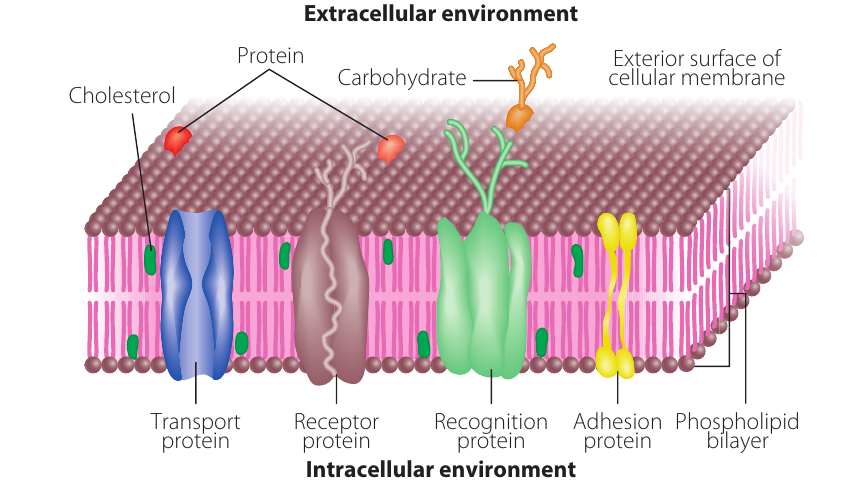
For example, nerve cells and cells in kidney tubules contain proteins embedded in the cell membrane that act as a sodium pump. This regulates the intake and output of sodium ions so the cell can function properly.
Cell signalling
Some proteins act as chemical messengers between cells:
- Hormones: Signal changes in cell functioning
- Neurotransmitters: Transmit signals between nerve cells
These messengers communicate information to cells about their environment and trigger responses in the target cell's functioning.
Biological recognition
Receptor proteins in cell membranes receive chemical signals. These proteins must have a shape that is exactly complementary to the shape of the molecule they bind with.
Some receptors on cell surfaces are genetically determined and act as markers. These allow the body to:
- Recognise its own cells
- Identify foreign invaders (pathogens) that need to be destroyed
Antibodies (also called immunoglobulins) are defence proteins. They recognise foreign invaders by detecting different proteins on the surfaces of pathogen cells (called antigens). Antibodies bind with these antigens to signal to other defence cells that the invading antigens need to be destroyed.
Transport and storage proteins
Some proteins bind to and carry or store chemicals in the body. These are termed ligand-binding proteins and must be able to bind easily with the chemical (ligand) and release it when and where needed.
Transport proteins
Haemoglobin is an excellent example. It has an affinity (attraction) for oxygen, particularly when oxygen is present in high concentrations (such as in the lungs). Haemoglobin loses its affinity for oxygen in areas with low oxygen concentration and high carbon dioxide concentration (such as in active tissues).
The protein changes shape when oxygen levels change, and this adaptive ability is essential to its functioning.
Storage proteins
Some proteins store chemicals for use by the organism:
- Ferritin: Stores iron
- Albumin: Found in egg whites, stores amino acids
- Casein: Found in milk, stores amino acids
Sensory proteins - responding to stimuli
Some proteins change their shape or biochemical activity in response to stimuli (changes in the environment).
Example - Light Detection:
Opsins are proteins in the retina of the eye that detect light. When light is absorbed by cells in the retina, these proteins undergo a change in molecular arrangement. This starts a series of reactions in which light energy is transformed into electrical and chemical signals that the brain can interpret.
Technology and protein research
Proteomics and genomics
Today, we can predict expected amino acid sequences in proteins because we understand how nucleotide sequences in DNA are decoded.
Genomics is the field of sequencing the nucleotides of complete sets of genes in organisms.
Proteomics is the large-scale study of sets of proteins produced in an organism or biological system. This research aims to understand how proteins in different cell types work together.
The genome of an organism remains relatively constant across different cells over time, but the proteome is more complex. Different cell types produce different sets of proteins, and these are often modified after translation. Different genes are expressed in different cell types, creating an enormous number of proteins to identify and study.
Bioinformatics
Bioinformatics is the field that combines computer science and genetics. People working in this field develop methods and software for analysing and interpreting biological data. It combines:
- Biology
- Computer science
- Mathematics
- Statistics
- Engineering
Advances in computer technology have enabled rapid storage and manipulation of big data (extremely large volumes of data), greatly accelerating the study of proteomics.
X-ray crystallography and synchrotrons
X-ray crystallography has been used for over a century to study molecular structures. Over the past years, it has been applied to study biological macromolecules like proteins.
Initially, progress was slow because X-ray diffraction technology needed to evolve to produce sufficiently strong X-ray beams. Collecting data for large molecules like proteins took many days or weeks.
The first complete protein structure (myoglobin) was published in . By , the Protein Data Bank (PDB) was created, containing seven protein structures determined using X-ray diffraction.
Because one protein may have between and different modified forms (each with a different function), progress was slow. What was needed were:
- High-brilliance X-ray radiation sources to speed up diffraction measurements
- Methods to analyse data quickly
The development of the synchrotron (a particle accelerator) brought about this change. A synchrotron emits electromagnetic radiation by accelerating charged particles to speeds close to the speed of light. By the s, third-generation synchrotrons provided the high-resolution images required.
This technology, combined with high-performance computing and recombinant methods for protein production, enabled molecular biologists to record and model thousands of protein structures in a relatively short time.
Today, detailed analysis of protein structure is assisted by 3D visualisation computer technology. Programs can sort protein structures by:
- Arrangement of particular chemical elements
- Bonding patterns
- Sequence of atoms
- Overall structure and shape
This is extremely useful for exploring how changes in amino acid sequences affect protein structure. Many proteins targeted in cancer research are currently being studied using these methods and big data analysis.
Remember!
Key Points to Remember:
-
Proteins are polymers made from chains of amino acids linked by peptide bonds. They contain carbon, hydrogen, oxygen, nitrogen and sometimes sulfur.
-
Proteins have four levels of structure: primary (amino acid sequence), secondary (alpha helix or pleated sheet), tertiary (3D folding), and quaternary (multiple polypeptide chains combined).
-
The amino acid sequence determines the overall shape of a protein, which determines its function. Even a single amino acid change can significantly alter protein function.
-
Fibrous proteins (like collagen and keratin) are long, stringy and structural. Globular proteins (like haemoglobin and enzymes) are spherical, soluble and typically functional.
-
Proteins have diverse functions including structural support, movement (actin and myosin), catalysing reactions (enzymes), cell communication (hormones and receptors), transport and storage (haemoglobin, ferritin), and sensory responses (opsins).