Human Immune System: The Fundamentals (HSC SSCE Biology): Revision Notes
Human Immune System: The Fundamentals
Introduction to the human immune system
We are constantly surrounded by pathogens, yet we rarely spend our lives continuously infected with disease. This remarkable protection comes from the integrated actions of the innate and adaptive immune systems working together to recognise and eliminate potential invaders.
The human immune system can recognise patterns and alert other cells when these patterns are perceived as 'non-self'. This triggers a chain of events aimed at eliminating infection. Scientists believe the elements of our immune system evolved around 500 million years ago in jawed vertebrates called gnathostomes.
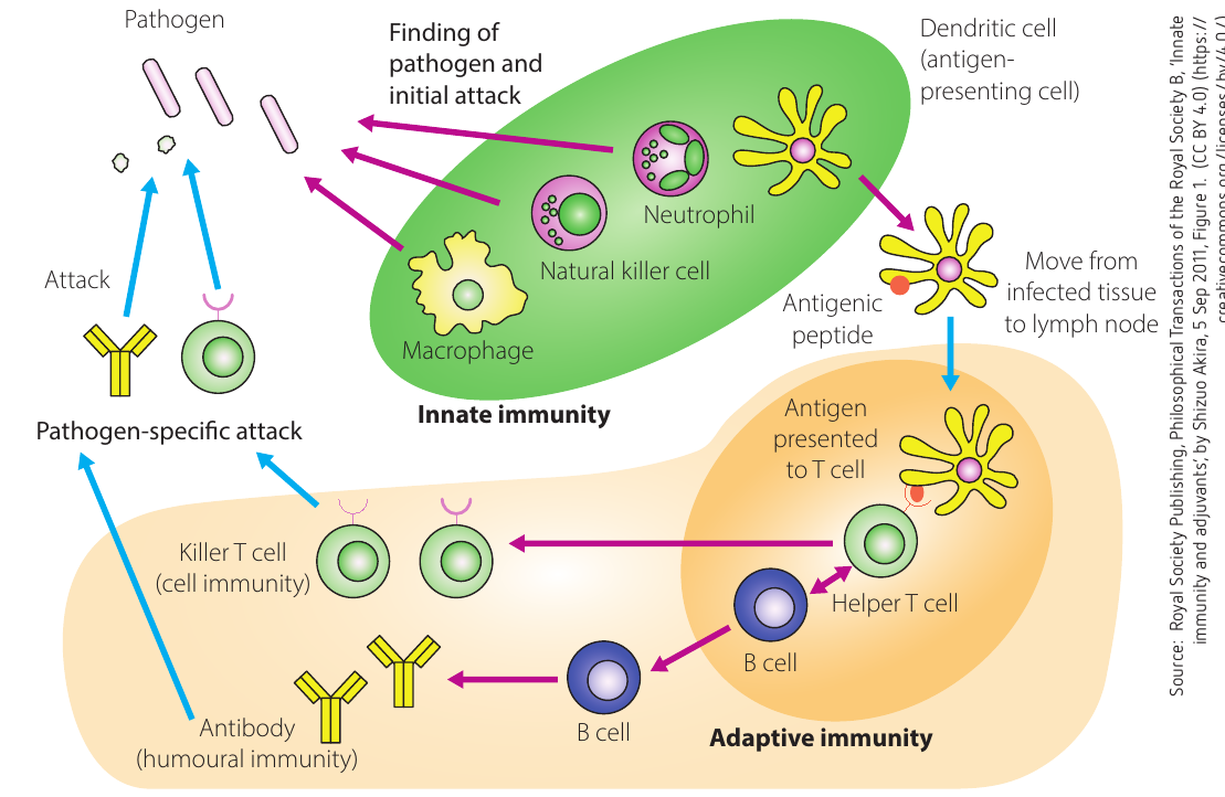
The diagram above shows how the innate and adaptive immune systems communicate and work together. The green-shaded area represents the innate response, whilst the buff-coloured area shows the adaptive response.
The innate immune system: first and second lines of defence
The innate immune system provides the body's first and second lines of defence against pathogens through:
- Physical barriers (e.g. skin)
- Body fluids (e.g. urine, mucus)
- Non-specific cellular responses (e.g. phagocytosis)
- Non-specific biochemical responses (e.g. complement activation, endogenous pyrogens)
The innate immune response is genetically pre-programmed and present from birth. With repeated exposure to the same pathogen, the body responds in exactly the same way. It involves no 'learning' or 'memory formation'.
The adaptive immune system: third line of defence
The adaptive immune response swings into action when the innate immune response fails to clear the pathogen from the body. It is often referred to as the third line of defence or acquired immunity.
The two arms of adaptive immunity
The adaptive immune response has two branches:
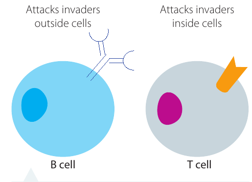
- Humoral response - effective against pathogens in body fluids (extracellular)
- Cell-mediated response - effective against intracellular pathogens
Key cells of the adaptive immune system
The cells responsible for generating the adaptive immune response are B and T lymphocytes (or B and T cells):
- B lymphocytes (B cells) develop into plasma cells, which produce antibodies (also called immunoglobulins) against pathogens. This is the humoral response. The term 'humoral' refers to the body's main fluids - the humoral response occurs in blood and tissue fluids.
- T lymphocytes (T cells) transform into cytotoxic T cells (also known as 'killer' T cells) and seek out infected body cells, binding to them and destroying them. This is the cell-mediated response.
Four key features of adaptive immunity
The adaptive immune response differs from the first two lines of defence in the following ways:
- It is specific - only responds to particular pathogens
- It involves great diversity - can respond to millions of possible pathogens
- It has memory - remembers previous infections for faster future responses
- It is capable of self-tolerance - does not attack the body's own cells
These four features - specificity, diversity, memory, and self-tolerance - are what make the adaptive immune system so effective at protecting us from repeated infections.
Antigens and antibodies: the specific response
What are antigens?
Antigens are components of bacteria, viruses, fungi, worms, tumour cells and other non-cellular materials. Antigens are generally molecules made of protein or polysaccharides found on cell membranes and in viral coats.
Antigen molecules have regions known as epitopes that can be recognised by specific lymphocytes in the immune system. Particular lymphocytes recognise the specific chemistry of each epitope and can then target these antigens.
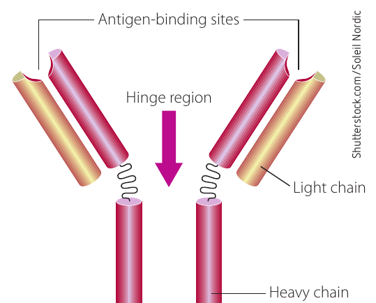
Antibody structure
Antibodies are Y-shaped molecules that consist of four chains of protein:
- Two larger heavy chains
- Two smaller light chains
There are five types of heavy chains, which give rise to the five classes of antibodies: , , , and .
A specific region of the antibody molecule serves as a binding site for antigens via the epitopes. An antibody molecule has two binding sites, each specific for a particular antigen. When antibodies and antigens bind, the resulting molecule is known as an antigen-antibody complex.
Antigen receptors
B and T cells have thousands of antigen receptors on their cell membranes. For a single B or T cell, each receptor can recognise only one of the millions of possible antigens in the world. However, each B and T cell is different, meaning the human immune system has millions of combinations of receptor types available.
Diversity of pathogens and clonal selection
The clonal selection theory states that all the B cells for all possible antigens are already present in very small amounts in the immune system at birth. When an antigen is present in the body, the B cell specific for that antigen is activated and then cloned.
Once the antigen is destroyed, these cloned B cells remain as memory cells, ready for the next time this specific antigen presents itself. Think of an army that has been shown an image of the enemy and now lies in wait - it is primed and ready to respond. It has 'adapted'.
This explains why the adaptive immune response is so specific. If a particular cold virus enters your body, only the B and T cells that recognise that pathogen will respond to it.
Primary and secondary immune responses
Primary immune response
When the adaptive immune system is first exposed to a pathogen, the response by B and T lymphocytes is called a primary immune response. Antibody-producing plasma cells and cytotoxic T cells are produced, and these work to clear the infection.
The body stores pre-made cells for the next time it encounters the same pathogen. The person will possibly now be immune to infection by this pathogen. This immunity is therefore acquired or adaptive.
The primary response is fairly fast but short-lived. The total number of antibodies produced in this initial response is less than for subsequent infections.
Secondary immune response
When the adaptive immune system is exposed to the same pathogen on subsequent occasions, there is a more rapid response and the production of antibodies is much greater. This is called a secondary immune response.
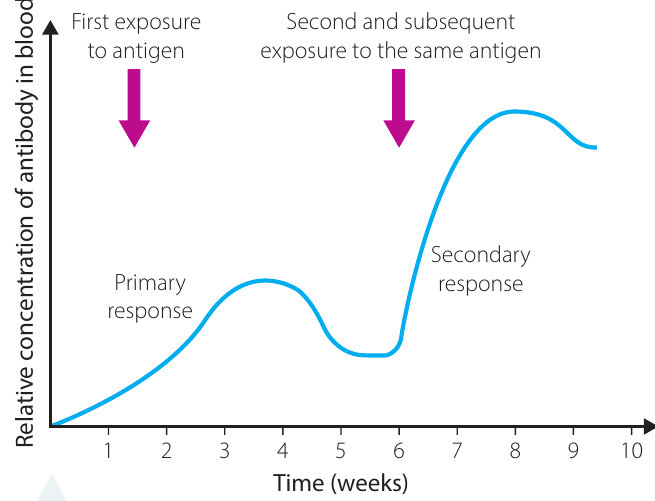
The secondary immune response occurs because of the existence of memory T and B cells that were produced during the first infection with the pathogen. This is why booster vaccinations are often recommended for various diseases.
Vaccination and Memory Cells
When you receive a vaccine, your body is exposed to a weakened or inactive form of a pathogen. This triggers the primary immune response, creating memory cells without causing the disease.
If you later encounter the actual pathogen:
- Memory cells recognise it immediately
- A rapid secondary response occurs
- Antibody production is much greater and faster
- The pathogen is eliminated before symptoms develop
This is why booster shots are important - they maintain high levels of memory cells in your system.
Self-tolerance: do no harm
Despite being genetically pre-programmed to respond to millions of possible antigens, the immune system does not normally react to the body's own tissues. How is this achieved?
B and T cells have a period in which they 'learn' not to harm the body. When they start to mature in the bone marrow (B cells) and thymus gland (T cells), they are examined for possible tendencies to cause harm to the body.
Cells that have a genetic tendency to identify self-molecules as foreign are identified and neutralised, so they never mature and cause harm. Only lymphocytes capable of reacting to 'other' and not to 'self' remain.
Scientists think autoimmune diseases may occur when this self-tolerance process has not been fully effective. In these conditions, the immune system mistakenly attacks the body's own healthy tissues.
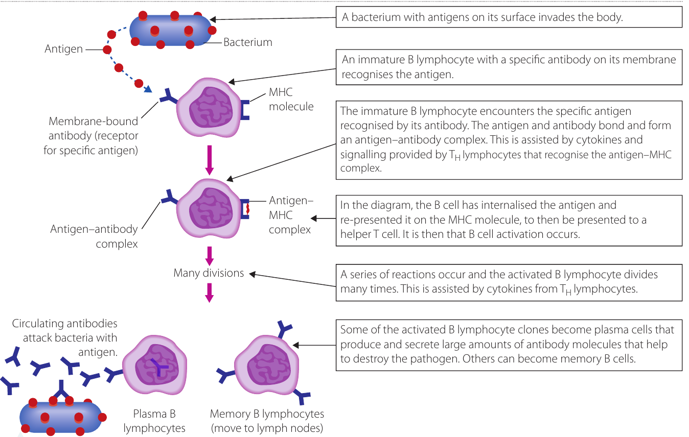
The humoral response to pathogens in body fluids
After primary exposure to a pathogen outside body cells (extracellular pathogen), the adaptive immune response targets and inactivates the pathogen through the humoral immune response.
B lymphocytes and plasma cells
B lymphocytes are the cells primarily responsible for the adaptive immune response outside cells. Mature B lymphocytes are stored in lymph nodes and peripheral lymphoid tissues, and circulate in the blood. Lymph nodes are special glands located at key points around the body where tissue fluids drain from major sites.
Helper T cells release substances such as cytokines that are involved in the activation of B cells. Two adaptive responses follow:
- The B cell multiplies, making many copies of itself with the same specificity as the original B cell
- These B cells differentiate into two possible cell types:
- Plasma cells - short-lived antibody 'factories' or effector cells (secrete up to 2000 antibody molecules per second)
- Memory cells - long-lived and stored for subsequent infections
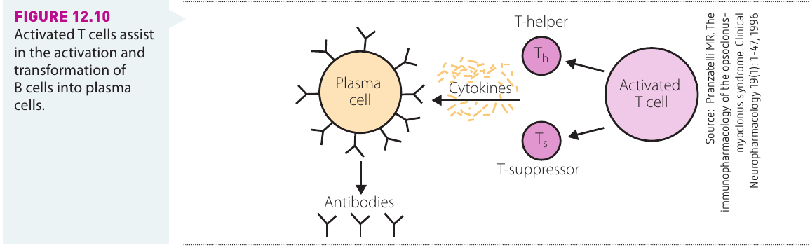
Activation of B lymphocytes
When protein-containing antigens are present, specific T cells called helper T cells are activated when they bind to the antigen. The T cells release special molecules known as cytokines. Examples include the molecule interleukin-2 (IL-2). When B lymphocytes are exposed to IL-2, they proliferate and differentiate into memory cells and plasma cells.
Not all antigens promote the production of memory cells. Some bacteria have antigens composed of long polysaccharide chains (not protein), which bind directly to B lymphocytes, causing them to multiply and differentiate into plasma cells. However, no memory cells are made in this process. These kinds of antigens are known as thymus-independent antigens.
Antibody classes and their functions
There are five classes of antibodies, also known as immunoglobulins (Ig):
| Antibody Class | Properties | Location and Function |
|---|---|---|
| Found in mucus, saliva, tears and breast milk | Protects against pathogens at body surfaces | |
| Part of the B cell receptor | Activates basophils and mast cells | |
| Protects against parasitic worms | Responsible for allergic reactions | |
| Secreted by plasma cells in the blood | Able to cross the placenta into the foetus | |
| May be attached to B cell surface or secreted into blood | Responsible for early stages of immunity |
How antibodies destroy pathogens
The different classes of antibodies use five strategies to ensure pathogens are stopped from harming the host:
- Neutralisation - antibodies bind to and coat pathogens, blocking their activity
- Agglutination - neutralised pathogens clump together and are surrounded by thousands of antibodies
- Precipitation - precipitation of dissolved antigens
- Complement activation - leads to lysis of infected cells
- Opsonisation - enhanced phagocytosis by natural killer cells (NK cells)
These five mechanisms work together to ensure pathogens are effectively neutralised and eliminated from the body. Different antibody classes may be more effective with certain strategies depending on the type and location of the pathogen.
The cell-mediated response to intracellular pathogens
The adaptive immune system has another branch responsible for eliminating pathogens located inside host cells (intracellular pathogens). Because antibodies have no direct access to these pathogens, another strategy is required. This is known as cell-mediated immunity, where special types of T lymphocytes target and destroy the entire infected host cell, along with the pathogens inside them.
What does cell-mediated immunity defend against?
T lymphocytes control cell-mediated immunity, which is effective in defending the body against:
- Protozoa, bacteria and viruses that are inside the host's body cells
- Macroparasites such as fungi, flatworms and roundworms
- Cancer cells and transplanted tissues
Unlike the humoral response which targets extracellular pathogens with antibodies, cell-mediated immunity destroys the entire infected cell to eliminate intracellular pathogens that are hidden from antibodies.
T cells and MHC molecules
Major histocompatibility complex (MHC) molecules
T cells recognise fragments of antigens only (not whole antigens like antibodies do). These antigen fragments are carried to and displayed on the cell surface by special protein molecules called major histocompatibility complex molecules (MHC).
MHC molecules are the 'self-identity' molecules of organisms. There are over a hundred genes that code for an individual's MHC marker molecules. Each person has a different set of MHC marker molecules on their cells.
There are two classes of MHC molecules:
- MHCI - found in every nucleated cell in vertebrates (including platelets, but excluding red blood cells). Infected cells present a fragment of antigen bound to an MHCI molecule.
- MHCII - found in antigen-presenting cells such as macrophages, dendritic cells and activated B cells. Macrophages present a fragment of antigen bound to an MHCII molecule (called an antigen-MHCII complex).
The unique MHC molecules on each person's cells are why organ transplants can be rejected - the recipient's immune system recognises the donor's MHC molecules as 'non-self' and attacks the transplanted tissue.
T lymphocyte development
T lymphocytes are produced in the primary lymphoid tissue of the bone marrow and mature in the thymus gland, which is situated in the thoracic (chest) cavity. After they mature, T cells are released into the blood and migrate to the spleen, tonsils, Peyer's patches of the intestines and lymph nodes.
The four main types of T cells
1. Helper T cells
Helper T cells have a surface protein receptor that recognises MHCII molecules bound to an antigen fragment on the surface of phagocytes ('antigen-presenting cells'). The helper T cells are activated to secrete cytokines, such as IL-2 (interleukin-2), which activate both the cell-mediated and humoral responses.
IL-2 activates cytotoxic T cells and B cells that are specific for this antigen. In this way, helper T cells are the 'bridge' between the two arms of the adaptive immune response.
2. Cytotoxic T cells (Tc cells)
Cytotoxic T cells are activated by helper T cells and respond by producing many copies (clones) of themselves. They form an 'army' of identical killer cells that move to the site of infection, bind with infected cells and release chemicals (cytokines) that destroy the infected cell.
Cytotoxic T cells use their surface receptor to recognise the MHCI-antigen complex typical of cells infected by a virus. Through a series of reactions, the cytotoxic T cell kills the infected cell by producing cytokines that cause lysis of the infected cell.
3. Memory T cells
Memory T cells are T cells that remain in the body to respond to future exposure from pathogens. All T cells make clones of themselves that persist in lymph nodes.
On re-exposure to the same antigen-containing pathogen, memory T cells cause the rapid production of more cytotoxic T cells specific to that antigen. This prevents the body from developing the symptoms of the disease again.
4. Suppressor T cells
Suppressor T cells (Ts cells) are responsible for stopping the adaptive response when the infection has been defeated. They release chemicals to stop the production and action of cytotoxic T cells once pathogens have been eliminated.
Summary of cell-mediated immunity
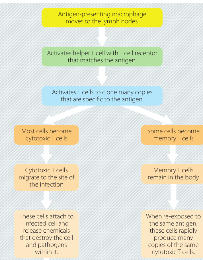
The process of cell-mediated immunity follows these steps:
- Foreign material is engulfed by macrophages, which display the antigen attached to their MHCII molecules
- The antigen-presenting macrophages move to the lymph nodes, where they are inspected by helper T cells with the corresponding T cell receptor
- These helper T cells activate the cloning of millions of cytotoxic T cells and memory T cells specific for this antigen
- The cytotoxic T cells leave the lymph nodes and migrate to the site of infection, where their antigen receptors bind with the antigen displayed on the infected cell
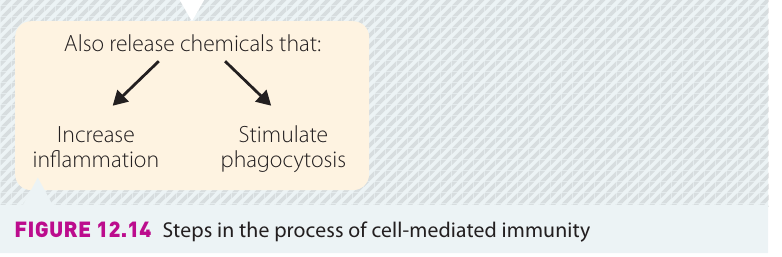
- These T cells release chemicals that destroy the cell and any pathogens within it
- These chemicals also increase inflammation and attract more macrophages to carry out phagocytosis
- Some cytotoxic T cells produce interferon, which protects healthy cells around an infected cell from viral invasion
- Once the infection is defeated, suppressor T cells release chemicals to stop the production and action of cytotoxic T cells
- Memory T cells remain in the lymph nodes for rapid response upon re-exposure to the same antigen
Step-by-Step: How Cell-Mediated Immunity Defeats a Viral Infection
Imagine a cell in your body becomes infected with a virus:
Step 1: The infected cell displays viral antigen fragments on its surface using MHCI molecules (like waving a red flag saying "I'm infected!")
Step 2: A macrophage engulfs some viral particles and presents antigen fragments using MHCII molecules
Step 3: The macrophage travels to a lymph node and activates helper T cells with matching receptors
Step 4: Helper T cells release IL-2, triggering the production of millions of cytotoxic T cells
Step 5: The cytotoxic T cells circulate through the body, find infected cells by recognising their MHCI-antigen complexes, and release chemicals that cause the infected cells to self-destruct
Step 6: Some cytotoxic T cells become memory T cells, ready for future encounters with the same virus
Interaction of B and T cells in the adaptive immune system
The two branches of the adaptive immune system do not act independently - they interact via helper T cells.
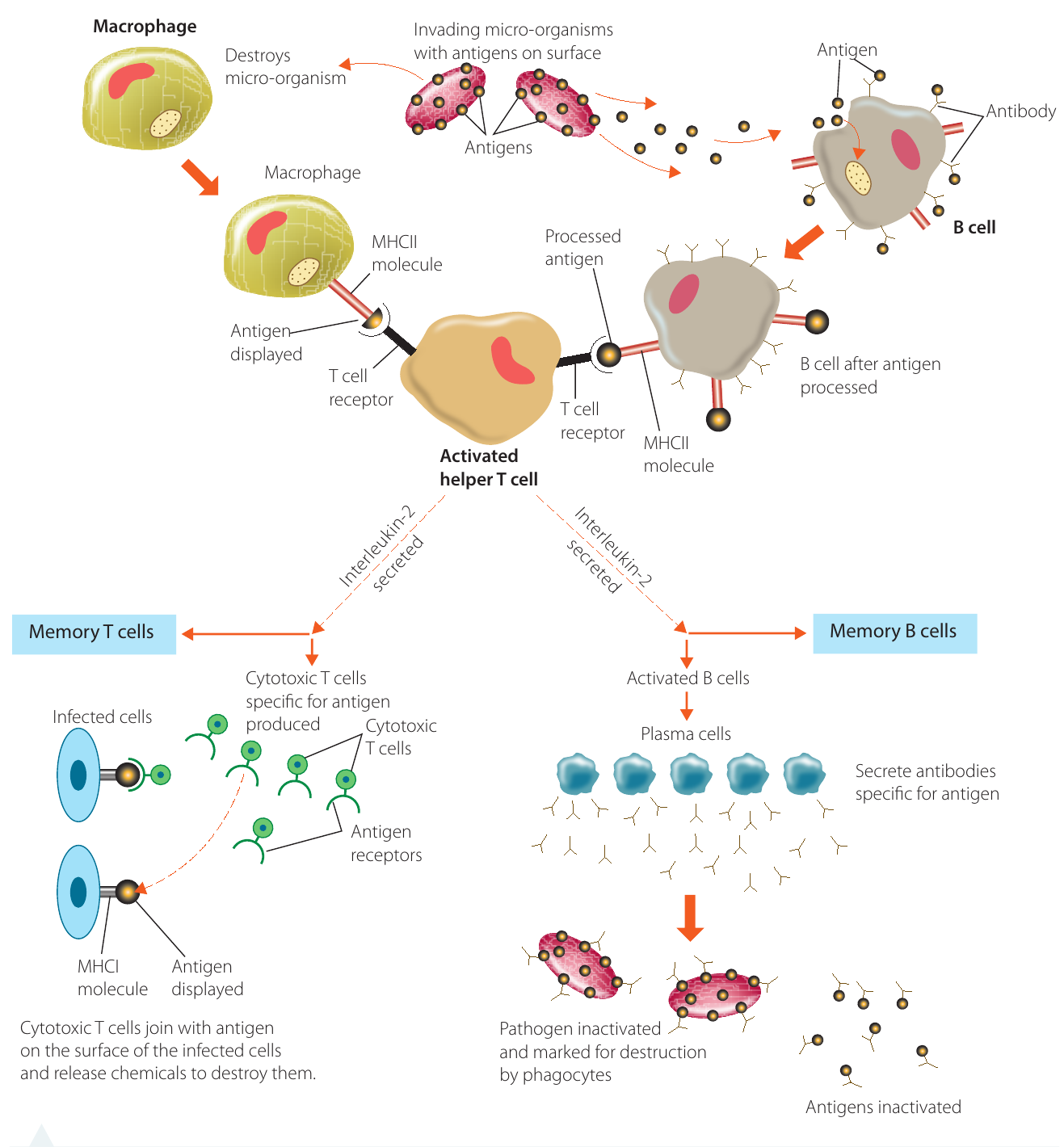
How macrophages initiate the response
When a macrophage encounters a foreign particle with an antigen attached to its surface, it surrounds and engulfs it through phagocytosis. During destruction of the foreign material, the antigen present on its surface is moved to the surface of the macrophage, which then transports it to the lymph nodes.
The antigen-presenting macrophage is presented to the helper T cell that has the T cell receptor corresponding to that particular antigen. This activates the helper T cell.
How B cells activate helper T cells
Helper T cells can also be activated by B cells. When a B cell encounters the antigen that corresponds to its surface antibodies, it binds the antigen. It then processes the antigen, attaches it to its surface molecules and presents this to the helper T cells that have the matching T cell receptors.
Chemical signalling coordinates the response
Chemical signals in the form of cytokines are secreted by helper T cells to:
- Activate more helper T cells and macrophages
- Activate production of B cell clones specific to that antigen (via interleukin-2)
- Activate production of cytotoxic T cell clones with that particular antigen receptor
When the immune response has successfully defeated the infection, suppressor T cells are responsible for suppressing the activity of B cells and cytotoxic T cells.
The interaction between B and T cells shows how the immune system works as an integrated whole. Helper T cells act as coordinators, ensuring both the humoral and cell-mediated responses are activated when needed, and that they work together effectively to eliminate the pathogen.
Remember!
Key Points to Remember:
-
The human immune system has two main components: the innate immune system (genetically pre-programmed, present from birth) and the adaptive immune system (responds to specific pathogens, has memory).
-
The adaptive immune system has two arms: humoral immunity (B cells and antibodies against extracellular pathogens) and cell-mediated immunity (T cells against intracellular pathogens).
-
B cells produce antibodies that neutralise pathogens in body fluids, whilst T cells destroy infected body cells containing pathogens.
-
The primary immune response is the first exposure to a pathogen (slower, produces fewer antibodies), whilst the secondary immune response is faster and stronger due to memory cells.
-
Helper T cells are the bridge between the two arms of adaptive immunity, activating both B cells and cytotoxic T cells through chemical signals called cytokines.