Genes and Chromosomes (VCE SSCE Biology): Revision Notes
Genes and Chromosomes
Introduction
Inheritance is the process by which characteristics pass from parents to offspring. Have you ever noticed similarities between yourself and your family members? Perhaps you share your father's height or your grandmother's smile. These similarities exist because of genes - the basic biological units of inheritance found in every cell of your body.
Understanding genes and chromosomes helps us explain how traits are passed down through generations and why each person is unique.
The striking resemblances you share with family members aren't coincidental - they're the result of genes passed down through generations, creating a biological connection that links you to your ancestors and descendants.
DNA structure
Before exploring genes in detail, we need to understand the structure of deoxyribonucleic acid (DNA).
Deoxyribonucleic acid (DNA) is a double-stranded nucleic acid chain made up of nucleotides. DNA carries the instructions for proteins which are required for cell and organism survival.
Components of DNA
DNA is built from smaller units called nucleotides. A nucleotide is the monomer unit of nucleic acids, made up of three components:
- A phosphate group
- A deoxyribose sugar molecule
- A nitrogen-containing base
There are four types of nitrogen-containing bases in DNA:
- Adenine (A)
- Thymine (T)
- Guanine (G)
- Cytosine (C)
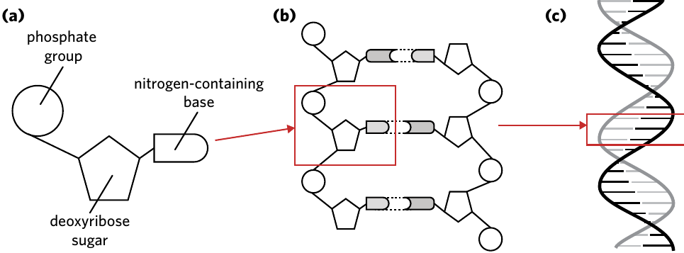
These nucleotides join together through base pairing to form a longer, double-stranded chain. The bases pair in a specific way: adenine always pairs with thymine (A-T), and guanine always pairs with cytosine (G-C). This creates the characteristic double helix structure of DNA.
Base Pairing Rules
Remember the fundamental pairing rules in DNA:
- Adenine (A) always pairs with Thymine (T)
- Guanine (G) always pairs with Cytosine (C)
This complementary base pairing is essential for DNA replication and genetic information transfer.
From DNA to genes
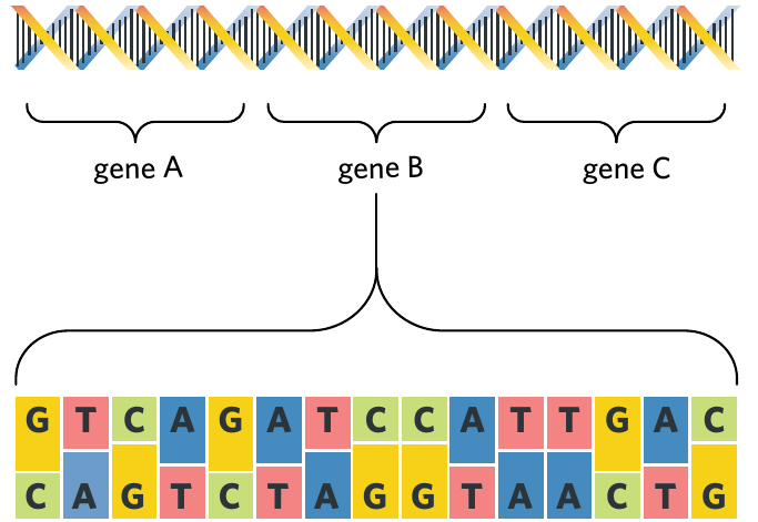
A gene is a section of DNA that carries the code to make a protein. Think of genes as instruction manuals that tell your cells how to build specific proteins needed for various functions like growth and tissue repair.
The order of bases in a gene determines which protein gets made. For example, the sequence 'ATG' provides different instructions compared to 'GGG'. Different sequences produce different proteins, which in turn create different characteristics in your body.
Example: How Base Sequences Determine Proteins
Consider two different base sequences:
- Sequence 1: ATG-CCG-TAA
- Sequence 2: GGG-ATC-CGA
Even though both sequences contain the same types of bases, their different orders result in completely different proteins being produced, leading to different traits in the organism.
Each human has approximately 25,000 different genes distributed throughout their DNA. You inherit two copies of each gene - one from your mother and one from your father.
The human genome
Your genome is the complete set of DNA contained within an organism's chromosomes. It represents all the genetic information needed to build and maintain you as a complex organism.
The human genome contains:
- Approximately 25,000 different genes
- Around 3 billion individual base pairs (in a haploid set)
Haploid describes a single set of chromosomes (n), containing one copy of each chromosome.
Alleles and genetic variation
Almost all genes are identical across every human being. However, a small proportion (less than 1%) show slight differences between individuals. These variations are what make each person unique.
Alleles are alternate forms of a gene. They are different versions of the same gene with small differences in their base sequence.
How alleles create variation
Consider eye colour as an example. Everyone has genes responsible for eye colour, but the specific allele you inherit determines whether you have brown, blue, green, or hazel eyes.
Example: Eye Colour Inheritance
The OCA2 gene on chromosome 15 influences eye colour:
- High protein P levels → Brown eyes
- Low protein P levels → Blue eyes
- Intermediate protein P levels → Green or hazel eyes
Each person inherits two alleles of this gene (one from each parent), and the combination determines the final eye colour.
While there may be multiple possible alleles for a particular gene in the human population, each individual only possesses two alleles for any given gene - one inherited from each parent. These alleles are found at the same locus (the fixed position on a chromosome where a particular gene is located).
The allele that is ultimately expressed in your phenotype (the observable trait) depends on how the two alleles interact with each other. Some alleles are dominant over others, meaning they mask the effect of other alleles.
Understanding Dominance
Not all alleles have equal influence on your traits. Dominant alleles can mask the effects of recessive alleles, which is why you might carry a gene for a trait without actually displaying that trait in your phenotype.
Genome, gene, and allele distinctions
Understanding the relationship between these three concepts is essential:
- Genome: The complete haploid set of chromosomes within an organism, including all genes. The human genome contains around 25,000 different genes and more than 3 billion base pairs.
- Gene: A section of DNA that codes for protein production. Genes form the basis of inheritance and are found at specific locations (loci) on chromosomes. For example, the OCA2 gene is located on chromosome 15 and codes for protein P, which influences eye colour.
- Allele: An alternative form of a gene. Individuals typically have two alleles (one from each parent) located at the same gene locus on corresponding chromosomes. For instance, different alleles of the OCA2 gene produce varying levels of protein P, resulting in different eye colours (brown eyes from high protein P levels, blue eyes from low levels).
Think of it this way: Your genome is like a complete library of instruction manuals, each gene is a specific instruction manual for building a particular protein, and alleles are different editions of the same manual with slight variations in the instructions.
Chromosomes
Your genome exists inside the nucleus of each somatic cell as DNA. However, this DNA doesn't float freely - it is organised into structures called chromosomes.
Somatic cells are any cells that are not reproductive cells (such as sperm and egg cells). Somatic cells are diploid (2n), meaning they contain two sets of chromosomes - one inherited from each parent.
What is a chromosome?
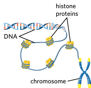
With approximately 25,000 genes and 3 billion base pairs, the genetic information in each cell is enormous. To fit inside the tiny nucleus, DNA molecules are coiled tightly around histone proteins and packaged into thread-like structures called chromosomes.
DNA Packaging
If you could stretch out all the DNA from a single human cell, it would measure approximately 2 meters long! Histone proteins act like spools, allowing this massive amount of DNA to be compressed into a nucleus that's only about 10 micrometers in diameter.
Histone proteins are highly basic proteins that associate with DNA inside the nucleus and help it condense into a chromosome structure, allowing it to fit inside the nucleus.
Human somatic cells contain 46 chromosomes, giving them a diploid number of 2n = 46. This means there are 46 individual chromosomes organised into 23 pairs.
Chromosome structure
A chromosome is the structure made of protein and nucleic acids that carries genetic information.
Chromosomes vary in size depending on how many nucleotides they contain. For example:
- Chromosome 3 spans about 198 million base pairs and contains roughly 1,000 genes
- Chromosome 14 spans approximately 107 million base pairs and contains around 800 genes
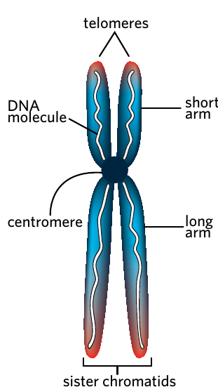
Features of a chromosome
| Feature | Description |
|---|---|
| Telomeres | A region of repetitive base sequences found at the end of every chromosome. Telomeres protect chromosome ends from fusing with other nearby chromosomes in the nucleus. |
| DNA molecule | Each chromosome is composed of a long DNA molecule that has been coiled tightly around histone proteins. |
| Centromere | A specialised DNA sequence that holds together the two chromatids. The centromere is very important for cell division processes. |
| Sister chromatids | The identical daughter strands of a replicated chromosome. |
| Short arm | Also known as the 'p arm' - the shorter section of the chromosome. |
| Long arm | Also known as the 'q arm' - the longer section of the chromosome. |
Understanding chromosome representations
Chromosomes are often shown in an X-shape, which can be confusing. It's important to understand that:
- A chromosome with two connected strands (the X-shape) represents a duplicated chromosome after DNA replication during cell division. This consists of two sister chromatids joined at the centromere.
- A chromosome can also exist as a single strand during normal cellular function (between the end of cell division and before DNA replication).
Chromosome Representations
Both the X-shaped and single-stranded forms are still considered one chromosome. The key difference is that the X-shape shows the chromosome after it has been duplicated during the S phase of the cell cycle. Don't confuse a duplicated chromosome (X-shape with two chromatids) with two separate chromosomes!
Chromatids are one half of a replicated chromosome. Prior to cell division, chromosomes are duplicated and two copies join together at their centromeres (joined chromatids are known as sister chromatids).
Homologous chromosomes
The 46 chromosomes in human cells are organised into 23 pairs. We call these pairs homologous chromosomes - pairs of chromosomes of similar length, gene position, and centromere location. One chromosome in each pair is inherited from the mother (maternal chromosome) and the other from the father (paternal chromosome).
Criteria for homologous chromosomes
For two chromosomes to be considered homologous, they must meet three criteria:
- They are the same in size and length
- They have the same centromere position
- They share the same genes at the same gene loci
Homologous vs. Identical
While homologous chromosomes carry the same genes at the same locations, they are not identical. The specific alleles at each gene locus may differ, which is why you might inherit your mother's allele for brown eyes on one chromosome and your father's allele for blue eyes on its homologous partner.
These homologous pairs are matched together because they carry the same genes, even though the specific alleles may differ. For example:
- The OCA2 gene (associated with eye colour) is located on chromosome 15
- The MC1R gene (associated with hair colour) is located on chromosome 16
Each gene occupies the same locus on both chromosomes in a homologous pair. This consistent positioning allows scientists to map the exact location of every gene in the human genome.
Example: Gene Locations on Homologous Pairs
Consider a pair of homologous chromosome 15s:
- Maternal chromosome 15: Contains the OCA2 gene at a specific locus (position)
- Paternal chromosome 15: Contains the OCA2 gene at the exact same locus
Both chromosomes carry the OCA2 gene in the same location, but one might have the "brown eye" allele while the other has the "blue eye" allele.
A homologue is another term for a homologous chromosome.
Karyotypes
Scientists can visualise and analyse chromosomes using a tool called a karyotype.
A karyotype is a visual representation of an individual's entire genome organised into homologous pairs.
How karyotypes are organised
In a karyotype, chromosomes are arranged according to size, from largest to smallest. Scientists use karyotypes to check for possible genetic abnormalities by verifying that:
- The correct number of chromosomes are present
- The size and length of each chromosome are correct
Karyotypes are typically created from cells in metaphase (a stage of cell division) when chromosomes are most condensed and visible. This makes them easier to photograph and analyse under a microscope.
Autosomes and sex chromosomes
Chromosomes can be classified into two categories:
Autosomes are any chromosomes (numbered 1-22 in humans) that are not sex chromosomes.
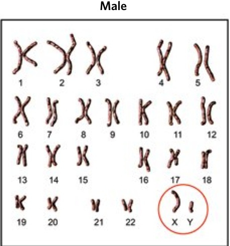
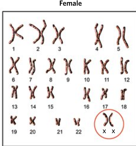
Sex chromosomes are chromosomes responsible for determining the biological sex of an organism. In humans, sex chromosomes can be either an X or Y chromosome.
In human karyotypes:
- Pairs 1-22 are autosomes, containing genetic information for thousands of genes
- Pair 23 consists of sex chromosomes (X and Y)
Sex determination in humans:
- Two X chromosomes (XX) = female
- One X chromosome and one Y chromosome (XY) = male
Chromosomal variation
Chromosome numbers vary widely across different species, and karyotypes help us understand these differences.
Chromosome numbers across species
The diploid chromosome number in human somatic cells is 2n = 46. However, different species have vastly different diploid numbers:
| Species | Diploid number (2n) |
|---|---|
| Animals | |
| Jack jumper ant (Myrmecia pilosula) | 2 |
| Housefly (Musca domestica) | 12 |
| Cat (Felis catus) | 38 |
| Chimpanzee (Pan troglodytes) | 48 |
| Dog (Canis familiaris) | 78 |
| Butterfly (Lysandra nivescens) | 190 |
| Plants | |
| Garden pea (Pisum sativum) | 14 |
| Cabbage (Brassica oleracea) | 18 |
| Corn (Zea mays) | 20 |
| Coconut tree (Cocos nucifera) | 32 |
| Pineapple (Ananas comosus) | 50 |
| Fern (Ophioglossum reticulatum) | 1,440 |
Chromosome Numbers Don't Indicate Complexity
Notice that chromosome number doesn't correlate with organism complexity. Humans have 46 chromosomes, but dogs have 78 and some ferns have over 1,000! What matters is the genetic information contained within those chromosomes, not how many chromosomes there are.
Scientists use karyotypes to represent these differences in chromosome number and to identify genetic differences between species.
Detecting genetic abnormalities
One of the most important uses of karyotypes is detecting chromosomal abnormalities within species. These abnormalities fall into two main categories: aneuploidy and polyploidy.
Aneuploidy
Aneuploidy occurs when a cell or organism varies in the usual number of chromosomes in its genome by the addition or loss of a chromosome. In humans, this means having more or fewer than the usual 46 chromosomes.
Types of Aneuploidy
Different forms of aneuploidy have specific names based on how many chromosomes are affected:
- Monosomy (2n-1): One missing chromosome
- Trisomy (2n+1): One extra chromosome
- Tetrasomy (2n+2): Two extra chromosomes
These conditions typically arise from errors during cell division, particularly during the formation of egg or sperm cells.
Examples of aneuploidy conditions
Turner syndrome (Monosomy)
Turner Syndrome
- Chromosomal pattern: Single X chromosome (often presented as XO)
- Incidence rate: 1 in 2,000 births
- Common symptoms: Infertility, short stature, fused neck and head (webbed neck)
This condition demonstrates how the loss of even a single chromosome can have significant effects on development and health.
Down syndrome (Trisomy 21)
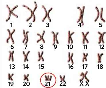
Down Syndrome
- Chromosomal pattern: Extra copy of chromosome 21
- Incidence rate: 1 in 1,000 births
- Common symptoms: Delayed physical growth, possible heart defects, flattened facial profile, mild to moderate intellectual disability
Down syndrome is one of the most common chromosomal abnormalities and demonstrates the impact of having an extra chromosome on development.
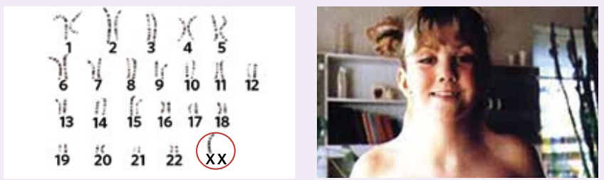
Klinefelter syndrome (Trisomy)
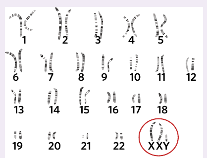
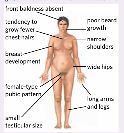
Klinefelter Syndrome
- Chromosomal pattern: Extra X chromosome in males (XXY)
- Incidence rate: 1 in 650 males born
- Common symptoms: Accelerated growth and taller height, small testes and reduced testosterone levels, delayed or incomplete puberty, decreased muscle mass, learning and intellectual disabilities, infertility, breast development, female-pattern pubic hair, narrow shoulders, wide hips, long arms and legs
This condition only affects males and results from having an additional X chromosome alongside the typical XY pattern.
Tetrasomy X (XXXX)
Tetrasomy X
- Chromosomal pattern: Two extra copies of the X chromosome (totalling 4 X chromosomes)
- Incidence rate: Approximately 100-150 confirmed cases worldwide (likely underdiagnosed as females with this condition rarely show symptoms)
- Common symptoms: Mild delay in physical development, delayed speech development, slight to moderate learning difficulties
This rare condition demonstrates that having multiple extra chromosomes can sometimes result in relatively mild symptoms.
Polyploidy
Polyploidy occurs when an organism contains additional sets of chromosomes in its genome. Instead of being diploid (2n), the organism has three or more complete sets of chromosomes (3n, 4n, etc.).
Polyploidy in Humans vs. Other Organisms
In humans, polyploidy is typically lethal, meaning it is extremely rare for a foetus to survive to term. However, polyploidy is quite common in other organisms, especially plants, where it can even be advantageous.
Advantages of polyploidy in some organisms include:
- Increased size and hardiness in certain fruits
- Sterility and faster growth rates (e.g., farmed Atlantic salmon are often triploid)
Example: Polyploidy in Agriculture
Many of the fruits and vegetables we eat are polyploid:
- Seedless watermelons are triploid (3n)
- Commercial strawberries are octoploid (8n)
- Many wheat varieties are hexaploid (6n)
These polyploid varieties often have larger fruits, better yields, and improved resistance to environmental stress compared to their diploid relatives.
Key Points to Remember:
-
DNA is organised into genes: DNA consists of nucleotides (phosphate group, deoxyribose sugar, and nitrogen-containing base). Genes are specific sections of DNA that code for proteins.
-
Genome, gene, and allele are distinct concepts: The genome is the complete set of DNA in an organism (about 3 billion base pairs and 25,000 genes in humans). A gene is a section of DNA coding for a protein. Alleles are different forms of the same gene.
-
Chromosomes package DNA efficiently: DNA wraps around histone proteins to form chromosomes. Humans have 46 chromosomes (23 pairs) in somatic cells - 22 pairs of autosomes and 1 pair of sex chromosomes.
-
Homologous chromosomes share key features: They have the same size and length, same centromere position, and same genes at the same loci. One chromosome comes from each parent.
-
Karyotypes reveal chromosomal abnormalities: Karyotypes visually organise all chromosomes and can detect aneuploidy (incorrect number of individual chromosomes like monosomy or trisomy) and polyploidy (extra complete sets of chromosomes).