Fertilisation and the Stages of Prenatal Development (VCE SSCE Health and Human Development): Revision Notes
Fertilisation and the Stages of Prenatal Development
Introduction
Understanding how pregnancy begins and how a baby develops before birth is crucial for health and human development studies. Currently, approximately one in six Australian couples face fertility challenges, making this knowledge particularly relevant.
The prenatal period refers to the time from conception until birth. This stage of development is remarkable because it represents the most rapid period of growth and change in the entire human lifespan. During these approximately 38 weeks, a single cell transforms into a fully formed baby ready to survive in the outside world.
The Three Stages of Prenatal Development
Prenatal development is divided into three distinct stages:
- Germinal stage (0-2 weeks)
- Embryonic stage (3-8 weeks)
- Foetal stage (9-38 weeks)
Each stage has unique characteristics and developmental milestones that are important to understand.
While physical development is the most obvious change during this period, the foundations for social, emotional and intellectual development also begin at this time. The prenatal stage presents both tremendous opportunities for healthy development and significant risks if problems occur.
Sperm, ova and fertilisation
Understanding sex cells
Most cells in the human body contain a structure called the nucleus, which acts like the control centre of the cell. The nucleus holds genetic material that provides instructions for how cells should function and reproduce throughout life. Different types of cells have different abilities to regenerate (regrow and replace themselves).
Sperm and ova (singular: ovum, sometimes called eggs) are specialised sex cells, also known as gametes. These cells are unique because they each contain only half the genetic information needed to create a new human being.
Sperm production
In males, sperm production begins at puberty and continues throughout adult life. Sperm are produced in the testes at an extraordinary rate—over 12 billion per month. This continuous production ensures that males remain fertile throughout their adult years.
Ova production
The production of ova in females is very different from sperm production. Females are born with all the ova they will ever have—these form in the ovaries before birth. Once a girl reaches puberty, these ova begin to mature and are released in a monthly cycle.
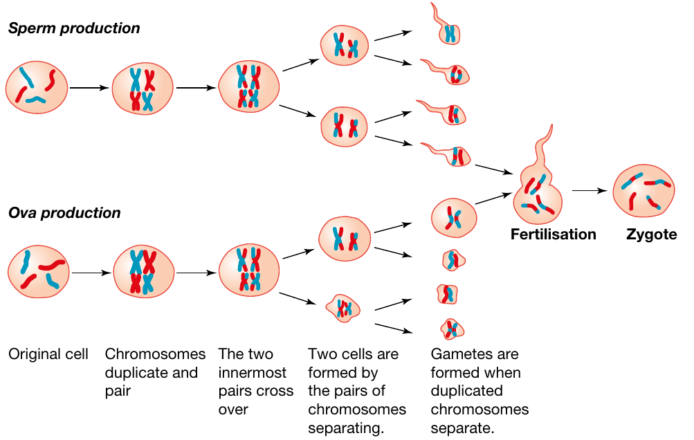
The diagram above illustrates how original cells divide in different ways each time a sperm or ovum is created. This process explains why siblings can look very different from each other, even though they share the same parents.
The process of fertilisation
Fertilisation (also called conception) is the moment when pregnancy begins. It occurs when a single sperm successfully penetrates an ovum, and the genetic materials from both parents fuse together.
Here's how fertilisation works step by step:
- During sexual intercourse, millions of sperm are deposited in the vagina
- The sperm swim through the cervix and uterus towards the fallopian tubes
- If an ovum is present in one of the fallopian tubes, many sperm will reach it and compete to enter
- The sperm release enzymes that break down the outer protective barrier of the ovum
- Once a single sperm penetrates the ovum's membrane, the ovum immediately changes its outer surface
- This change blocks all other sperm from entering—this is crucial because if more than one sperm entered, the resulting cell would have incorrect genetic information and could not survive
Why Only One Sperm Can Enter
Once a single sperm penetrates the ovum's membrane, the ovum immediately changes its outer surface to block all other sperm. This mechanism is critical for survival because if more than one sperm entered, the resulting cell would have incorrect genetic information and could not develop properly.
The zygote
When sperm and ovum successfully combine, they create a single cell called a zygote. This zygote contains:
- 23 chromosomes from the sperm
- 23 chromosomes from the ovum
- A total of 46 chromosomes arranged in 23 pairs
Chromosomes are structures made of DNA that carry genetic information. Within chromosomes are genes—the specific instructions that determine the rate and timing of development, the sex of the child, and physical characteristics. Because the zygote receives genetic material from both parents, the resulting child will display a mix of characteristics from both mother and father.
Germinal stage (0-2 weeks)
The germinal stage represents the first two weeks of prenatal development. It begins at fertilisation and ends when the developing cell mass successfully implants into the wall of the uterus.
Journey to implantation
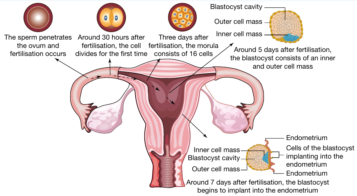
After fertilisation occurs in the fallopian tube, the newly formed zygote doesn't remain static—it immediately begins an important journey:
- Cell division begins: The zygote starts dividing within approximately 30 hours of fertilisation. This cell division will continue throughout the person's entire life.
- Travelling down the fallopian tube: While dividing, the zygote moves down the fallopian tube towards the uterus. This journey takes about three to four days.
- Morula formation: By about three to four days after fertilisation, when the cell cluster contains approximately 16 cells, it forms a solid ball-like structure called a morula.
- Blastocyst transformation: At around five days after fertilisation, when there are about 64 cells, the structure transforms into a blastocyst. This is a more complex, thin-walled hollow structure with three distinct parts:
- An outer cell mass (which will eventually become the placenta)
- An inner cell mass (which will become the embryo)
- A fluid-filled cavity in the centre
- Reaching the uterus: The blastocyst enters the uterus and prepares for implantation.
- Implantation: About seven days after fertilisation, the blastocyst begins to attach itself to the endometrium (the nutrient-rich lining of the uterine wall). This implantation process takes approximately a week to complete.
From blastocyst to embryo
Once implantation is complete, two important changes occur:
- The developing baby is now referred to as an embryo rather than a blastocyst
- The placenta begins to form from the outer cell mass
Characteristics of the germinal stage
Week-by-Week Development: Germinal Stage
| Week | Key developments |
|---|---|
| Week 1 | • Fertilisation creates a zygote when sperm and ovum combine • Cell division begins approximately 30 hours after fertilisation and continues for life • By day three, the zygote contains 16 cells • The developing cells travel down the fallopian tube towards the uterus |
| Week 2 | • Around seven days after fertilisation, the blastocyst (smaller than a grain of rice) begins implanting into the endometrium • The implantation process takes about a week to complete • Formation of the placenta begins |
Embryonic stage (3-8 weeks)
The embryonic stage is perhaps the most critical period of prenatal development. It begins when implantation is complete (around week 3) and continues until the end of week 8. During this short five-week period, the foundations for all major body systems are established.
Cell differentiation and organogenesis
The embryonic stage is characterised by two crucial processes:
Cell differentiation is when cells begin to take on specialised roles. Instead of all cells being identical, they start to become different types with specific functions. Some cells become heart cells, others become skin cells, brain cells, bone cells, and so on. This specialisation is essential for creating a functioning human body.
Organogenesis refers to the formation of organs. During the embryonic stage, the basic structures of all major organs and body systems begin to develop, including:
- The circulatory system (heart and blood vessels)
- The nervous system (brain and spinal cord)
- The digestive system (stomach and intestines)
- The respiratory system (lungs)
- The urinary system (kidneys)
Physical development during the embryonic stage
By the end of the embryonic stage (week 8), remarkable changes have occurred:
- The embryo is only about 2-2.5 centimetres in length
- The brain and spinal cord (neural tube) are almost complete in structure (though they will continue to grow in size and complexity for years)
- The circulatory system, powered by a beating heart, is the first organ system to function
- Limbs have developed from small buds into recognisable arms and legs
- Fingers and toes are beginning to form
- Facial features are taking shape
- The embryo begins to look distinctly human, though the head and neck account for about half of the total length
- The brain makes up nearly half of the embryo's body weight
- Bone tissue starts replacing cartilage
- Approximately 90 per cent of the structures found in an adult human can be identified in an eight-week-old embryo
The critical period and teratogens
The embryonic stage is the most vulnerable period of prenatal development. Because all major organs and systems are forming during this time, the developing embryo is extremely sensitive to harmful environmental influences.
Understanding Teratogens
Teratogens are substances or factors in the environment that can cause defects in development. Common teratogens include:
- Tobacco smoke
- Alcohol
- Prescription and non-prescription medications
- Illegal drugs
- Certain diseases (such as rubella)
- Environmental toxins
Exposure to teratogens during the embryonic stage can cause major structural abnormalities because this is when organs are forming. The impact is less severe during later stages of development.
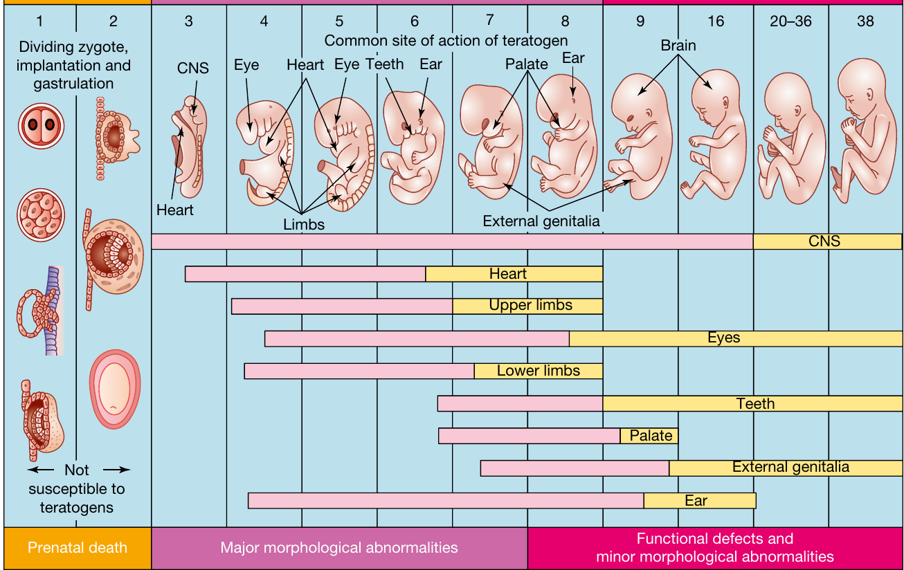
The diagram above shows the critical periods of development for different organs and body systems. Notice how the most sensitive periods (shown in pink and purple) occur during the embryonic stage. This is when exposure to teratogens can cause major structural abnormalities. Later exposure (shown in yellow) may cause functional problems or minor abnormalities, but the risk of severe malformation decreases after the embryonic stage.
Teratogens are thought to interfere with the complex process of forming connections between the brain, spinal cord, muscles, and outer parts of the developing body. For coordinated body systems to work properly, these connections must form correctly during the embryonic stage.
Week-by-week embryonic development
Week-by-Week Development: Embryonic Stage
| Week | Key developments |
|---|---|
| Week 3 | • Implantation is complete • The developing baby is now called an embryo • Cells divide rapidly and begin taking on specialised roles as organs start to develop |
| Week 4 | • The neural tube (which will become the brain and spine) starts to form • The embryo is approximately 3 millimetres long • The embryo secretes hormones that maintain the endometrium and prevent menstruation |
| Week 5 | • Small buds appear on each side of the embryo—these will become arms and legs • The heart begins to beat • The placenta is not yet fully functional, so the embryo relies on its own temporary systems • Brain cells are generated at a rate of 100 per minute |
| Week 6 | • The spinal cord has a tail-like appearance • The head is large compared to the rest of the body • The embryo measures approximately 1.3 centimetres in length |
| Week 7 | • Blood cells are being produced in the liver • Facial features (eyes and mouth) are forming • Tiny muscles have formed, allowing the embryo to move |
| Week 8 | • The embryo is approximately 2.5 centimetres long • Fingers and toes are starting to form • The brain becomes active • The embryo now has the basic structure of all major organs and systems |
Foetal stage (9-38 weeks)
The foetal stage is the final and longest stage of prenatal development, lasting from week 9 until birth at approximately week 38. Once the ninth week begins, the developing baby is referred to as a foetus rather than an embryo.
At the start of this stage, the foetus measures just 2.5 centimetres in length and weighs about 2 grams. By the end, at full term, the baby will typically measure around 50 centimetres and weigh approximately 3500 grams (3.5 kilograms).
Characteristics of the foetal stage
While rapid growth is the most obvious characteristic of the foetal stage, this period involves much more than just getting bigger. The foetal stage is marked by:
- Maturation of all organs and body systems
- Development of sensory abilities
- Preparation for life outside the uterus
- Increasing coordination and movement
- Development of fat deposits for temperature regulation
The role of the placenta
By 14 weeks of pregnancy, the placenta is fully developed and functioning. The placenta is a remarkable temporary organ that plays multiple vital roles during pregnancy.
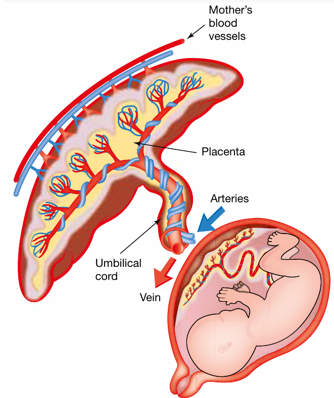
Functions of the Placenta
The placenta is a disc-shaped structure made largely of blood vessels. It connects the foetus to the mother's uterine wall through the umbilical cord. The placenta performs several crucial functions:
Exchange of substances: The placenta acts as the foetus's lung, digestive system, and kidney by:
- Supplying oxygen from the mother's bloodstream
- Providing nutrients needed for growth and development
- Transferring immune support (antibodies) from mother to foetus
- Removing waste products such as carbon dioxide and urine
Hormone production: The placenta produces important hormones, including progesterone, which:
- Help maintain the pregnancy
- Prevent the release of additional ova during pregnancy
- Support the development of the foetus
The Placenta Cannot Block All Harmful Substances
While the placenta allows beneficial substances to pass from mother to foetus, it cannot block all harmful substances. Teratogens such as alcohol, drugs, and tobacco can cross the placental barrier and affect the developing foetus. This is why avoiding harmful substances throughout pregnancy is so important.
Development during the foetal stage
Weeks 9-13: During this early foetal period, all body organs are formed but not all are fully functional yet. The foetus grows to approximately 7 centimetres by week 11. Teeth begin forming in the gums, though they won't emerge until after birth. The eyelids are fused shut over the developing eyes and will remain closed for several more weeks.
Weeks 14-18: The foetus grows to about 14 centimetres in length. Several important developments occur during this period. The tongue develops taste buds, allowing the foetus to sense different flavours in the amniotic fluid. The ears become fully functional, and the foetus can hear muffled sounds from the outside world. The sex of the foetus can now be distinguished through ultrasound examination.
Weeks 19-23: By week 22, the foetus measures approximately 33 centimetres in length. The foetus begins swallowing regularly, though it only takes in amniotic fluid. An important milestone occurs when the eyelids separate into upper and lower lids, and the foetus gains the ability to open and close its eyes.
Weeks 24-28: The foetus now measures around 37 centimetres long and weighs approximately 1 kilogram. Fingernails and toenails begin to grow. The foetus's body has grown more, making it more proportionate to the size of the head, though this process will continue until childhood. A crucial development for survival occurs around week 24 when the foetus begins producing surfactant—a substance that reduces surface tension in the lungs and prevents the small air sacs from collapsing during breathing. Babies born after 24 weeks may survive with intensive medical care, and by 28 weeks, most babies are likely to survive if born prematurely.
Weeks 29-33: During this period, the foetus spends most of its time sleeping. Eyebrows and eyelashes grow. Fat deposits form under the skin—this fat layer will help the baby regulate body temperature after birth. The foetus moves in a strong and coordinated way, and these movements are clearly noticeable to the mother.
Weeks 34-38: In preparation for birth, the foetus typically assumes a head-down position. The lungs develop rapidly during these final weeks, continuing to produce surfactant and preparing for breathing air. By the end of this stage, the foetus measures approximately 50 centimetres in length and is ready for birth.
Movement and sensory development
Throughout the foetal stage, movement increases and becomes more sophisticated. The foetus moves almost all parts of its body, and these movements become more noticeable as size increases. Important reflexes develop and strengthen, including:
- Sucking reflex (essential for feeding after birth)
- Grasping reflex
- Breathing movements (though the lungs are filled with amniotic fluid, not air)
The senses also develop during the foetal stage. Around 25 weeks after fertilisation, the foetus may respond to:
- Light
- Sound
- Touch
These senses become increasingly sensitive as development continues.
Other important developments
Sex organs: Sex organs begin taking shape early in the foetal stage. By around week 15, female foetuses will have produced millions of ova, though this number will decrease before birth. Male foetuses' testes begin producing testosterone.
Bones: Bones, which initially consist mainly of cartilage, begin to harden (ossify) during the foetal stage. This ossification process will continue until the end of puberty.
Teeth: Tooth buds form in the gums during the later part of the foetal stage, though teeth won't erupt through the gums until after birth.
Detailed characteristics of foetal development
Week-by-Week Development: Foetal Stage
| Weeks | Key developments |
|---|---|
| 9-13 | • The developing baby is now called a foetus • All body organs are formed but not all are functioning yet • Length reaches approximately 7 centimetres by week 11 • Teeth begin forming in the gums • Eyelids are fused over the eyes |
| 14-18 | • Length increases to approximately 14 centimetres • Taste buds develop on the tongue • Ears are fully functioning—the foetus can hear muffled sounds from outside • The sex of the foetus can be identified via ultrasound |
| 19-23 | • Length reaches approximately 33 centimetres by week 22 • The foetus swallows regularly, taking in amniotic fluid • Eyelids separate into upper and lower lids • The foetus can open and close its eyes |
| 24-28 | • Length reaches approximately 37 centimetres • Weight is approximately 1 kilogram • Fingernails and toenails grow • Body proportions become more balanced relative to head size • Production of surfactant begins in preparation for breathing |
| 29-33 | • Most time is spent sleeping • Eyebrows and eyelashes grow • Fat deposits form under the skin to help with temperature regulation after birth • Movement is strong and coordinated |
| 34-38 | • The foetus assumes a head-down position in preparation for birth • Lungs develop rapidly during this period • Length reaches approximately 50 centimetres • The baby is ready for birth |
Exam tips
Key Points for Exam Success
When answering questions about prenatal development in exams, remember to:
- Be accurate about the timing of each stage (germinal: 0-2 weeks, embryonic: 3-8 weeks, foetal: 9-38 weeks)
- Be specific about the characteristics of each stage—don't confuse what happens when
- Use correct terminology (zygote → morula → blastocyst → embryo → foetus)
- Understand that the embryonic stage is the most critical period for organ formation
- Remember that teratogens have the greatest impact during the embryonic stage
- Know the key functions of the placenta
- Be familiar with major developmental milestones at different points in prenatal development
Remember!
Key Takeaways: Fertilisation and Prenatal Development
-
Fertilisation occurs when a sperm penetrates an ovum, creating a zygote with 46 chromosomes (23 from each parent).
-
Prenatal development has three stages: the germinal stage (0-2 weeks), embryonic stage (3-8 weeks), and foetal stage (9-38 weeks).
-
The embryonic stage (3-8 weeks) is the most critical period because this is when all major organs and body systems begin to form through cell differentiation and organogenesis. This makes the embryo extremely vulnerable to teratogens.
-
The placenta is fully functional by week 14 and serves as the foetus's lung, digestive system, and kidney, exchanging nutrients, oxygen, and wastes between mother and baby.
-
The foetal stage (9-38 weeks) is characterised by rapid growth and the maturation of organs and systems, with the foetus growing from 2.5 centimetres and 2 grams to approximately 50 centimetres and 3500 grams by birth.