The Effect of Radiation on Humans (VCE SSCE Physics): Revision Notes
The Effect of Radiation on Humans
Introduction: The radium girls
In 1917, many young women worked at the United States Radium Company in New Jersey, painting clock faces with glowing radium paint. This historical case highlights the devastating effects of radiation exposure when safety precautions are inadequate.
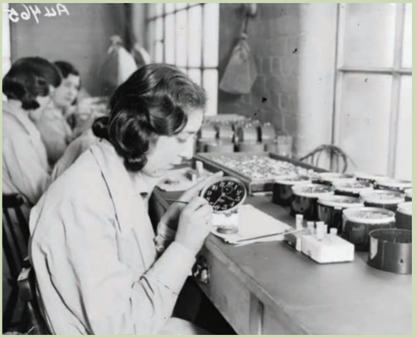
The workers were instructed to lick their brushes to create a fine point for detailed work. This practice caused them to ingest small amounts of radium hundreds of times per day. While men handling large quantities of radium wore lead aprons for protection, the women were told they were handling such small amounts that no protection was necessary.
The radium paint was marketed as "UnDark" and was considered safe at the time. Workers even used it playfully to paint their nails and teeth, completely unaware of the dangers they were exposing themselves to.
By 1927, more than 50 women had died from radium paint poisoning, suffering from severe symptoms including rotten teeth, huge tumours, and bones that glowed with radioactivity.

This tragedy demonstrates how internal radiation exposure can be extremely dangerous, even when external exposure to the same material might seem minimal. The route of exposure - whether external or internal - is a critical factor in determining radiation's harmful effects.
Discovering the effects of radiation on humans
Soon after radiation was discovered, researchers began experiencing the harmful effects of radiation exposure on living tissues. These early experiences helped establish our understanding of radiation safety.
Henri Becquerel discovered radiation's harmful effects firsthand when he placed a vial of radium in his pocket, later noticing severe skin burns. Marie Curie, who discovered both radium and polonium in 1898, eventually died from a malignant blood disease caused by prolonged radiation exposure. It is estimated that more than 300 early radiation researchers and workers died from radiation doses they received.
Marie Curie's work was pioneering but dangerous. Her laboratory notebooks are still stored in lead-lined boxes in France today because they remain highly radioactive and will continue to be so for many years.
Despite these dangers, the early 20th century saw radium marketed as a "miracle wonder drug". The idea of using radioactive elements to treat cancer began with Becquerel's accidental skin burn in 1901. By 1902, radium was successfully used to treat throat cancer. Subsequently, radium found its way into numerous products including tonic water, toothpaste, and cosmetics. Hundreds of thousands of people consumed radium-infused products, unaware of the dangers.
The capacity of alpha, beta and gamma radiation to cause cell damage
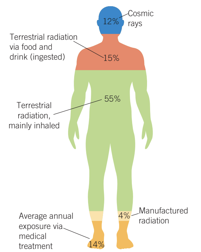
Given the prevalence of radiation in our environment from both natural and artificial sources, understanding how different types of radiation affect our health is crucial. Approximately 82% of the average annual radiation exposure comes from natural sources.
Our bodies cannot sense lethal doses of radiation, making it particularly dangerous. Without specialised radiation detection equipment such as a Geiger counter, we would be unaware of life-threatening radiation exposure.
How radiation damages cells
When ionising radiation strikes tissue, cells, or molecules, it significantly alters them. The damage can occur in several ways:
Direct damage: Radiation can break molecular bonds, altering the molecular structure so that damaged cells no longer carry out their proper functions. DNA molecules may become damaged and no longer carry correct genetic information.
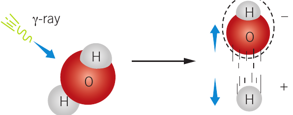
Indirect damage: Radiation can break water molecules surrounding DNA (approximately 60% of our body mass is water). When water molecules are broken, they produce chemically reactive ions such as unstable oxygen molecules that can damage cells and organs.
Once a cell is damaged by radiation, three outcomes are possible:
- Cell repair: If the damage is not too severe, the cell repairs itself and returns to normal function.
- Cell alteration: If the cell damage is not repaired or is incorrectly repaired, this change may eventually lead to cancerous tumour formation.
- Cell death: If there is too much damage, the cell dies.
Cell death is not always harmful. If a few radiation-damaged cells die, your body recovers without the risk of those cells turning into cancer. However, widespread cell death caused by high radiation doses can lead to organ failure and ultimately death.
Penetration and ionising power
The ability of radiation to damage tissue, cells, organs and molecules relates directly to its ionising power and penetration ability.
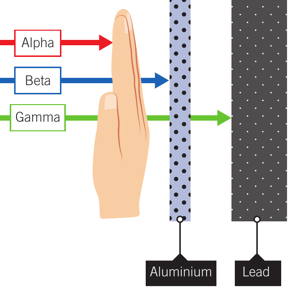
Alpha particles are highly ionising and have a short range, being stopped by skin or paper. They have very high ionising ability but very low penetration. Because of their limited penetration, alpha particles pose minimal threat from external sources located about a metre or more away. However, if alpha-emitters are inhaled or ingested, they become extremely dangerous as there is very little protection from internal sources.
Beta particles have intermediate ionising power and penetration. They are less ionising than alpha particles but can penetrate further, being stopped by approximately 2 mm of aluminium. Like alpha particles, the greatest danger occurs when beta-emitters are inhaled or taken in with food or water.
Gamma rays are very penetrating and easily pass through the human body. They have very low ionising ability but very high penetration. Lead approximately 5 cm thick or concrete approximately 1 m thick will absorb 97% of gamma rays. Many gamma rays may pass completely through a human body without striking anything, but when they do interact, they have enough energy to cause cell death.
| Particle | Symbol | Penetration | Ionising ability | Shielding |
|---|---|---|---|---|
| Alpha | Very low | Very high | Paper, skin | |
| Beta | Intermediate | Intermediate | Aluminium 2 mm | |
| Gamma | Very high | Very low | Lead 5 cm |
Protection from radiation
Lead aprons, lead thyroid shields and lead eyeglasses are essential protective equipment for medical personnel working with penetrating radiation. Gamma rays and X-rays are both highly penetrating forms of radiation that require such shielding.
The safest amount of radiation exposure to the human body is zero. However, since we are all subject to background radiation, the way to minimise risk is to:
- Limit the time of radiation exposure
- Increase distance from the radioactive source
- Use appropriate shielding
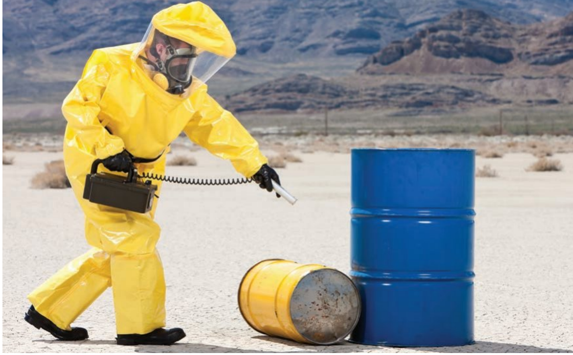
Events like nuclear explosions or accidents, where radioactive alpha-emitters are spread in the environment, are extremely dangerous to all life forms because the radioactive particles become part of the food chain.
Radiation doses
Our bodies cannot sense high-intensity doses of radiation. A dose of gamma radiation large enough to be lethal would only increase body temperature by about one thousandth of a degree Celsius. Without specialised radiation detection equipment, we would be unaware of life-threatening situations.
Measuring equipment
Geiger counters are devices for measuring radiation that provide real-time measurement of radioactive isotope activity. They are suitable for measuring alpha, beta and gamma radiation, with a maximum capacity of about 10,000 particles per second (10 kBq). They are useful for monitoring radiation in factories and hospitals, detecting radiation leaks, and searching for radioactive geological formations.
Dosimeters do not provide protection against radiation exposure but instead detect and measure radiation that you have already been exposed to. These are monitored to ensure people working in radiation environments do not exceed their annual limits. The optically stimulated luminescence (OSL) monitor measures potential occupational doses from gamma and X-rays.

Units of measurement
Physicists use three different units to quantify radioactivity and radiation:
Activity is measured in becquerels (Bq), which represents the number of nuclear transformations occurring per second.
Absorbed dose measures the radiation energy absorbed at the target site - the energy absorbed per kilogram of an irradiated object. The unit is the gray (Gy), where:
Dose equivalent quantifies the potential of radiation to damage and kill cells. The unit is the sievert (Sv), which measures the biological effect of ionising radiation.
The radiation weighting factor
The number of ionisations in a cell for a given dose of radiation is much greater for alpha radiation than for beta or gamma radiation. To account for the biological damage caused by different types of radiation, the radiation weighting factor is used.
For beta and gamma radiation, the radiation weighting factor is 1. An absorbed dose of 1 μGy of beta or gamma radiation has a dose equivalent of exactly 1 μSv.
For the more strongly ionising alpha radiation, the radiation weighting factor is 20. The same absorbed dose of 1 μGy of alpha radiation has a radiation dose equivalent of 20 μSv.
The relationship is:
This formula accounts for the fact that different types of radiation cause different amounts of biological damage even when the absorbed energy is the same.
Worked Example: Calculating Radiation Doses
A 60 kg adult absorbs 180 mJ of energy due to ionising radiation.
a) Calculate the absorbed dose in mGy:
b) Calculate the dose equivalent for different radiation types:
For gamma radiation (radiation weighting factor = 1):
For alpha radiation (radiation weighting factor = 20):
Notice that the same absorbed dose results in a 20 times greater dose equivalent for alpha radiation compared to gamma radiation.
Effective dose
Different parts of the human body have different risk-weighting factors. Organs in which many cell divisions occur, such as bone marrow or lungs, are more vulnerable to radiation damage. This gives rise to the concept of effective dose, which applies to specific organs.
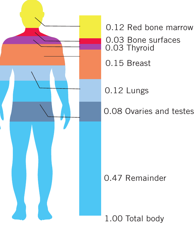
A given effective dose equivalent of radiation is four times more likely to cause a possibly fatal cancer in the lungs than in the thyroid gland (0.12:0.03). Our reproductive organs (ovaries and testes) are very sensitive to radiation damage, which has important medical implications when using radiation therapy.
Equivalent dose is calculated for the whole body by adding effective doses to all organs, each adjusted to account for the sensitivity of each organ to radiation. This is the most frequently used dose in radiological protection, given in sieverts (Sv) or millisieverts (mSv).
In the simplest cases, for uniform whole-body exposure to gamma or beta radiation, the radiation weighting factor is 1, and the tissue weighting factors add up to 1. Therefore, an absorbed dose of 1 mGy equals an equivalent dose of 1 mSv.
Short- and long-term effects of low and high doses
Somatic effects arise when ordinary body cells are damaged, and the severity depends on the dose size. The period between initial radiation exposure and the appearance of damage is called the latent period.
Short-term effects
If the dose exceeds 1 Sv (1000 mSv), symptoms of radiation illness can occur within a few days, including:
- Nausea
- Headaches
- Vomiting
- Diarrhoea
These are classified as short-term effects.
Acute radiation syndrome
Most acute effects of radiation become evident hours, days and months after a single-dose exposure to the entire body. Acute radiation syndrome (ARS), also known as radiation sickness or radiation poisoning, results from exposure to high amounts of ionising radiation in a short period.

Apart from damage to reproductive organs and the lens of the eye, there will be damage to:
- Bone marrow
- Gastrointestinal system
- Central nervous system
Dose equivalents greater than 8.0 Sv are normally lethal to humans.
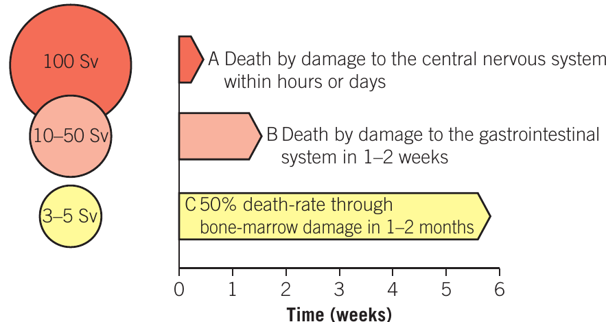
| Whole-body dose (Sv) | Symptoms |
|---|---|
| <1 | Non-fatal. Only minor symptoms such as nausea. White blood cell level drops. |
| 2 | Death unlikely in most cases. Radiation sickness (nausea, vomiting, diarrhoea). Skin burns, possible hair loss. Bone marrow damage. |
| 3–5 | 50% likelihood of death within 1–2 months. Severe radiation sickness. High probability of leukaemia and tumours for survivors. |
| 8 | Almost certain death within 1 or 2 weeks. Acute radiation sickness, convulsions, lethargy. |
Long-term effects
Lower doses may not show effects for several decades. These delayed or long-term effects include:
- Leukaemia
- Cancerous tumours
Radiation standards
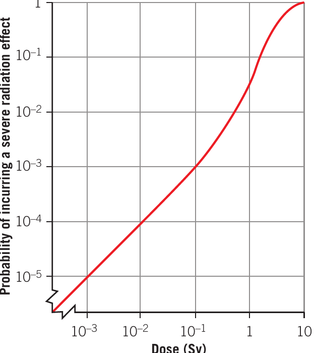
The relationship between radiation dose and its effects on humans is complex. The International Atomic Energy Agency (IAEA) has published a dose–risk relationship graph used for radiation protection purposes.
The International Commission for Radiological Protection (ICRP) currently recommends:
- General public: 1 mSv per year whole-body dose limit
- Radiation industry workers: 20 mSv per year averaged over 5 years, not exceeding 50 mSv in any single year
These limits are in addition to natural background radiation (approximately 2.3 mSv per year). Workers in radiation industries can receive 20 times the dose that the general public receives per year.
The ICRP argues these standards give a probability of:
- 1 in 100,000 for a member of the general public dying from radiation-induced illnesses over a 30-year period
- 1 in 5,000 for a radiation industry worker dying from radiation-induced illnesses over a 30-year period
Genetic effects
Accurately assessing genetic effects of radiation on humans is difficult. It may be many generations before effects become apparent (for humans, perhaps 50–200 years), reliable data is limited, and many genetic defects are hard to directly link to radiation exposure.
Two main known genetic effects of radiation are:
Chromosome aberrations: Changes in the actual number or structure of chromosomes
Genetic mutations: Alterations of the nucleotide sequence of a gene, which can lead to birth defects in future generations
Results from animal studies suggest that an accumulated 1 Sv dose of radiation on human populations will cause between 1,000 and 2,000 severe genetic mutations, and up to 1,000 severe effects due to chromosome aberrations, per million births.

Long-term studies of more than 27,000 children of parents exposed to radiation from nuclear bombs at Hiroshima and Nagasaki are inconclusive. Research into genetic effects from nuclear reactor accidents at Chernobyl and Fukushima is ongoing. Deformed butterflies have been found near Fukushima, raising concerns about similar effects in humans.
External and internal radiation
Radiation irradiates us in two distinct ways:
External radiation: The radiation source remains outside the body and irradiates from the outside.
Internal radiation: The radiation source is inhaled into the lungs or swallowed, irradiating from inside the body.
This distinction is important because different properties of various radiation types make them more or less dangerous depending on whether they are external or internal sources. Alpha particles, for example, are relatively harmless from external sources but extremely dangerous when inhaled or ingested.
Alpha particles, beta particles and gamma rays are all ionising. They have enough energy to break molecular bonds within cells. For example, a gamma ray can break the bonds of a water molecule, creating chemically reactive ions (OH⁻ and H⁺), which may lead to uncontrolled cell division resulting in cancerous tumours.
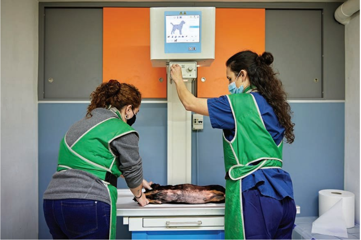
Nuclear medicine
Nuclear radiation is a powerful tool in modern medicine, both for diagnosis (finding the problem) and treatment.
Diagnostic strategy
In medical diagnosis, the strategy is to keep the radiation dose extremely small while gaining maximum information. Small amounts of short-lived radioactive isotopes are injected, taken orally, or inhaled by patients. The radioisotope then circulates through the body or is taken up only by certain tissues.
The emitted radiation can be captured by imaging techniques such as:
- Single photon emission computed tomography (SPECT)
- Positron emission tomography (PET)
Through such imaging, physicians can examine blood flow to specific organs and assess organ function or bone growth. The radioisotopes used typically have short half-lives and decay before their emitted radioactivity can damage the patient's body.
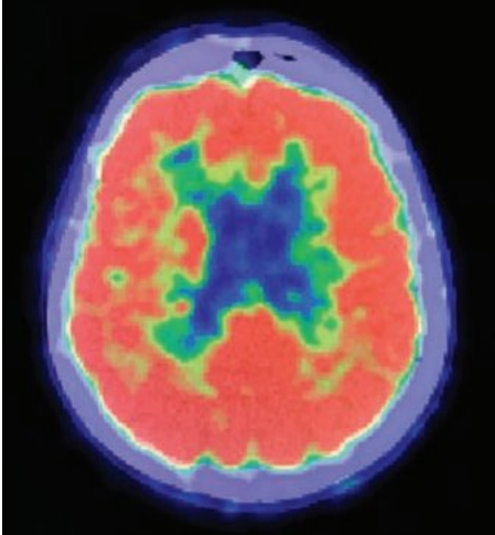
PET scans
When a positron meets an electron, there is complete annihilation and two gamma rays are produced. This forms the basis of PET scans, which are very useful for various medical brain scans.
Radioactive iodine-123 (diagnostic) and iodine-131 (therapy)
Two different isotopes of iodine are used for diagnosis and treatment of the thyroid gland:
Iodine-123 (relatively harmless to thyroid cells) is used for diagnosis Iodine-131 (destroys thyroid cells) is used for therapy
To diagnose thyroid function, iodine-123 (a gamma ray emitter with a half-life of 13 hours) is injected into the bloodstream. The thyroid has a great affinity for iodine. If after one hour:
- 98% of radioactive iodine is collected evenly in the thyroid: gland is functioning normally
- Iodine-123 is taken up more quickly: overactive thyroid condition
- Iodine-123 is taken up more slowly: underactive thyroid condition
- Iodine-123 is not evenly distributed: probably indicates a cancerous tumour
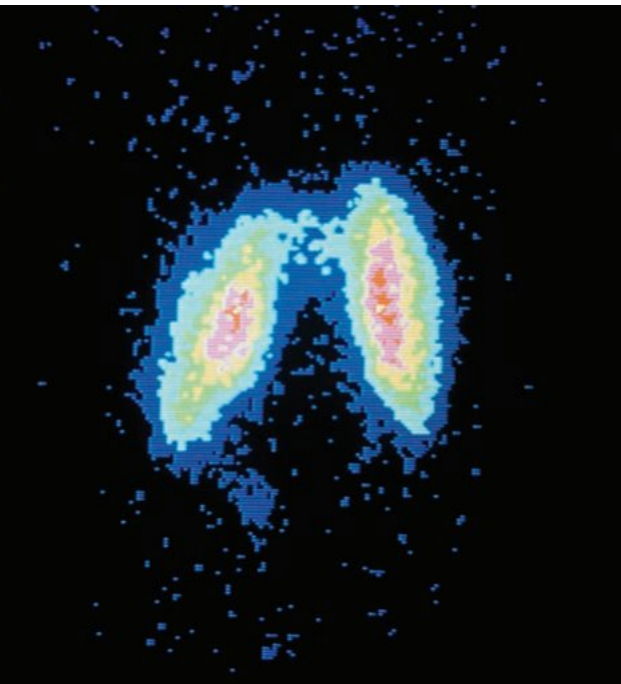
The diagnostic radiologist measures the uptake rate and distribution of radioactive iodine-123 by moving a gamma ray detector across the thyroid gland. The distribution is represented using false or enhanced colours.
In treating thyroid cancer, radioactive iodine-131 is taken orally. Most of this iodine ends up at the thyroid gland. Iodine-131 is both a beta and gamma emitter with a half-life of 8 days.
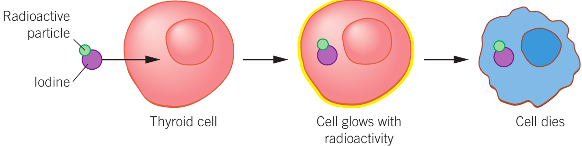
The beta particles, with their relatively low penetrating ability, irradiate only tissue within the thyroid gland and destroy cancerous cells. The gamma rays penetrate the patient's body, exposing healthy cells to some radiation but allowing external monitoring with a gamma ray camera.
Radiation therapy
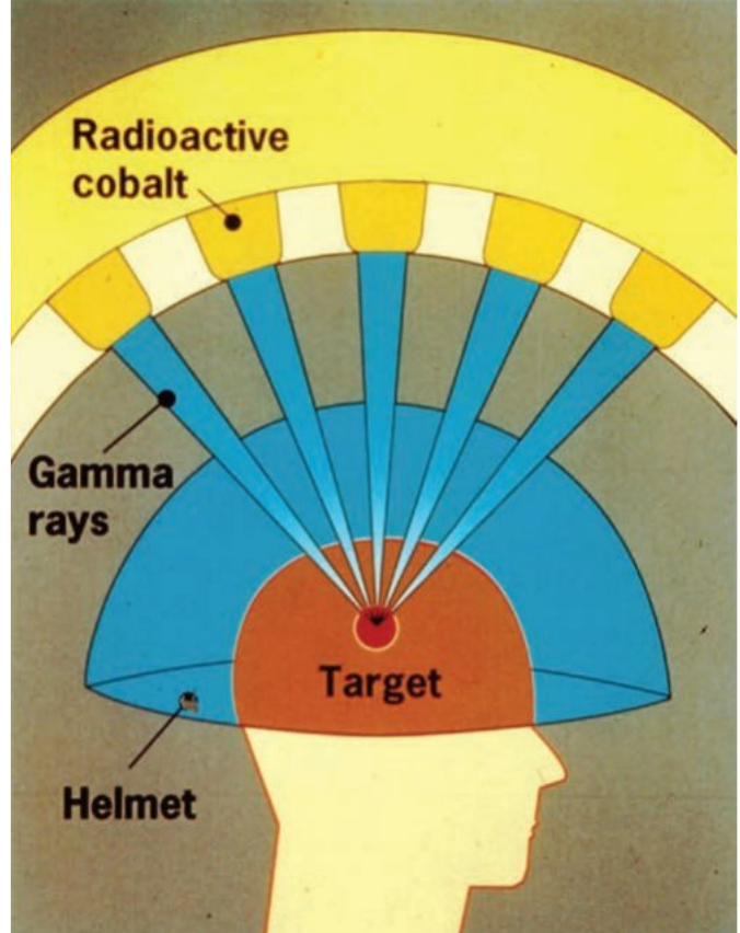
In radiation therapy, the strategy is to deliver a lethal radiation dose to a selected site while keeping the dose to surrounding areas at a minimum. Common methods involve selecting organ-specific radioactive isotopes or using shielding and rotation techniques to target specific sites.
In Australia, approximately 50,000 deaths each year are attributed to cancer-related causes. The probability of surviving cancer depends on early detection and the type of cancer. For example, early detection of breast cancer leads to an average 5-year survival rate of 91%.
Usual cancer treatment methods include:
- Surgery
- Chemotherapy (use of radiopharmaceuticals)
- Irradiation (use of radiation beams)
- Combination of these methods
In diagnostic applications, the objective is to minimise radiation damage. However, in cancer treatment, the purpose is to cause cell damage, reduce tumour cell growth, or kill them.
Medical radiographers plan high-dose treatments using the principle of acceptable risk, carefully weighing the advantages and disadvantages of radiation treatment. Although the dose may cause permanent damage (such as hair loss and reduced disease resistance), it may be the only realistic means of saving lives.
Cancerous cells do not perform required tasks. They are dysfunctional cells that reproduce rapidly and uncontrollably, usually forming lumps or tumours. As abnormal cell numbers increase, debilitating biological problems arise as the body struggles to function normally.
Fortunately, rapidly reproducing cells (such as cancerous cells) are much more sensitive to radiation than normal cells. Techniques that irradiate specific cancerous cell sites can administer doses effectively up to 1,000 times greater than the radiation dose received by neighbouring healthy cells.
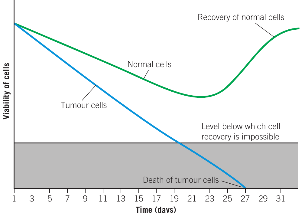
This graph shows the effect of a fractionated radiation treatment plan on both normal and tumour cells. The total radiation dose (e.g. 20 Gy) is split into ten doses of 2.0 Gy each, administered every second day for 10 days. Normal cells can recover between treatments, whilst tumour cells continue to decline until they reach the level below which recovery is impossible.
Key Takeaways:
-
Alpha radiation is highly ionising but has low penetration (stopped by paper/skin). It is most dangerous as an internal radiation source when inhaled or ingested.
-
Beta radiation has intermediate ionising power and penetration (stopped by 2 mm aluminium). Like alpha, it poses the greatest danger when taken internally.
-
Gamma radiation has low ionising power but very high penetration (requires 5 cm lead to shield). It can cause damage both externally and internally.
-
Radiation dose calculations use three key units: Gray (Gy) for absorbed dose (), Sievert (Sv) for dose equivalent, and the formula: dose equivalent (Sv) = absorbed dose (Gy) × radiation weighting factor (, , ).
-
Short-term effects of high doses (>1 Sv) include nausea, vomiting, and diarrhoea, whilst long-term effects can include leukaemia and cancerous tumours. Doses above 8 Sv are normally lethal.
-
Medical applications use radiation for both diagnosis (keeping doses extremely small) and therapy (delivering lethal doses to cancerous cells whilst minimising damage to healthy tissue). Cancerous cells are more sensitive to radiation than normal cells, making targeted radiotherapy effective.