Cell Structures & Organelles (Leaving Cert Biology): Revision Notes
Cell Structures & Organelles
Introduction to cell structures
Cells contain many different structures called organelles that work together to keep the cell functioning. These organelles can only be seen clearly using an electron microscope, which provides much greater magnification than a light microscope. Each organelle has a specific structure that enables it to carry out particular functions within the cell.
Why electron microscopes? Electron microscopes can magnify specimens up to 500,000 times, compared to light microscopes which only magnify up to 2,000 times. This incredible magnification reveals the intricate details of cellular organelles that would otherwise be invisible.
Cell membrane
Structure of the cell membrane
The cell membrane (also called the plasma membrane) forms the outer boundary of all cells. It has a unique structure that allows it to control what enters and leaves the cell.

The cell membrane is made up of a phospholipid bilayer - essentially two layers of phospholipid molecules arranged back-to-back. Each phospholipid molecule has:
- A phosphate head that is water-loving (hydrophilic)
- Lipid tails that are water-hating (hydrophobic)
The phospholipids arrange themselves so that the water-loving heads face outward towards the watery environments inside and outside the cell, while the water-hating tails face inward, away from water.
Self-Assembly Marvel The phospholipid bilayer forms automatically due to the chemical properties of phospholipids. This self-assembly process means that cell membranes can repair small holes and maintain their structure without external energy input.
Functions of the cell membrane
The cell membrane has several crucial functions:
- Controls entry and exit: It determines what substances can enter or leave the cell
- Recognition: Contains protein receptors that recognise specific molecules
- Selective permeability: Some molecules can pass through easily (like water and oxygen), while others (like large proteins) need special transport mechanisms
- Maintains cell shape: Provides structural support and maintains the cell's boundary
Nucleus
Structure of the nucleus
The nucleus is often called the control centre of the cell because it contains the cell's genetic material. It's usually the largest organelle in the cell and has several important parts:
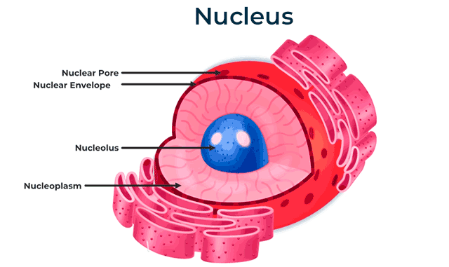
- Nuclear membrane: A double membrane that surrounds the nucleus
- Nuclear pores: Small openings in the nuclear membrane that allow molecules to move in and out
- Chromatin: Long, thin strands of DNA and protein that contain the genetic information
- Chromosomes: Condensed form of chromatin that becomes visible during cell division
Functions of the nucleus
The Nucleus: Master Controller The nucleus performs two critical functions that are essential for cell survival:
- Controls cell activities: Regulates what happens in the cell by controlling which proteins are made
- Contains genetic information: Houses the DNA that contains instructions for making proteins and controlling cell division
Cytoplasmic organelles
The cytoplasm is the jelly-like substance that fills the cell and surrounds the nucleus. Suspended within the cytoplasm are various organelles, each with specific functions.
Mitochondria
Mitochondria are known as the "powerhouses of the cell" because they produce most of the energy that cells need to function.
Structure of mitochondria

Mitochondria have a distinctive structure:
- Double membrane: An outer membrane and a highly folded inner membrane
- Cristae: The folds of the inner membrane that increase surface area
- Mitochondrial DNA: Small loops of DNA that allow mitochondria to reproduce themselves
Functions of mitochondria
- Energy production: Carry out aerobic respiration to produce ATP (the cell's energy currency)
- More active cells have more mitochondria: Muscle cells and liver cells contain many mitochondria because they need lots of energy
- Less active cells have fewer mitochondria: Storage cells like fat cells have fewer mitochondria
Worked Example: Mitochondria in Different Cell Types
Step 1: Consider a muscle cell during exercise
- High energy demand for contraction
- Contains thousands of mitochondria
- Result: Sustained energy production for movement
Step 2: Compare with a fat storage cell
- Low energy demand for storage
- Contains fewer mitochondria
- Result: Energy mainly used for basic cellular maintenance
Ribosomes
Ribosomes are tiny structures responsible for making proteins in the cell.
Structure and function of ribosomes
Ribosomes are made of ribosomal RNA (rRNA) and protein. They appear as small, bead-like structures under an electron microscope. Their main function is protein synthesis - they read the genetic instructions from DNA and use them to build proteins that the cell needs.
Chloroplasts (plant cells only)
Chloroplasts are organelles found only in plant cells and some algae. They're responsible for photosynthesis.
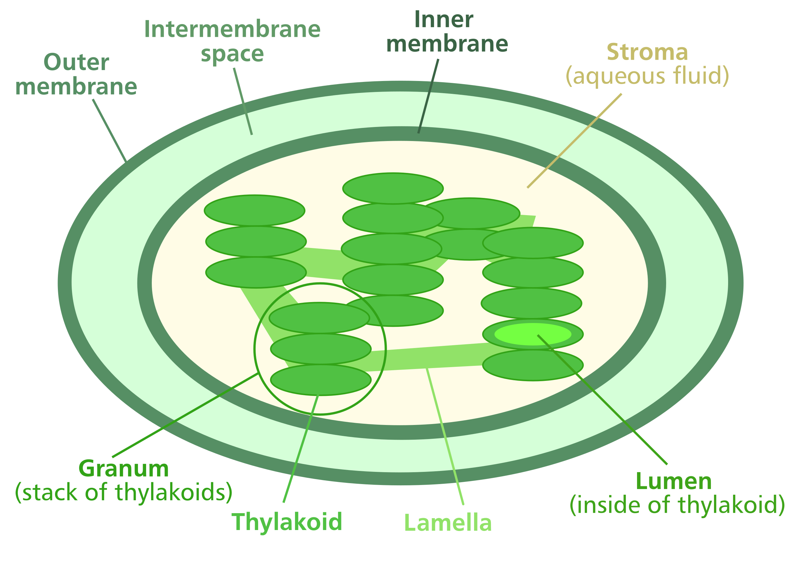
Structure of chloroplasts
Chloroplasts have several key features:
- Double membrane: Similar to mitochondria, with outer and inner membranes
- Stacks of chlorophyll: Called thylakoids, these contain the green pigment chlorophyll
- Chloroplast DNA: Like mitochondria, chloroplasts have their own DNA loops
Function of chloroplasts
The primary function of chloroplasts is to carry out photosynthesis - the process by which plants convert light energy, carbon dioxide, and water into glucose and oxygen.
Why Only Plants? Chloroplasts contain chlorophyll, the green pigment that captures light energy. This is why plants are green and why they can make their own food through photosynthesis, while animals must obtain energy by consuming other organisms.
Endoplasmic reticulum
The endoplasmic reticulum (ER) is a network of membrane-bound tubes and sacs that extends throughout the cytoplasm.
Types of endoplasmic reticulum
There are two types of ER:
- Smooth ER: Does not have ribosomes attached to it
- Makes lipids and hormones
- Stores calcium
- Breaks down harmful substances
- Rough ER: Has ribosomes attached to its surface, giving it a "rough" appearance
- Produces and transports proteins
- Works closely with ribosomes to make proteins that will be secreted from the cell
Golgi apparatus
The Golgi apparatus is the cell's processing and packaging centre.
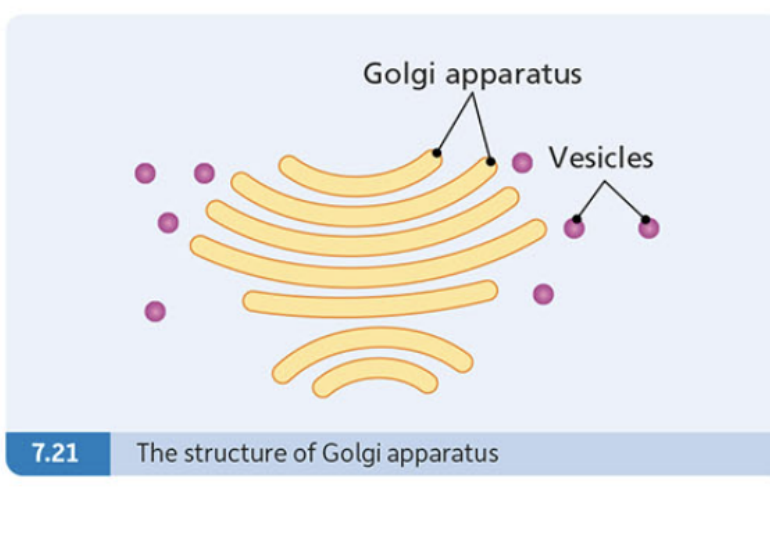
Structure of the Golgi apparatus
The Golgi apparatus consists of:
- Flattened membrane sacs: Stacked like pancakes
- Vesicles: Small membrane-bound sacs that transport materials to and from the Golgi
Function of the Golgi apparatus
The Golgi apparatus receives proteins from the endoplasmic reticulum and then:
- Modifies proteins: Adds chemical groups or changes protein structure
- Packages proteins: Wraps proteins in membrane vesicles
- Transports proteins: Sends packaged proteins to their final destinations in the cell
Cell wall (plant cells only)
Plant cells have an additional structure that animal cells lack - the cell wall.
Plant-Exclusive Structure The cell wall is one of three structures found ONLY in plant cells (along with chloroplasts and large vacuoles). This rigid structure is what allows plants to grow tall and maintain their shape without a skeleton like animals have.
Structure of cell walls
Cell walls are made of interwoven fibres of a carbohydrate called cellulose. These fibres form a mesh-like structure that leaves large gaps, making the cell wall fully permeable to water and small molecules.
Function of cell walls
Cell walls serve several important purposes:
- Provide shape: Give the plant cell a defined, rigid shape
- Provide strength: Support the plant and prevent it from collapsing
- Protection: Shield the cell from damage
Prokaryotic vs eukaryotic cells
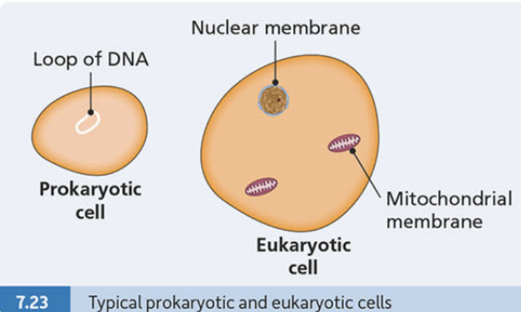
There are two main types of cells based on how their genetic material is organised:
Fundamental Cell Classification
Eukaryotic cells:
- Have a nucleus: Genetic material is enclosed within a nuclear membrane
- Have membrane-bound organelles: Such as mitochondria, ER, and Golgi apparatus
- Examples: Plant cells, animal cells, fungi cells
Prokaryotic cells:
- No nucleus: Genetic material exists as a loop of DNA floating freely in the cytoplasm
- No membrane-bound organelles: Much simpler internal structure
- Examples: Bacterial cells
Summary of cell structures
Structures found only in plant cells:
- Cell wall
- Chloroplasts
- Large permanent vacuole
Structures found in both plant and animal cells:
- Cell membrane
- Cytoplasm
- Nucleus and nuclear pores
- Chromosomes
- Mitochondria
- Ribosomes
- Endoplasmic reticulum
- Golgi apparatus
Key Points to Remember:
- Cell membranes are made of phospholipid bilayers that control what enters and exits cells
- The nucleus is the control centre containing DNA and controlling cell activities
- Mitochondria are the powerhouses that produce energy through respiration
- Plant cells have three unique structures: cell wall (strength), chloroplasts (photosynthesis), and large vacuole (storage)
- Prokaryotic cells lack a nucleus and membrane-bound organelles, while eukaryotic cells have both