Human Responses to the Environment (Grade 12 NSC Matric Life Sciences): Revision Notes
The Human Ear
The human ear is a remarkable sensory organ that allows us to hear sounds and maintain our balance. Understanding its structure and function is essential for comprehending how we respond to our environment through hearing and spatial orientation.
The ear is unique among sensory organs because it serves two distinct functions - hearing and balance - both of which are crucial for survival and daily functioning.
Overview of ear structure
The human ear is divided into three main sections, each with specific roles in the hearing and balance processes. These sections work together as a coordinated system to convert sound waves into nerve impulses that our brain can interpret.
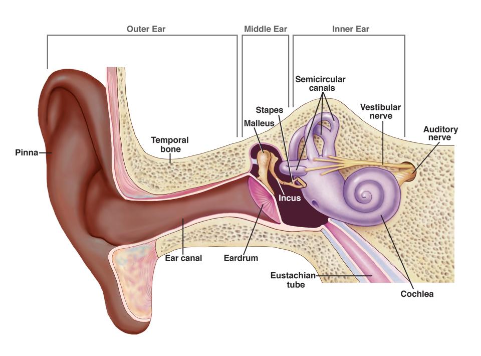
The three main sections are:
- Outer ear: Collects and directs sound waves
- Middle ear: Amplifies and transmits vibrations
- Inner ear: Converts vibrations to nerve impulses and maintains balance
Coordinated System
The ear functions as an integrated system where each section depends on the others. A problem in any one section can affect overall hearing ability, which is why understanding the complete pathway is important.
The outer ear
The outer ear serves as the initial sound collection system of our auditory apparatus. Its design is perfectly adapted to gather sound waves from the environment and channel them efficiently towards the middle ear.
Structure and components
The outer ear consists of two main parts that work together to capture and direct sound waves. The pinna (also called the auricle) is the visible part of the ear made from cartilage flaps. Its curved shape and ridges are specifically designed to collect sound waves from different directions and funnel them into the ear canal.
The auditory canal (also called the ear canal) is a tube-like passage that connects the pinna to the eardrum. This canal is approximately 2.5 cm long and serves as a protected pathway for sound waves to travel deeper into the ear.
Functions of the outer ear
The pinna's complex shape allows it to direct sound waves efficiently into the auditory canal. Different frequencies and directions of sound are captured and funnelled towards the eardrum. The auditory canal not only transmits these sound waves but also provides important protective functions.
Protection is Key
Inside the auditory canal, tiny protective hairs act as philtres to prevent foreign objects, dust, and insects from entering deeper into the ear. Additionally, the canal produces earwax (cerumen), which serves as a natural moisturiser to prevent the eardrum from drying out and becoming brittle.
The middle ear
The middle ear is an air-filled cavity located within the skull bone, positioned between the outer and inner ear. This remarkable chamber acts as an amplification system that increases the strength of sound vibrations before they reach the inner ear.
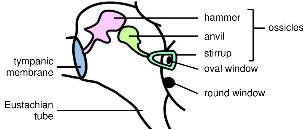
The tympanic membrane
The tympanic membrane (commonly known as the eardrum) forms the boundary between the outer and middle ear. This thin, delicate membrane is crucial for hearing as it vibrates in response to sound waves travelling through the auditory canal.
When sound waves reach the tympanic membrane, they cause it to vibrate at the same frequency as the original sound. These vibrations are then transmitted to the middle ear structures, beginning the process of sound amplification.
The ossicles - tiny bones with a big job
The middle ear contains three tiny bones called the ossicles, which form a chain that transmits vibrations from the eardrum to the inner ear. These are the smallest bones in the human body, yet they play a crucial role in our ability to hear.
The three ossicles are:
- Hammer (malleus): The largest ossicle, directly connected to the tympanic membrane
- Anvil (incus): The middle bone that connects the hammer to the stirrup
- Stirrup (stapes): The smallest ossicle, which connects to the oval window of the inner ear
Exam tip: Remember the ossicles with "Have An Itch" - Hammer, Anvil, Stirrup!
Other middle ear structures
The oval window is a membrane that separates the middle ear from the inner ear. When the stirrup vibrates against it, these vibrations are transmitted into the fluid-filled inner ear.
The round window is located below the oval window and serves as a pressure release valve. It absorbs excess pressure waves from the inner ear, preventing harmful vibrations from being echoed back.
The Eustachian tube connects the middle ear to the back of the throat. This tube is essential for equalising air pressure on both sides of the eardrum, which is why you might feel your ears "pop" when flying or changing altitude.
Pressure Equalisation
The Eustachian tube's role in pressure equalisation explains why yawning, swallowing, or chewing gum can help relieve ear discomfort during flights - these actions help open the tube and balance pressure.
The inner ear
The inner ear is the most complex part of the auditory system, containing organs responsible for both hearing and balance. Located within the bones of the skull, it consists of fluid-filled chambers and canals that convert mechanical vibrations into electrical nerve impulses.

The inner ear contains two important fluid systems: the bony labyrinth filled with perilymph, and the membranous labyrinth filled with endolymph. These fluids play crucial roles in both hearing and balance detection.
Semi-circular canals - detecting head movement
The semi-circular canals are three fluid-filled loops arranged at right angles to each other. This arrangement allows them to detect head movements in all three dimensions of space - up and down, side to side, and rotational movements.
Each canal has an enlarged area called an ampulla at one end, which contains sensory receptors called cristae. When your head moves or rotates, the fluid inside the canals moves, causing the cristae to bend. This bending generates nerve impulses that are sent to the cerebellum in the brain, helping you maintain balance and spatial orientation.
Common misconception: Many students think the semi-circular canals are involved in hearing, but they are purely balance organs!
The vestibule - detecting head position
The vestibule is the central chamber of the inner ear, located between the semi-circular canals and the cochlea. It contains two fluid-filled sacs called the sacculus and utriculus, which together detect changes in head position relative to gravity.
Inside these sacs are specialised receptors called maculae. The maculae contain tiny hair cells covered with a jelly-like substance and small calcium carbonate stones called otoliths. When your head tilts or when you experience linear acceleration (like in a lift), gravity causes the otoliths to shift, bending the hair cells and generating nerve impulses that inform your brain about your head's position.
Gravity Detection
The vestibule's ability to detect gravity and linear acceleration is why you can sense when a lift is moving up or down, even with your eyes closed. The otoliths respond to these gravitational changes.
The cochlea - the organ of hearing
The cochlea is the spiral-shaped structure responsible for converting sound vibrations into nerve impulses that the brain interprets as sound. Its snail-shell shape maximises the surface area available for sound detection within the limited space of the skull.
The cochlea is divided into three fluid-filled chambers. The upper and lower chambers contain perilymph, while the middle chamber contains endolymph and houses the organ of Corti - the actual hearing receptor.
The organ of Corti contains thousands of tiny sensory hair cells embedded in a membrane. When sound vibrations travel through the cochlea's fluid, these hair cells bend, converting the mechanical energy of sound waves into electrical nerve impulses. These impulses travel along the auditory nerve to the brain, where they are interpreted as the sounds we perceive.
The Final Conversion
The organ of Corti is where the mechanical energy of sound finally becomes electrical energy that the nervous system can process. This conversion is the culmination of the entire hearing process.
Key terminology review
Understanding these key terms is essential for NSC exams:
- Organ of Corti: The specialised hearing receptor located in the cochlea
- Crista/Cristae: Balance receptors that detect changes in head speed and direction
- Maculae: Balance receptors that detect changes in head position relative to gravity
- Ampulla: Enlarged areas of the semi-circular canals containing the cristae
- Vestibule: Structure containing the sacculus and utriculus with their maculae
- Grommet: A medical device inserted into the eardrum to allow air flow (used to treat certain ear conditions)
Exam Success Tip
These terms frequently appear in NSC examinations. Make sure you can both define them and explain their functions within the context of hearing and balance.
Remember!
Key Points to Remember:
-
The ear has three main sections: outer ear (collects sound), middle ear (amplifies vibrations), and inner ear (converts to nerve impulses and maintains balance)
-
The ossicles amplify sound: The hammer, anvil, and stirrup work together to increase the strength of vibrations by about 20 times before they reach the inner ear
-
Balance and hearing are separate functions: The semi-circular canals and vestibule detect movement and position, while the cochlea is responsible for hearing
-
Fluid movement is key: Both hearing and balance depend on the movement of fluids (perilymph and endolymph) within the inner ear structures
-
The organ of Corti converts vibrations: This specialised structure in the cochlea transforms mechanical sound vibrations into electrical nerve impulses that the brain can interpret