DNA Structure: The Watson and Crick Model (HSC SSCE Biology): Revision Notes
DNA structure: The Watson and Crick model
Introduction
Today we understand that cells store their biological information in a molecule called DNA (deoxyribonucleic acid). However, the actual chemical nature and structure of this hereditary material remained a mystery until the middle of the 20th century. The discovery of DNA's structure revolutionised our understanding of genetics and heredity.
The journey from discovering DNA as a molecule to understanding its structure took nearly a century and involved scientists from around the world working with different technologies and approaches.
A brief history of DNA discovery
Early discoveries (1869-1944)
The journey to understanding DNA began over 150 years ago:
1869 - Friedrich Miescher: A Swiss scientist discovered a chemical he called 'nuclein' in the nuclei of cells. This substance turned out to be DNA mixed with proteins. Miescher was the first scientist to identify DNA as a distinct molecule.
1910 - Discovery of chromosomes: Scientists discovered that chromosomes were made of a mixture of DNA and protein, and that they carried units of heredity called genes (a term coined by William Johannsen in 1909).
1930s-1940s: Most scientists expected that proteins would hold the secret of heredity because they are built from 20 different amino acids, compared to DNA's four bases. However, some researchers began to suspect DNA was the key.
Common misconception: In the early 20th century, most scientists believed proteins, not DNA, carried genetic information because proteins seemed more complex with their 20 amino acids compared to DNA's four bases.
1944 - Oswald Avery: Working with his team at Rockefeller University hospital in New York, Avery provided the first experimental proof that DNA, not protein, carries genetic information. Many biologists remained sceptical, suspecting his DNA samples might have been contaminated with protein.
The race to discover DNA's structure (1951-1953)
By 1951, several research teams were competing to solve the puzzle of DNA's structure:
British teams:
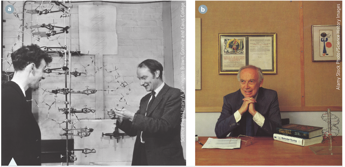
- James Watson and Francis Crick worked at the Cavendish laboratory, Cambridge University, under Lawrence Bragg's leadership
- Maurice Wilkins and Rosalind Franklin worked at King's College, London, under John Randall's leadership
American team:
- Linus Pauling led research in the United States
There was intense rivalry between these groups. Lawrence Bragg and Linus Pauling (who had won a Nobel Prize for his work on chemical bonds) had a longstanding competitive relationship.
In early 1953, Linus Pauling proposed that DNA was the heredity molecule with a triple helix structure. However, this model proved to be incorrect.
The breakthrough announcement
Watson and Crick used chemical evidence and X-ray crystallography data to construct their model of DNA's molecular structure. Their model revealed a stable molecule that could store vast amounts of information. They described DNA as a two-stranded molecule with paired bases twisted into a spiral ladder, which they called a 'double helix'.
A critical feature of their model was that it could explain how the molecule could copy itself (self-replicate).
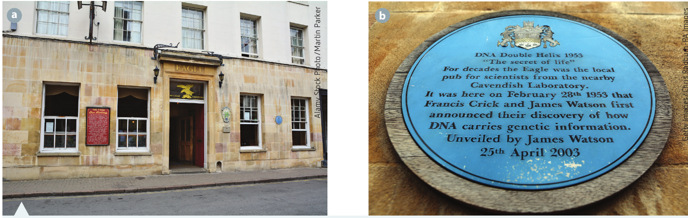
On 28 February 1953, Watson and Crick won the race and announced at a pub called The Eagle that they had discovered 'the secret of life'.
Collaborative scientific work
Watson and Crick worked collaboratively, evaluating the research and conclusions of other scientists. They asked questions of colleagues like Maurice Wilkins, who showed Watson one of Rosalind Franklin's X-ray crystallography photographs of DNA (unfortunately without first asking her permission).
They applied critical and creative thinking to analyse primary and secondary chemistry and crystallography data. By considering the quality of all available evidence and using careful reasoning, they built and rigorously tested their model. Their innovation, attention to scientific detail, and collaborative approach led to a valid conclusion that won them the Nobel Prize in 1962.
Recognition and tragedy: Sadly, both Oswald Avery and Rosalind Franklin died before they could be awarded Nobel Prizes for their contributions to this discovery. The Nobel Prize is not awarded posthumously, and Franklin's critical X-ray crystallography work was essential to Watson and Crick's breakthrough.
The Watson and Crick model of DNA structure
The double helix
Watson and Crick's model showed DNA as a large linear molecule arranged as a double helix or 'twisted ladder'. The key features of this structure are:
- Four nitrogenous bases held in pairs by hydrogen bonds
- The genetic information is stored in the sequence or order of these bases
- Two strands that run parallel to each other but in opposite directions (antiparallel)
Key structural features
The antiparallel arrangement:
Crick realised that each DNA strand, made of a sugar-phosphate backbone, runs antiparallel to the other. This means the twisting strands run parallel but in opposite directions. X-ray crystallography showed a set distance between the 'backbones' of the molecule.
Think of antiparallel strands like a two-lane road where traffic flows in opposite directions. The strands are side by side but oriented in opposite ways, with one running 5' to 3' and the other running 3' to 5'.
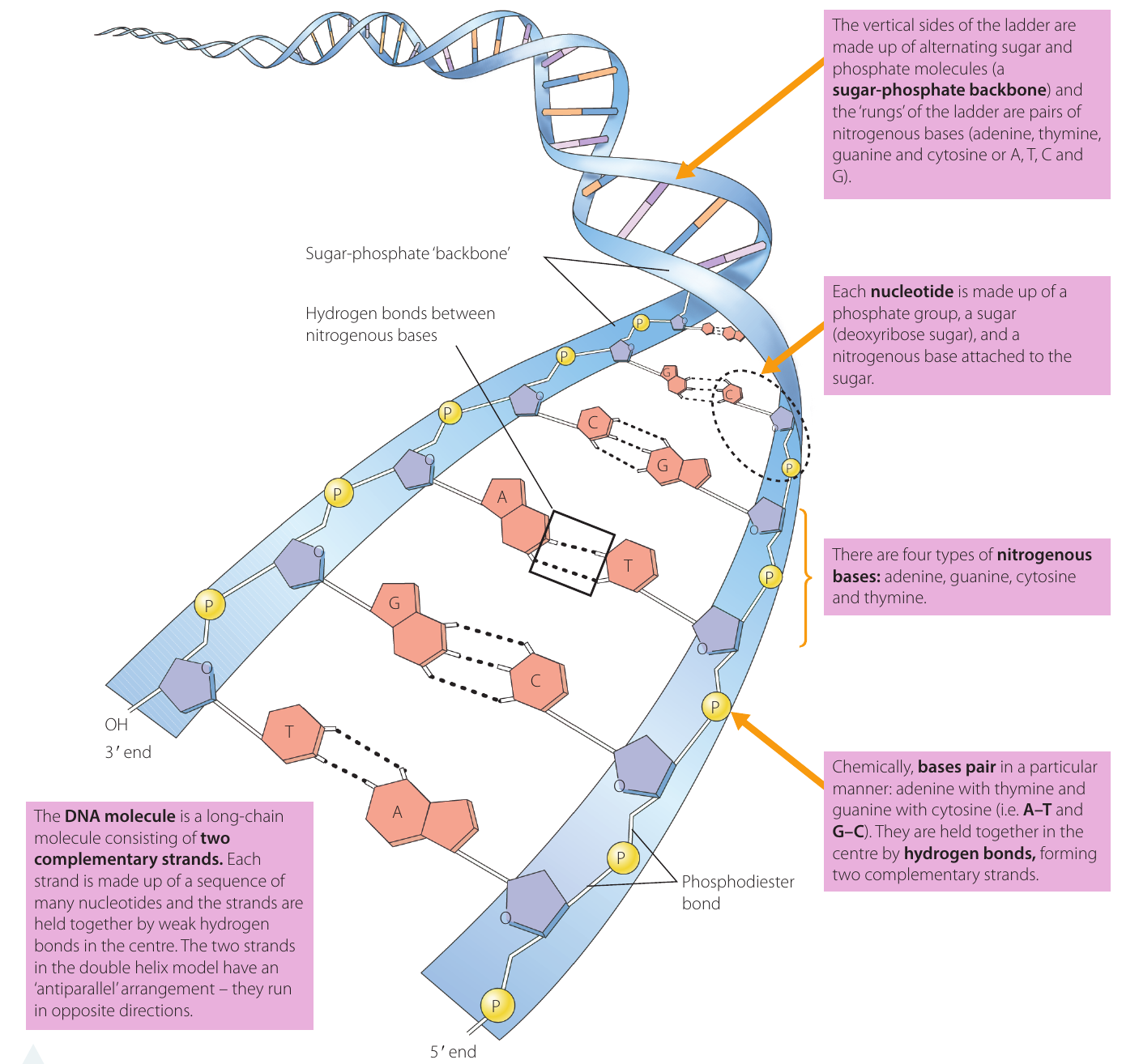
The ladder structure:
Think of DNA as a twisted ladder:
- The vertical sides are made of alternating sugar (deoxyribose) and phosphate molecules, forming a sugar-phosphate backbone
- The rungs are pairs of nitrogenous bases that line up between the backbones
- Each base has one end attached to the backbone and the other end hydrogen-bonded to another base
Nucleotide pairing and bonding
Complementary base pairing:
Watson and Crick discovered that to keep the DNA backbones at equal distances from each other, a double-ringed purine base must bond with a single-ringed pyrimidine base:
- Adenine (A) pairs with thymine (T) - forms two hydrogen bonds
- Guanine (G) pairs with cytosine (C) - forms three hydrogen bonds
This is called complementary base pairing.
Critical concept: The pairing is always A–T and G–C, never A–G or C–T. This specific pairing ensures the DNA strands remain at a constant distance from each other and is essential for accurate DNA replication.
Base classification:
- Purines (larger, double-ringed): adenine (A) and guanine (G)
- Pyrimidines (smaller, single-ringed): cytosine (C) and thymine (T)
Evidence for base pairing:
Watson and Crick found support for this pairing in Chargaff's rule. In 1951, Austrian biochemist Erwin Chargaff discovered that in DNA, the ratio of A
and C is always 1:1.Worked Example: Applying Chargaff's Rule
If a DNA sample contains 30% adenine (A), we can determine the percentages of all other bases:
Step 1: Since A pairs with T, the percentage of thymine must equal adenine
- T = 30%
Step 2: Calculate the remaining percentage for G and C
- Total for A + T = 30% + 30% = 60%
- Remaining = 100% - 60% = 40%
Step 3: Since G pairs with C in equal amounts, divide the remaining percentage equally
- G = 20%
- C = 20%
Answer: A = 30%, T = 30%, G = 20%, C = 20%
DNA serves as a template for replication
Watson and Crick's model explained how DNA meets all requirements of hereditary material:
-
Information storage: DNA can carry all the instructions for cell formation and function in coded form, using sequences of just four nitrogenous bases
-
Self-replication: The structure allows DNA to copy itself. Each strand serves as a template for enzymes to build a new complementary strand
-
Inheritance: DNA can be transferred from one generation to the next, packaged as chromosomes and carried by gametes (sex cells)
In their 1953 research paper, titled 'Molecular structure of nucleic acids', Watson and Crick wrote: "It has not escaped our notice that the specific pairing we have postulated immediately suggests a possible copying mechanism for the genetic material." This understated sentence hinted at one of the most important implications of their discovery.
Validation of the Watson and Crick model
Watson and Crick's findings were supported by multiple lines of evidence:
| Finding | Evidence-based reasoning |
|---|---|
| The whole DNA 'ladder' molecule spirals and is known as the 'double helix' | Evidence: X-ray crystallography suggested a helix measuring nm for every turn. This fitted the model where exactly 10 base pairs would measure nm in length and make up one twist of the helix |
| The four nitrogenous bases (A, G, C, T) always pair as A–T and C–G | Evidence: Chargaff's rule stated that DNA has a 1:1 ratio of pyrimidine and purine bases. Specifically, the amount of guanine equals cytosine and the amount of adenine equals thymine. Watson and Crick found that complementary base pairing (A–T and G–C) was essential to keep the DNA backbones equidistant |
| The two complementary strands are held together by hydrogen bonds between the bases | Reasoning: Chemically, when A bonds to T, a double hydrogen bond forms. When G bonds to C, a triple hydrogen bond forms |
| The strands of the backbone are identical and run in opposite directions (antiparallel) | Evidence: DNA crystal images generated by Rosalind Franklin looked the same when turned upside down or backwards. Reasoning: The backbones are made of sugar-phosphate molecules, based on the ratio of these components in chemical analyses |
| Each DNA strand serves as a template for producing a complementary strand, allowing self-replication | Reasoning: The two complementary strands could 'unzip' if the hydrogen bonds break between base pairs, allowing them to replicate |
Model building approach:
Watson and Crick built physical models to test how atoms fit together. They sometimes used cardboard cut-outs to represent the four nitrogenous bases and other DNA components, arranging them like a jigsaw puzzle on a desk. They initially made errors in the configuration of thymine and guanine rings. Based on a suggestion from American scientist Jerry Donahue, they adjusted the atomic arrangements and found the perfect fit: complementary base pairing with the correct ratios to reflect Chargaff's rule and hydrogen bonding between purines and pyrimidines.
Hands-on science: The use of physical models was crucial to Watson and Crick's success. By building 3D representations, they could test whether different atomic arrangements were chemically possible and consistent with the X-ray crystallography data.
The structure of nucleic acids
Components of a nucleotide
All nucleic acids are polymers (long chains) made up of simple repeating units called nucleotides. Each nucleotide consists of three parts:
- A phosphate group
- A sugar (deoxyribose in DNA, ribose in RNA)
- A nitrogenous base attached to the sugar
Nucleotides link together to form single chains (as in RNA) or double strands (as in DNA).
Remember the three components: Every nucleotide has phosphate + sugar + base. Think of it as the fundamental building block of all genetic material.
DNA strand length
DNA strands can be extremely long. For example, human chromosome 1 contains 249 million base pairs of DNA, representing approximately 8% of the total DNA in a human cell.
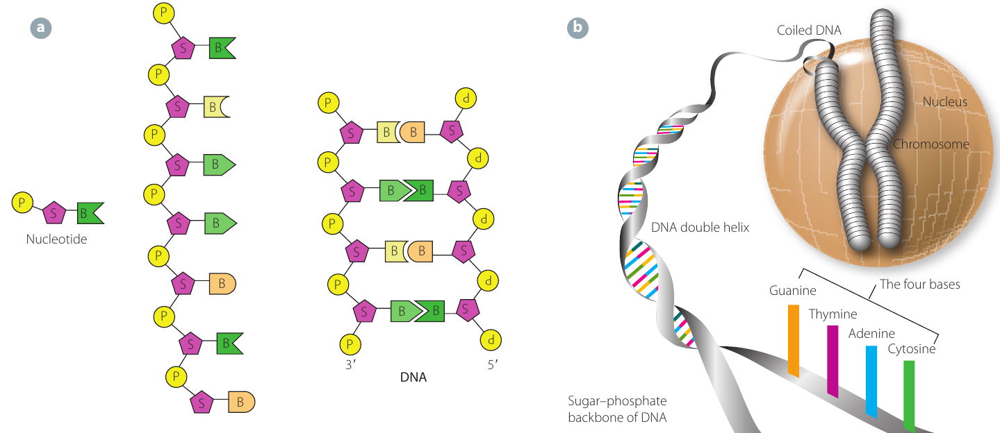
DNA packaging with histones
DNA is packaged efficiently in chromosomes with the help of proteins called histones. DNA coils around these proteins, similar to how cotton thread winds around a cotton reel. This packaging:
- Keeps long DNA threads compact
- Makes DNA easier to sort and separate during cell division
- Allows efficient transport of genetic material between generations
- May play a role in exposing sections of DNA so genes can be expressed
Without histone packaging, the DNA in a single human cell would stretch about 2 metres if laid end to end. Histones help compress this enormous length into a nucleus that's only about 6 micrometres in diameter.
The genetic code
The genetic code is created by the consecutive sequence of bases along the DNA strand. These base sequences differ in each gene, providing the unique 'genetic code' for cells to build specific proteins.
Comparing DNA and RNA
While DNA and RNA are both nucleic acids made of nucleotides, they have distinct roles and characteristics:
DNA (deoxyribonucleic acid):
- Stores genetic information that controls the cell and organism
- Main chemical component of chromatin in the nucleus
- Small amounts also found in mitochondria and chloroplasts
- Transmits inherited information during cell division and reproduction
- Contains the base thymine (T)
- Forms a double-stranded double helix
RNA (ribonucleic acid):
- Found in small amounts in the nucleus and larger amounts in the cytoplasm
- Usually associated with ribosomes
- Contains the base uracil (U) instead of thymine (T)
- Forms single strands
- Three main types with different functions:
- Messenger RNA (mRNA): carries information from DNA to the cytoplasm
- Ribosomal RNA (rRNA): associated with proteins in ribosomes
- Transfer RNA (tRNA): assists in translating the mRNA message into proteins
Key difference: The main structural difference is that DNA is double-stranded with thymine, while RNA is single-stranded with uracil. This reflects their different roles: DNA for stable long-term storage, RNA for temporary working copies.
Remember!
Key Points to Remember:
-
DNA has a double helix structure, like a twisted ladder, with two antiparallel strands running in opposite directions
-
The four nitrogenous bases are adenine (A), thymine (T), guanine (G), and cytosine (C), which always pair as A–T and G–C
-
Each nucleotide consists of three parts: a phosphate group, a deoxyribose sugar, and a nitrogenous base
-
The two DNA strands are held together by weak hydrogen bonds between complementary bases, allowing easy separation for replication
-
Watson and Crick's 1953 model was validated by multiple sources of evidence, including X-ray crystallography and Chargaff's base-pairing rules, earning them the Nobel Prize in 1962