Homeostasis (HSC SSCE Biology): Revision Notes
Mechanisms of Homeostasis
Introduction
Organisms use numerous mechanisms to maintain their internal environment within acceptable limits, which is essential for optimal metabolic efficiency and overall health. These mechanisms include various adaptations and internal coordination systems, particularly the nervous and endocrine systems. Both systems play crucial roles in homeostasis by coordinating and providing communication pathways for negative feedback systems that operate throughout the body.
Homeostasis is the process of maintaining a stable internal environment despite changes in external conditions. Without effective homeostatic mechanisms, organisms cannot survive or function optimally.
Internal coordination systems
The nervous and endocrine systems work together, either independently or collaboratively, to ensure homeostasis is maintained. Their primary role is to coordinate communication pathways that relay messages from receptors to control centres and then to effectors, initiating responses that counteract stimuli and keep internal conditions within tolerance limits.
Differences in message transmission
The two systems transmit messages in very different ways:
- Nervous system: Nerve impulses travel very rapidly along nerves to specific body locations, providing quick responses to stimuli
- Endocrine system: Chemical substances called hormones are transported through the bloodstream at a much slower rate than nerve impulses
The speed difference between these systems is crucial: the nervous system provides rapid, short-term responses (milliseconds to seconds), while the endocrine system produces slower but longer-lasting effects (minutes to hours or days). This allows the body to respond appropriately to both immediate threats and gradual changes.
Receptors
Receptors are responsible for detecting stimuli in the form of any changes from the set point that fall outside tolerance limits. They contain sensory cells and can take numerous forms depending on the stimuli that activate them.
Types of receptors
In more complex forms, receptors are concentrated in particular areas, forming sense organs such as the eye, ear, and tongue. In many animals, including humans, receptors in sense organs detect stimuli in the external environment.
Interoceptors are receptors located within the body that detect internal stimuli related to homeostasis.
Receptors may be named according to the type of energy or molecules they detect:
- Thermoreceptors detect changes in temperature. Thermoreceptors in the skin are nerve endings sensitive to heat or cold, sending information to the brain about external temperature. Internal thermoreceptors in the hypothalamus detect the temperature of blood flowing through the brain
- Chemoreceptors detect the concentration of certain chemicals inside the body. These receptors are located in certain blood vessels and detect pH levels as well as concentrations of chemicals such as carbon dioxide and oxygen
- Osmoreceptors detect changes in osmotic pressure and are located in the hypothalamus. Osmotic pressure in the blood is determined by the concentration of substances dissolved in blood plasma. Small changes in osmotic pressure cause the body to implement processes that regulate water levels, keeping them within tolerance limits
Each receptor type is specialized to detect specific stimuli. This specialization allows the body to monitor multiple parameters simultaneously and respond to each appropriately. The diversity of receptor types ensures comprehensive monitoring of both internal and external environments.
The nervous system
The nervous system provides neural pathways through which messages travel in the body. It also acts as a control centre to coordinate activities that maintain homeostasis.
Structure of the nervous system
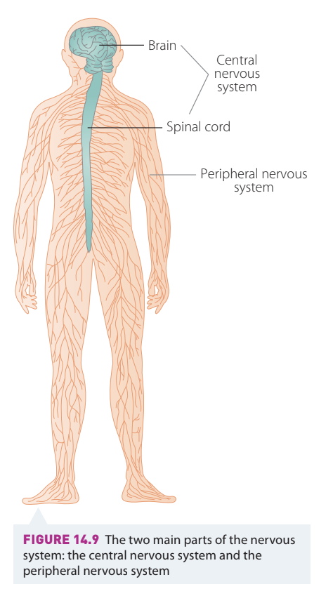
The nervous system has two main parts:
- Central nervous system (CNS): Composed of the brain and spinal cord
- Peripheral nervous system (PNS): Comprises all other nerves throughout the body that are not part of the CNS
The peripheral nerves carry information to and from the CNS. Information carried by nerves consists of messages transmitted in the form of electrochemical impulses.
Some actions involving the nervous system may occur voluntarily, but all those involved in homeostasis take place without conscious thought. They are involuntary, and many are innate, unconditioned reflexes in response to particular stimuli.
Neurons
The millions of units that make up the nervous system are called nerve cells or neurons. Although no two neurons are exactly alike in size, shape, and function, they all contain three common structures:
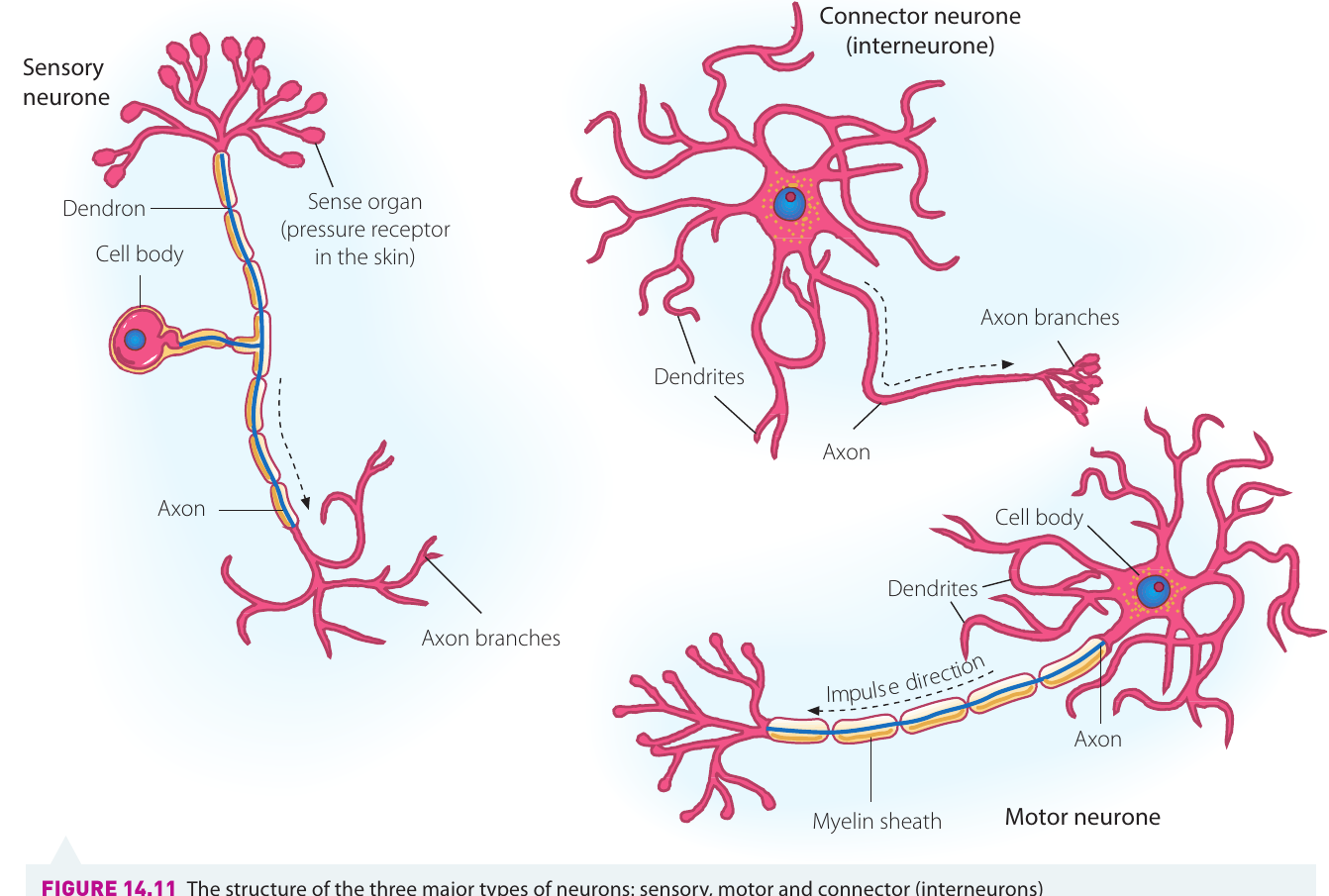
- A cell body that contains a nucleus and many organelles found in other cells. These form the grey matter of the CNS
- One or more fine branching extensions called dendrites, which are extensions of the cytoplasm of the cell body. Dendrites receive messages in the form of impulses from other axons and conduct these nerve impulses towards the cell body. In sensory neurons, the single elongated dendrite is called a dendron
- One single, very long extension of the cytoplasm of the cell body called an axon. Axons conduct messages away from the cell body and form the white matter of the CNS
Types of neurons
Neurons are classified according to their function and the direction in which nerve impulses are carried:
- Sensory neurons carry impulses from sensory cells in the peripheral nervous system to the CNS. They usually have the cell body at the side, one long dendron, and short axons
- Motor neurons transfer messages from the CNS to effectors such as muscles or glands. The dendrites are usually short and the axon quite long
- Interneurons (also known as association or connector neurons) are located within the CNS and are the link between sensory and motor neurons. They have short dendrites and either long or short axons
The three types of neurons form a complete pathway for homeostatic responses: sensory neurons detect changes, interneurons process the information in the CNS, and motor neurons deliver commands to effectors. This arrangement allows for coordinated, rapid responses to maintain homeostasis.
Nerve structure
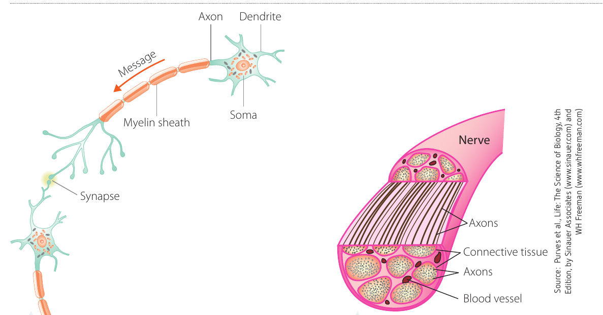
The fibres of neurons are gathered into bundles held together by a connective tissue sheath to form nerves. This provides a structured pathway for the transmission of electrochemical impulses along the axons of neurons.
When a nervous impulse is transferred from the axon of one neuron to the dendrites of an adjacent neuron, it must cross a small gap called a synapse, as adjacent neurons do not actually touch.
Transmission of nerve impulses
The messages transmitted by neurons as nerve impulses are in the form of electrochemical impulses. This is the quickest way to initiate responses by the body to stimuli in order to coordinate and maintain homeostasis.
The action potential
Electrochemical impulses involve a change in the electrical potential of the cell membrane of the axon. This temporary change is known as an action potential. It is brought about by a change in the concentration of electrically charged chemicals called ions on either side of the cell membrane of the axon. In other words, the change in the concentration of chemicals by movement across the cell membrane causes an electrical impulse.
Sodium ions (), potassium ions (), and chloride ions () are important for the transmission of electrochemical messages in the nervous system. These ions are on both sides of the cell membrane, but their concentrations differ. Also inside the cell are large negatively charged organic ions ().
A factor that influences the movement of these ions across the cell membrane is that the membrane is selectively permeable. It allows some substances to pass through easily, such as ions, but hinders the passage of and ions. The large ions are unable to move through the cell membrane.
Resting potential
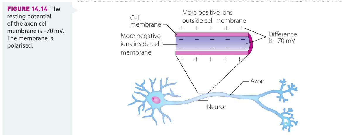
A neuron is said to be at rest if it is not transmitting any electrochemical messages. In this state, ions can only move through the cell membrane through special ion channels, which are closed when the neuron is at rest. There are a large number of ions outside the cell compared to the number of ions inside the cell. Also, there are many organic ions trapped inside the cell.
Because of this, there are more total negative charges on the inside of the cell than on the outside, and there is said to be a potential difference across the membrane. The value of this potential difference, called the resting membrane potential, is mV (millivolts). The inside of the membrane is said to be negative in relation to the outside of the membrane – the membrane is polarised.
Key values to remember:
- Resting membrane potential: mV
- Threshold value: mV
- The neuron must reach the threshold value for an action potential to occur
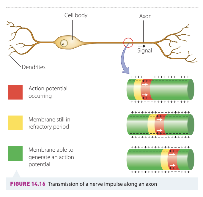
Action potential – depolarisation and repolarisation
When a stimulus is detected by a neuron, it causes sodium channels in the cell membrane to open. Because there are many more ions outside the neuron than inside, the ions move into the neuron and reduce the overall negative charge.
If the stimulus is strong enough to open enough Na channels to allow ions to move into the neuron and change the resting potential to its threshold value of mV, the movement of ions into the neuron will continue independently of the stimulus. This causes the inside of the membrane to become more positive in relation to the outside of the neuron, and the potential to move to mV and beyond. The membrane has been depolarised.
Just as quickly, the potassium channels open and ions move out of the neuron, causing the repolarisation of the membrane. The potassium channels stay open a little longer and the action potential goes past mV (hyperpolarisation) before returning to its original resting state. This rapid depolarisation and repolarisation is called the action potential.
Worked Example: The Action Potential Sequence
Step 1: Resting state - Membrane potential at mV (polarised)
Step 2: Stimulus applied - Sodium channels begin to open
Step 3: Threshold reached - At mV, action potential is triggered
Step 4: Depolarisation - ions rush in, potential reaches mV
Step 5: Repolarisation - ions move out, potential returns toward negative
Step 6: Hyperpolarisation - Potential briefly drops below mV
Step 7: Return to resting state - Membrane returns to mV
The all-or-none principle
The action potential is only activated if the stimulus is strong enough to cause the potential to reach a threshold value of mV. Once this value is reached, the action potential goes ahead automatically. If the threshold value is not reached, there is no action potential and therefore no nerve impulse is generated. Either the threshold potential is not reached or a full action potential is fired. This is the all-or-none principle.
The all-or-none principle means that action potentials do not vary in strength – they either happen completely or not at all. This ensures consistent signal transmission regardless of stimulus strength. Stronger stimuli result in more frequent action potentials, not stronger ones.
Each action potential causes another action potential in the neighbouring region of the neuron. This series of action potentials along the neuron is the nerve impulse.
Synapses
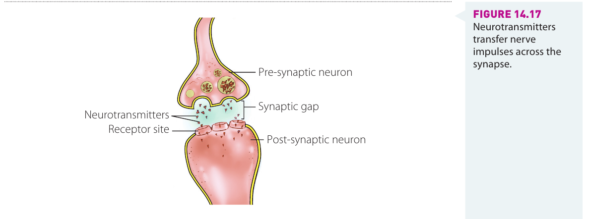
When a nerve impulse reaches the axon terminal, it must move across a small gap, the synapse, to the dendrites of the next neuron. The action potential triggers the release of chemicals called neurotransmitters from the synaptic vesicles. These chemicals move across the synapse to receptors in the dendrites of the adjacent neuron, initiating an action potential to continue the nerve impulse.
Synapses allow for one-way transmission of nerve impulses and provide points where signals can be modulated or blocked. This is crucial for complex neural processing and allows for the integration of multiple signals from different neurons.
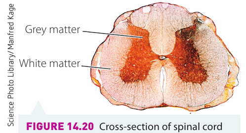
The central nervous system
The central nervous system (CNS) is made up of the brain and spinal cord, and is integral to the maintenance of homeostasis. Both the brain and spinal cord are made up of two types of nervous tissue: grey matter and white matter. Grey matter consists mainly of neuron cell bodies, while white matter is nerve fibres surrounded by myelin sheaths, which cause the white appearance. In the brain, grey matter tends to be on the outside, whereas in the spinal cord it is in the centre.
The brain
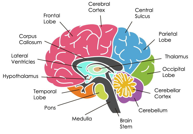
The brain is the main control centre of the body and is therefore a very complex organ. It consists of numerous parts that all work together to ensure the efficient functioning of the body. The brain largely controls the maintenance of homeostasis.
The major parts of the brain include:
- Cerebrum: The largest part of the brain
- Cerebellum: Located at the back of the brain
- Medulla oblongata: Connects the brain to the spinal cord
- Corpus callosum: Provides a pathway for messages between the two sides of the brain
- Hypothalamus: Small area located centrally, close to the pituitary gland
- Pituitary gland: Located just below the hypothalamus
The hypothalamus
The hypothalamus is a small area of the brain located centrally and close to the pituitary gland. It is the control centre for the regulation of many activities in the body that are required to maintain a stable internal environment (homeostasis). It achieves this by directing effectors to carry out a response either by sending messages via neural pathways or by chemical messages (hormones). Some of the conditions that need to be regulated are heart rate, body temperature, blood pressure, and the concentration of oxygen and carbon dioxide in the blood.
The hypothalamus is the most critical structure for homeostasis. It acts as the main link between the nervous system and the endocrine system, allowing for coordinated responses using both rapid neural signals and longer-lasting hormonal messages.
The hypothalamus is also the main link between the nervous system and the endocrine system. It is responsible for hormone secretions and directs the actions of the pituitary gland in coordinating other glands to secrete hormones in order to maintain homeostasis.
The spinal cord
The spinal cord extends from the medulla oblongata down through the vertebral column to the waist area. It contains the nerve fibres that provide the link for the pathway of nerve impulses between the peripheral nervous system and the brain.
The two main functions of the spinal cord are:
- To act as a conduction pathway for nerve impulses from the receptors around the body to the brain, and for nerve impulses from the brain to the effectors. This is essential for the efficient functioning of all areas of the body, including the maintenance of homeostasis
- To coordinate reflex actions, such as removing your hand quickly when you touch something hot, before you feel the pain
Reflex actions coordinated by the spinal cord occur without conscious thought and are faster than responses requiring brain processing. This provides rapid protection from harmful stimuli and demonstrates the efficiency of the nervous system's organization.
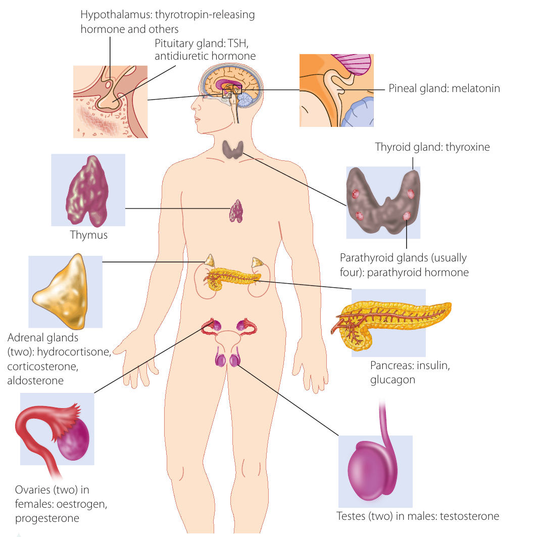
The endocrine system
The endocrine system, along with the nervous system, regulates the activity of the body. Its main components are hormones, which are chemical messenger molecules secreted by endocrine glands. The word 'endocrine' comes from endo meaning 'from within' and krine meaning 'to secrete'.
These hormones are transported by the bloodstream to cells possessing the receptors for the particular hormone. The hormones cause these cells to change their activity in a way that will maintain homeostasis within the body. Hormones achieve this by influencing the activity of particular enzymes or the concentration of these enzymes in the cells, known as target cells.
Glands can be stimulated to secrete hormones by:
- Messages from the nervous system
- Other hormones
- Receptors located in the particular gland
The pituitary gland
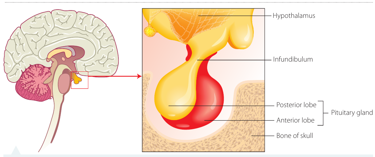
The pituitary gland, situated just below and working in close collaboration with the hypothalamus, is often referred to as the master gland. It releases hormones, often on direction from the hypothalamus, to regulate the activity of other glands.
There are two distinct regions of the pituitary gland: the front (anterior) and the back (posterior). Hormones released by the hypothalamus control the anterior area of the pituitary gland, while the posterior section is controlled by nerve impulses.
Anterior pituitary
One hormone secreted by the anterior section controls growth. Other hormones secreted act on other glands to control the activity of the thyroid, the adrenal gland, and the gonads (ovaries and testes).
Posterior pituitary
One of the hormones secreted by the posterior section of the pituitary gland is antidiuretic hormone (ADH), which helps to regulate the concentration of water in the body. If receptor cells in the hypothalamus detect that the levels of water in the body are too low, the hypothalamus stimulates the pituitary gland to release ADH. This acts to conserve water in the body by promoting its reabsorption by the kidney tubules. The opposite occurs when the level of water in the blood is too high – the hypothalamus detects this and directs the pituitary gland to reduce its production of ADH. This leads to less water being absorbed in the kidneys and the increased excretion of water.
Worked Example: ADH Regulation of Water Balance
Scenario: A person becomes dehydrated after exercising without drinking water.
Step 1: Water levels in blood decrease
Step 2: Osmoreceptors in hypothalamus detect increased blood concentration
Step 3: Hypothalamus sends nerve impulses to posterior pituitary
Step 4: Posterior pituitary releases more ADH into bloodstream
Step 5: ADH travels to kidneys
Step 6: Kidney tubules increase water reabsorption
Result: Less urine produced, water conserved, blood concentration returns to normal
Thyroid and parathyroid glands
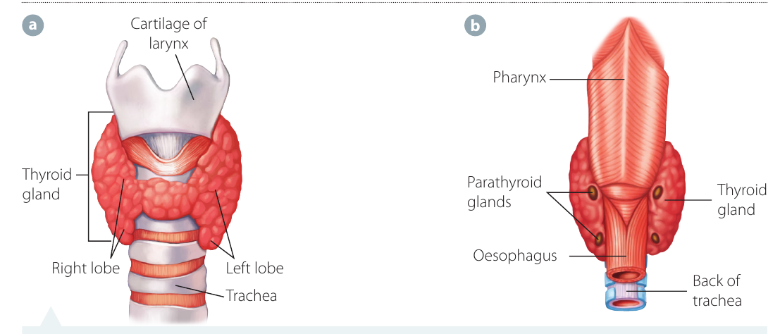
Thyroid gland
The thyroid gland consists of two lobes located on either side of the neck. The follicles in this gland predominantly produce an iodine-containing hormone called thyroxine. The amount of thyroxine produced is controlled by thyroid-stimulating hormone, which is released by the anterior pituitary gland.
The release of thyroxine is controlled by the hypothalamus when it releases a regulatory hormone into the pituitary gland. One of the reasons this may occur is that receptors in the hypothalamus detect a drop in body temperature to below the tolerance limit.
Thyroxine is the means by which the message is transferred to cells to increase their metabolic rate. In this process, energy is released for a number of purposes, one of which is to provide heat to maintain body temperature.
Parathyroid gland
The parathyroid gland is made up of a number of small glands embedded in the surface of the thyroid gland. Its function is to maintain the level of calcium in the blood. Calcium is required for the successful transfer of nerve impulses in the nervous system and allows muscles to contract properly.
The parathyroid gland monitors the blood flowing through it. When it detects that the calcium level is too low, it secretes parathyroid hormone (PTH). This hormone travels to the following effectors:
- Bones: Release calcium into the blood
- Small intestines: Absorb more calcium from digested food
- Kidneys: Reabsorb more calcium in the tubules
The parathyroid gland demonstrates a classic negative feedback mechanism. When calcium levels drop, PTH is released to raise them. Once calcium levels are restored to normal, PTH secretion decreases. This precise regulation is essential for proper nerve and muscle function.
The net result is to increase calcium levels, thus returning them to normal levels. If the calcium levels detected are too high, the parathyroid gland stops secreting PTH.
Adrenal glands
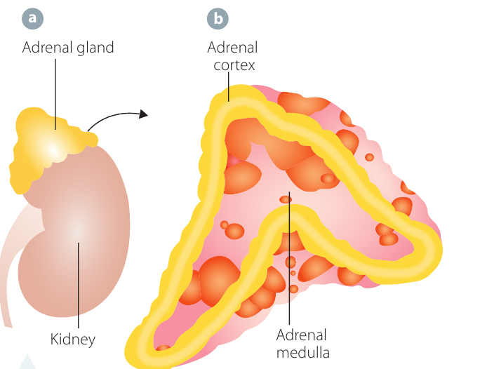
The adrenal glands are located on the top of each kidney and are made up of two distinct parts. The outer portion of the adrenal gland is known as the adrenal cortex and the inner region is the adrenal medulla. These regions have very different functions and secrete different hormones.
The hypothalamus regulates activities in both regions. Some of the hormones produced by the cortex are regulated by negative feedback involving the hypothalamus and hormones produced by the pituitary gland. The medulla is regulated by nerve impulses from the hypothalamus.
Adrenal cortex
When the hypothalamus is stimulated by receptors, it produces a hormone that in turn stimulates the pituitary gland to produce another hormone that stimulates the production of hydrocortisone (cortisol) and corticosterone by the cortex region of the adrenal glands.
- Cortisol regulates how the body manages stress and how it converts carbohydrates, fats, and proteins to energy. It also plays a role in regulating cardiovascular function and blood pressure
- Cortisol and corticosterone work together to regulate the immune response and suppress inflammatory reactions
The adrenal cortex hormones are essential for the body's response to long-term stress. Unlike the rapid "fight or flight" response from the adrenal medulla, cortisol provides sustained metabolic support during extended periods of stress or illness.
Another hormone secreted by the adrenal cortex is aldosterone, which increases the reabsorption of sodium ions and decreases the reabsorption of potassium ions in the kidney. If receptor cells in the kidneys detect low levels of sodium ions, they stimulate the adrenal cortex to produce more aldosterone, which causes greater reabsorption of sodium ions and decreased reabsorption of potassium ions. This results in sodium and potassium levels returning to values within their tolerance limits.
The levels of sodium ions in the blood are linked to the maintenance of blood volume and blood pressure. A low level of sodium ions in the blood will reduce blood volume and therefore blood pressure.
Pancreas
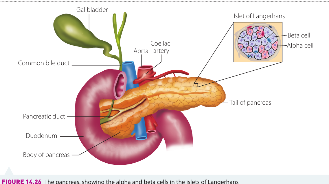
The endocrine portion of the pancreas consists of structures called pancreatic islets or the islets of Langerhans, where the hormones insulin and glucagon are produced. These islets contain two types of cells:
- Beta cells: Produce insulin
- Alpha cells: Produce glucagon
Insulin
Chemoreceptors in the beta cells detect high levels of glucose in the blood and stimulate the production of insulin. Insulin causes glucose to be removed from the blood in a number of ways:
- In the liver, glucose is converted into glycogen and fat
- Skeletal muscles convert glucose into glycogen
- Glucose is converted into fat in fat storage tissue
When the levels of glucose decrease, the production of insulin decreases.
Glucagon
Alpha cells in the islets of Langerhans produce glucagon in response to low levels of glucose in the blood. Glucagon causes the levels of glucose in the blood to increase by stimulating the production of glucose through:
- The breakdown of glycogen in the liver
- The breakdown of fat in the liver and the fat storage tissues
When the level of glucose increases back to normal, the production of glucagon is reduced.
Blood Glucose Regulation: A Perfect Balance
Insulin and glucagon work as antagonistic hormones – they have opposite effects. This allows for precise regulation of blood glucose levels:
- When glucose is high → Insulin released → Glucose stored
- When glucose is low → Glucagon released → Glucose released from storage
This dual control system ensures blood glucose remains within narrow tolerance limits, which is essential for brain function and overall metabolism.
Worked Example: Blood Glucose Regulation After a Meal
Scenario: A person eats a carbohydrate-rich meal.
Step 1: Blood glucose levels rise above normal (set point)
Step 2: Chemoreceptors in beta cells of pancreas detect high glucose
Step 3: Beta cells secrete insulin into bloodstream
Step 4: Insulin travels to target cells (liver, muscles, fat tissue)
Step 5: Target cells respond:
- Liver converts glucose → glycogen
- Muscles convert glucose → glycogen
- Fat tissue converts glucose → fat
Step 6: Blood glucose levels decrease
Step 7: As glucose returns to normal, insulin secretion decreases
Result: Blood glucose returns to set point, homeostasis maintained
Key Points to Remember:
Nervous System:
- The nervous and endocrine systems work together to maintain homeostasis by coordinating communication pathways between receptors, control centres, and effectors
- Receptors (thermoreceptors, chemoreceptors, and osmoreceptors) detect changes from the set point that fall outside tolerance limits
- The nervous system transmits messages rapidly via electrochemical impulses, while the endocrine system uses chemical messengers (hormones) transported more slowly through the bloodstream
- Neurons consist of a cell body, dendrites, and an axon. The three types are sensory neurons, motor neurons, and interneurons
- Action potentials involve depolarisation and repolarisation of the neuron membrane. The threshold value is mV, and the resting potential is mV. The all-or-none principle states that an action potential either occurs fully or not at all
- Neurotransmitters bridge the synapse between neurons, allowing nerve impulses to continue from one neuron to the next
Central Nervous System:
- The hypothalamus is the main control centre for homeostasis and provides the crucial link between the nervous and endocrine systems
- The spinal cord acts as a conduction pathway and coordinates reflex actions
Endocrine System:
- Major endocrine glands include the pituitary (master gland), thyroid, parathyroid, adrenal glands, and pancreas
- Each gland secretes specific hormones that regulate various aspects of homeostasis such as metabolism, calcium levels, stress response, and blood glucose levels
- Hormones work through negative feedback mechanisms to maintain conditions within tolerance limits
- Insulin and glucagon work as antagonistic hormones to precisely regulate blood glucose levels