Causes and Effects of Non-Infectious Diseases (HSC SSCE Biology): Revision Notes
Causes and Effects of Non-Infectious Diseases
Introduction to non-infectious diseases
A disease is any condition that disrupts or has the potential to disrupt the normal functioning of the body. Non-infectious diseases (also called non-communicable diseases) are conditions that cannot be passed from one person to another. Unlike infectious diseases that are caused by pathogens such as bacteria or viruses, non-infectious diseases have different origins.
The main causes of non-infectious diseases include:
- Genetic mutations inherited from parents
- Poor nutrition and dietary deficiencies
- Lack of physical activity
- Exposure to harmful substances in the environment
- Malfunctions in the immune system
Many non-infectious diseases develop when the body's homeostatic mechanisms fail to maintain balance. The endocrine system, which produces hormones, plays a critical role in regulating the internal environment. Hormones act as chemical messengers that control vital functions including growth, metabolism, reproduction and tissue repair. When the endocrine system malfunctions, homeostatic disruption can lead to disease.
Homeostasis refers to the body's ability to maintain a stable internal environment despite changes in external conditions. When homeostatic mechanisms fail, the delicate balance required for normal body function is disrupted, potentially leading to disease.
Genetic diseases
Genetic diseases result from mutations that alter normal gene expression and protein production. When a person inherits a faulty gene, their cells may not produce the correct proteins needed for normal body function. The severity of genetic diseases varies greatly depending on the specific mutation involved.
Phenylketonuria (PKU)
Phenylketonuria is a genetic condition caused by a mutation in the gene that codes for the enzyme phenylalanine hydroxylase. This enzyme normally breaks down the amino acid phenylalanine into another amino acid called tyrosine.
Inheritance pattern: PKU is an autosomal recessive trait, meaning a person must inherit two copies of the mutated gene (one from each parent) to develop the condition. In NSW, approximately 1 in 10,000 babies are born with PKU each year.
How PKU affects the body:
- The body cannot convert phenylalanine to tyrosine
- Phenylalanine accumulates to dangerous levels in the blood
- High levels can cause mental retardation
- Affected individuals often have reduced skin and hair pigmentation
- Stunted head growth may occur
- Seizures can develop
During pregnancy, a mother's enzymes help remove excess phenylalanine from an affected fetus. After birth, however, breastfed babies with PKU will accumulate phenylalanine from digesting milk proteins.
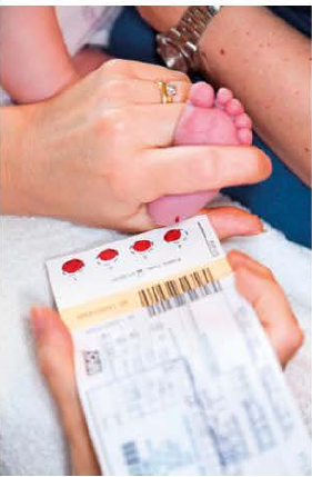
Diagnosis and management: PKU can be detected using a simple Guthrie blood test performed within the first 24 hours after birth. If diagnosed with PKU, patients must follow a strict low-phenylalanine diet throughout their lives. This means:
- Consuming low-protein foods such as fruits and vegetables
- Avoiding high-protein foods including dairy products, fish and red meat
- Avoiding foods containing the artificial sweetener aspartame (found in chewing gum and many soft drinks)
Albinism
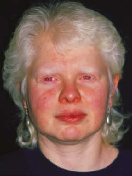
Albinism is characterised by the complete absence of melanin pigment from the skin, hair and eyes. People with albinism have pale skin, white hair and pale-coloured eyes. Their eyes may appear red because light reflects off blood vessels at the back of the eye, which become visible through the pale iris.
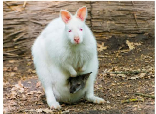
Albinism occurs in many vertebrate species, including birds, reptiles and other mammals.
Genetic cause: In humans, mutations in the TYRP1 gene cause oculocutaneous albinism type 3. This gene codes for the enzyme tyrosinase, which is essential for melanin production. Without functional tyrosinase, the body cannot produce melanin pigment.
Effects and management:
- Babies with albinism have normal mental development
- High sensitivity to ultraviolet (UV) light due to lack of skin pigment
- Require strict sun protection including sunscreens and minimal UV exposure
- Nystagmus (rapid involuntary eye movements)
- Reduced visual acuity (sharpness of vision)
- Photophobia (discomfort in bright lights)
- Prescription glasses can help manage visual problems
Cystic fibrosis
Cystic fibrosis is a genetic disorder that causes thick, sticky mucus to accumulate in the lungs, leading to breathing difficulties and frequent infections. Although respiratory problems are most common, other organs can also be affected.
Prevalence: Cystic fibrosis is one of the most common genetic disorders in people of European descent. In Australia, 1 in 2,500 babies is born with the condition each year.
Inheritance: Cystic fibrosis is an autosomal recessive condition, meaning both males and females are equally affected and they must inherit two copies of the faulty gene (one from each parent). In Australia, 1 in 25 people carry one copy of the cystic fibrosis gene without showing symptoms. If two carriers have a child together, there is a 25% chance their child will have cystic fibrosis.
The CFTR gene: The gene responsible for cystic fibrosis is called the CFTR (cystic fibrosis transmembrane conductance regulator) gene, located on chromosome 7. The normal gene produces the CFTR protein, which forms a channel in cell membranes. This channel is crucial for secreting sweat, mucus and digestive fluids. When mutations disrupt CFTR protein production, sodium ions move into cells, drying out the mucus layer. The mucus becomes thick and sticky, preventing cilia from clearing debris from airways, leading to infections. Over 1,500 different mutations in the CFTR gene have been identified, causing varying degrees of disease severity.
Symptoms of cystic fibrosis: Most babies with cystic fibrosis are diagnosed within the first two months through a routine heel prick blood test. Symptoms vary between individuals and may include:
- Persistent coughing
- Shortness of breath
- Frequent respiratory infections
- Either excessive or no appetite
- Poor weight gain and small stature
- Late onset of puberty
- Reflux and constipation
- Tiring easily
- Sinusitis
Because the CFTR gene affects sweat secretion, people with cystic fibrosis have high salt levels in their sweat and frequently produce sticky mucus in their lungs, blocking airways and increasing infection risk. The pancreas often doesn't work efficiently, and some individuals develop liver problems.

Management of cystic fibrosis: In the past, babies born with cystic fibrosis rarely survived beyond childhood. Today, with improved understanding, treatments and medications, most babies born with cystic fibrosis in Australia can live well into adulthood.
Management strategies include:
- Daily physiotherapy to clear thick mucus from airways
- Inhaled medications delivered via nebuliser
- Intermittent positive pressure breathing (IPPB) machines that deliver controlled gas pressure to help ventilate and expand the lungs
In severe cases, lung transplants may be performed. Transplanted lungs contain normal CFTR genes, greatly enhancing quality of life and life expectancy. However, transplants don't cure non-respiratory symptoms such as digestive difficulties and male sterility. Recipients must take immunosuppressant drugs lifelong to prevent tissue rejection.
Gene therapy research: Gene therapy may offer a future treatment for lung disease caused by cystic fibrosis. Clinical trials began in 1993 to test delivery methods and dosages. Researchers used viral vectors to deliver normal CFTR genes into patients' airways. Recent research has successfully used pigs with cystic fibrosis to identify effective virus-based vectors for restoring functional CFTR protein.
Genetic markers
Each human genome is unique, with small differences arising from mutations and genetic recombination during reproduction. While mutations create healthy genetic variation, some disrupt normal functioning and cause disease.
Genetic markers are DNA sequences that can identify if someone carries alleles associated with genetic disease and their likelihood of developing symptoms. The most abundant genetic markers are single nucleotide polymorphisms (SNPs). Humans have approximately 10 million SNPs throughout their genome.
By comparing genetic marker variations in large population samples, scientists can identify patterns linking markers to diseases and other physiological differences.
Genetic markers serve several purposes:
- Tracing inheritance patterns of genetic diseases
- Identifying specific disease-causing genes
- Determining modes of inheritance
- Helping develop appropriate treatments
Diseases caused by environmental exposure
Some non-infectious diseases result from mutations or reactions caused by environmental factors rather than inheritance. Exposure to radiation or certain chemicals can damage DNA. Agents that damage DNA are called mutagens. If damage occurs to proto-oncogenes or tumour-suppressor genes, cell division control can be disrupted.
Other environmentally-caused diseases result from immune system overreactions to environmental antigens, such as those in grass pollen, poison ivy and certain medications. This is known as a hypersensitivity reaction.
Hypersensitivity reactions
The immune system normally eliminates invading pathogens. However, when it malfunctions, it can react to normally harmless antigens, such as those on pollen grains. This causes a hypersensitivity reaction. While often tolerated, some reactions can be severe or even fatal.
Hypersensitivity reactions have both genetic and environmental components, as many triggering antigens are found in the environment.
These reactions occur when the immune system responds to harmless antigens, causing cell damage and disease. They are classified into four types based on the antigens and immune mechanisms involved:
- Type I (immediate) hypersensitivity
- Type II (cytotoxic) hypersensitivity
- Type III (immune complex) hypersensitivity
- Type IV (delayed-type) hypersensitivity
Type I hypersensitivity is commonly called allergy. Autoimmune diseases can cause types II, III and IV hypersensitivity reactions, though these aren't the only causes.
Type I hypersensitivity—allergic reactions
Immediate hypersensitivity reactions (allergic reactions) result from rapid, vigorous immune system overreactions to normally harmless antigens. These antigens are called allergens. Common allergenic substances include pollen, animal fur, house dust, latex, and foods such as peanuts, lobster and monosodium glutamate (MSG).
Reactions range from mild to life-threatening. Severe, potentially fatal allergic reactions are called anaphylaxis.
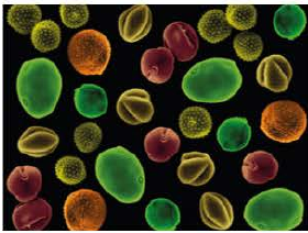
Hay fever (allergic rhinitis) is triggered by pollen particles carrying allergenic antigens. Grass and tree pollens are the most common causes in Australia and New Zealand. Pollen sensitivity follows seasonal patterns, with pollen most abundant during spring and early summer.
The mechanism of allergic reactions: Allergic reactions are mediated by immunoglobulin E (IgE) antibodies produced by plasma cells. When IgE contacts mast cells (common in epithelial and mucosal tissues), the IgE tail binds to cell surface receptors. Upon subsequent allergen exposure, the allergen binds to adjacent IgE molecules, bridging them together. This triggers cellular signals causing mast cells to release histamine and other inflammatory mediators.
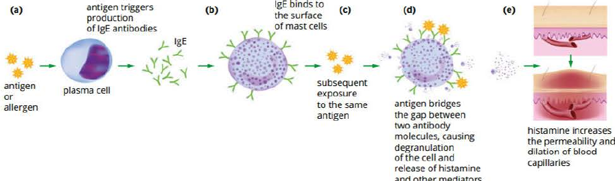
How histamine affects the body: Histamine is involved in immune responses, working to eliminate foreign particles. When an allergen is detected, histamine is released from mast cells and basophils (white blood cells) and binds to histamine receptors on cell surfaces. This causes:
- Blood vessel dilation and decreased blood pressure
- Increased blood vessel permeability, allowing white blood cells to access the antigen site
- Fluid leaking from capillaries into tissues, causing runny nose and watery eyes
- Smooth muscle contraction in airway linings, making breathing difficult
- Sensory stimulation triggering sneezing to expel foreign antigens

Treatment for allergic reactions:
Medications: Antihistamines block histamine effects by binding to the same receptors as histamine, preventing histamine from binding. Other medications (like cortisone) suppress the immune system more broadly.
For severe anaphylaxis, immediate intramuscular adrenaline (epinephrine) injection is needed. Adrenaline auto-injectors (commonly known as EpiPens) counter histamine actions by:
- Constricting blood vessels (decreasing swelling and increasing blood pressure)
- Relaxing airway muscles (opening airways)
- Increasing heart rate and blood flow to the heart (preventing cardiovascular collapse)
Allergen immunotherapy: Desensitisation treats hypersensitive reactions by injecting extremely small allergen amounts multiple times in increasing doses over months. This causes specific immunoglobulin G (IgG) antibodies to form against the allergen. If IgG antibodies react with the allergen before it binds to IgE antibodies, the allergic response is prevented. The individual gradually becomes less sensitive to the allergen.
Type II hypersensitivity (cytotoxic)
Type II cytotoxic hypersensitivity reactions involve immunoglobulin M (IgM) and IgG antibodies directed against cell surface or extracellular matrix antigens. These reactions can take hours to develop, unlike type I reactions which occur within minutes.
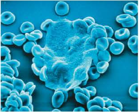
An example is antibody-mediated destruction of red blood cells (haemolytic anaemia) occurring when a mother produces antibodies against rhesus antigen on fetal red blood cells. Type II reactions can also result from certain medications. For example, penicillin binds to red blood cells, and if anti-penicillin antibodies are present, they bind to the drug and trigger destruction of the red blood cells.
Type III hypersensitivity (immune complex)
Type III immune complex hypersensitivity reactions also involve IgM and IgG, but antibodies target soluble antigens rather than cell surface antigens. When antibodies bind to soluble antigens, they form antigen-antibody immune complexes that deposit in tissues, causing inflammation and damage. These reactions can take hours or days to develop.
An example is serum sickness, an immune response to foreign antigens in medications or antiserum (such as snake venom antiserum). Penicillin is the most common cause of serum sickness.
Type IV hypersensitivity (delayed-type)
Unlike types I, II and III which are antibody-mediated, type IV hypersensitivity is mediated by helper T lymphocytes. These activate macrophages and eosinophils to produce inflammatory responses, and cytotoxic T lymphocytes, which directly attack and kill cells. Delayed-type reactions take days to develop.
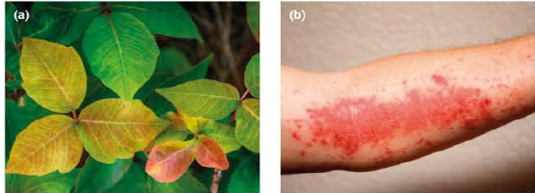
An example is the rash caused by contact with poison ivy. The plant's sap contains a lipid-soluble compound that crosses cell membranes. Inside cells, it causes new peptides to be produced and delivered to the cell surface, where cytotoxic T lymphocytes recognise them. The T-cell mediated immune response causes a blistering, itchy rash and, in severe cases, anaphylaxis.
Thunderstorm asthma
Australia has the highest asthma incidence globally, affecting 9% of adults and 12% of children. Climate significantly affects asthma attack risk. Thunderstorms can trigger attacks under specific conditions.
Thunderstorm asthma occurs when high pollen levels (usually during late spring and early summer) combine with certain thunderstorms. Grass pollen grains are swept into clouds as storms form. When pollen absorbs moisture, it bursts open, releasing large amounts of much smaller allergen particles. These particles can enter airways and reach the lungs. In susceptible people, this irritates the lungs, causing swelling, airway narrowing and mucus secretion, making breathing difficult and potentially causing asthma attacks.
On 21 November 2016, a thunderstorm swept across Melbourne, causing a thunderstorm asthma event that hospitalised 8,500 people and caused nine deaths. During such storms, people should avoid pollen exposure by staying indoors.
Autoimmune diseases
Many diseases result from a combination of genetic and environmental factors. For autoimmune diseases, causes are largely unknown, though research suggests both genetic and environmental factors play roles in development and severity. Over 80 autoimmune diseases are currently known, including Crohn's disease, systemic lupus erythematosus, rheumatoid arthritis and type 1 diabetes.
All autoimmune diseases involve the body triggering an immune response against its own cells, leading to tissue and organ inflammation and damage. Symptoms vary extremely between individuals, making diagnosis difficult. Researchers are working to understand causes, risk factors and the rise of autoimmune diseases in industrialised countries.
Autoimmunity
When functioning properly, the immune system targets foreign cells (non-self antigens), not the body's own cells (self-antigens or autoantigens). This is called self-tolerance. Normally, T and B lymphocytes reactive against the body's own cells are destroyed.
Autoimmune diseases result from self-tolerance failure, leading to adaptive immune responses directed against specific self-antigens. An antibody acting against a self-antigen is called an autoantibody.
In autoimmune diseases, cytotoxic T lymphocytes directly attack tissues and B lymphocytes act indirectly by secreting antibodies. Mast cells (white blood cells playing key immune roles) can be activated to release histamines, causing inflammation around affected tissues.
Autoimmune diseases can be:
- Organ-specific: localised to particular body parts (e.g., multiple sclerosis affects only the brain and spinal cord)
- Generalised: occurring widely throughout the body (e.g., systemic lupus erythematosus affects various organs and tissues including joints, skin, kidneys and brain)
Autoimmune haemolytic anaemia
This is an autoimmune disease causing type II hypersensitivity reactions because it involves autoantibodies directed against self-antigens on red blood cell surfaces.
Rheumatoid arthritis
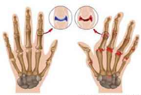
Rheumatoid arthritis is an autoimmune disease causing type III hypersensitivity reactions. It involves deposition of antigen-antibody immune complexes in tissue, resulting in inflammation and damage. Rheumatoid arthritis mainly affects joints, commonly in knees and hands. It's also thought to cause type IV hypersensitivity reactions, with T lymphocytes attacking unidentified antigens in joints.
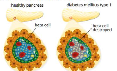
Type 1 diabetes
Type 1 diabetes is an autoimmune disease where T lymphocytes attack and destroy beta cells in the pancreas. Beta cells produce insulin, which regulates blood glucose levels. People with type 1 diabetes must inject insulin to maintain glucose balance.
There are two diabetes types:
- Type 1: genetic, possibly triggered by some viruses
- Type 2: often late-onset, related to lifestyle
Type 1 diabetes is common in the developing world. People with type 1 diabetes must maintain blood glucose values between 4.0 and 6.0 mmol/L for normal bodily functioning.
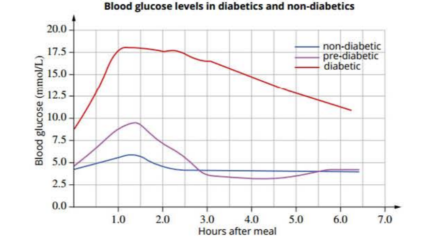
Blood glucose regulation: When body sensors detect blood glucose levels outside normal limits, the body releases hormones insulin and glucagon:
- When blood glucose is too high, the body secretes insulin, which stimulates glucose uptake into muscle and liver cells and converts it into stored glycogen
- When blood glucose is too low, the body produces glucagon, which stimulates glycogen breakdown into glucose in the liver, releasing glucose into the blood
Cause of type 1 diabetes: Scientists are unsure what causes beta cell destruction in the islets of Langerhans. Evidence suggests links between Coxsackie A and B4 viruses (common in children) and autoimmune disease onset. Other childhood viruses including enterovirus, mumps, polio and rubella have also been suggested as triggers.
Without functioning beta cells, the body cannot secrete insulin to convert glucose to glycogen in the liver and stimulate glucose uptake into muscle and fat. This causes dangerously high blood glucose levels.
Symptoms of type 1 diabetes: Insulin deficiency results in hyperglycaemia (high blood glucose) and accelerates fat breakdown for energy. Symptoms include:
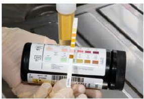
- Glucose in urine
- Increased urine production
- Excessive thirst and hunger
- Ketoacidosis
- Weight loss and fatigue
- Blurred vision
- Irritability and muscle cramps
- Skin infections
- Delayed wound healing
- Tingling or numbness in feet
Longer-term consequences include kidney and eye disease. These symptoms occur because elevated blood glucose exceeds the kidneys' filtration capacity (normally kidneys prevent glucose from entering urine). Glucose escaping into nephron tubules draws in more water by osmosis, increasing urine volume. Frequent urination leaves the body dehydrated and thirsty. Glucose in urine is a simple diabetes test.
Dehydration can blur vision as the lens loses moisture and blood vessels are damaged, potentially causing blindness if untreated. Raised glucose levels cause chemical reactions with molecules on neuron surfaces and cells lining small blood vessels. Nerve damage can lead to limb sensation loss, while capillary damage contributes to kidney malfunction and diabetic retinopathy (eye disease).
Management of type 1 diabetes: Type 1 diabetes can be managed throughout life, allowing patients to take health responsibility and live fully. Besides diet management, people with type 1 diabetes must receive artificial insulin.
Artificial insulin: This is usually administered by injection. Patients monitor blood glucose by pricking a finger and testing a small blood drop with a glucose meter or chemical strip.
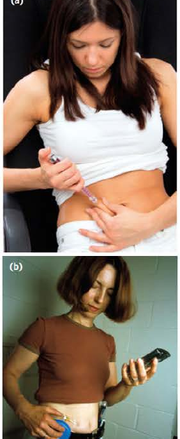
Alternatively, an electrode placed under the skin connects to a continuous glucose monitoring device, warning when glucose levels reach high or low levels. The monitor can couple to an electronic pump delivering insulin when blood glucose reaches predetermined levels. An improved 'artificial pancreas' system uses monitoring and feedback to deliver insulin as the body requires, mimicking natural pancreas function.
Transplants: Pancreas transplants from deceased donors are usually given to patients with serious diabetes complications. Human pancreas cells can also be transplanted into a patient's liver, where they produce insulin (pancreatic islet transplantation). Though still experimental, this may become widely available soon. Recipients must take immunosuppressant drugs lifelong to prevent organ rejection. These drugs can cause side effects including high blood pressure, fatigue and increased infection risk.
Gene therapy: Gene therapy, inserting the insulin-coding gene into patient cells, is a potential future treatment. US trials have succeeded in diabetic rats, targeting the liver for its regenerating ability. A major benefit is that patients wouldn't require immunosuppressant drugs.
Multiple sclerosis
Neurons relay signals along their axons. The central nervous system contains several cell types besides neurons. Oligodendrocytes produce myelin, composed mostly of lipids and some protein. Myelin forms an insulating sheath around axons. Though some peripheral nervous system nerves also have myelin sheaths, multiple sclerosis (MS) generally only affects central nervous system myelin sheaths.
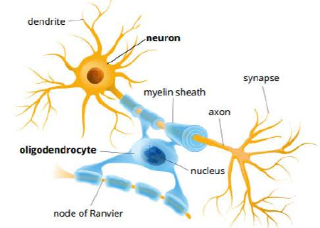
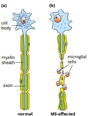
In MS, nerves lose myelin sheaths, impairing signal conduction along nerves and eventually damaging them.
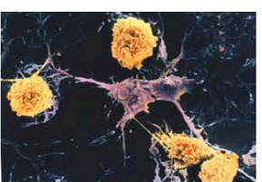
Both helper and cytotoxic T lymphocytes are involved in MS, and plasma cells produce antibodies targeting myelin sheath proteins and lipids. Mitochondria in oligodendrocytes are damaged, releasing signalling molecules inducing apoptosis (programmed cell death) in these cells. Macrophages called microglia, specific to the central nervous system, are also involved in oligodendrocyte destruction.
MS causes are not fully understood, but it resembles type IV hypersensitivity because it's mediated by T lymphocytes and involves macrophage activation, resulting in inflammation.
Oligodendrocyte damage and death lead to myelin sheath destruction (demyelination). Once myelin sheaths are damaged, nerve axons themselves are also damaged.
Symptoms vary between individuals and may include:
- Visual, motor and sensory problems such as double vision, tiredness, numbness, muscle weakness, heat sensitivity, and balance and coordination difficulties
- Mental problems including memory lapses, mood swings, depression and epilepsy
There is no MS cure. However, certain medications can manage symptoms and delay disease progression. Standard treatments include medications reducing inflammation (steroids) and suppressing immune response (immunosuppressants).
Nutritional diseases
Many nutrients are essential for human health and survival. The body cannot synthesise these molecules and must obtain them from a balanced diet. Essential nutrients are grouped into:
- Minerals (such as calcium, magnesium)
- Vitamins (such as vitamins A, C, D and K)
- Amino acids (such as tryptophan and histidine)
- Fatty acids (such as omega-3 fatty acids)
Not all diets contain required nutrients for health. Many nutritional diseases result from deficiency, imbalance or over-consumption of certain nutrients, leading to different malnutrition types. For example, insufficient protein causes kwashiorkor, a disease causing fluid retention, anorexia, ulcerating skin and enlarged liver. Lack of other nutrients causes diseases such as scurvy, rickets and coronary heart disease.
Scurvy
Citrus fruits such as oranges and grapefruits are good vitamin C sources. Vitamin C deficiency can cause scurvy, leading to exhaustion, anaemia and swelling.

Scurvy was common among sailors in past centuries due to lack of fresh fruits and vegetables in diets. Many sailors on long voyages died from scurvy. In countries where sufficient fruits and vegetables are unavailable, scurvy outbreaks still occur. Scurvy was common during the Irish potato famine in 1845 and in Afghanistan in 2002 following war and drought. However, modern scurvy cases are rare, especially where vitamin C-enriched breads and cereals are available.
Vitamin C (L-ascorbic acid) cannot be synthesised by humans and some other primates. It's required to produce collagen, found in skin, connective tissues, blood vessels and tendons. Animals with GLO gene mutations (coding for an enzyme needed in L-ascorbic acid synthesis) cannot synthesise their own vitamin C. The recommended daily intake (RDA) of vitamin C for humans is approximately 50 mg per day.
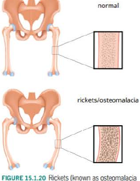
Rickets
Rickets is a disease associated with vitamin D deficiency in children. In adults, this condition is called osteomalacia and is milder. Vitamin D is necessary to absorb calcium in the digestive system. Because calcium is required for healthy bones, vitamin D deficiency leads to bone softening. Children with rickets appear to have bowed legs.
Vitamin D is essential for:
- Maintaining calcium balance by promoting calcium absorption in intestines
- Maintaining calcium and phosphate levels for bone formation
A diet high in vitamin D foods (including fish, avocados, egg yolks and dairy products such as cheese, milk and yogurt) helps prevent low vitamin D levels. Exposing human skin to low UV light levels triggers vitamin D production in skin. If skin is exposed to open air and light briefly each day, enough vitamin D is supplied. For this reason, vitamin D doesn't fit the vitamin definition as it's one of few vitamins the body can make. The World Health Organization recommends 5 to 15 minutes of sun exposure of hands, face and arms two to three times weekly in summer. Closer to the equator where UV light levels are greater, even shorter exposure periods are recommended.
In winter and at very northerly and southerly latitudes, UV light availability is limited and there's insufficient sunlight to trigger vitamin D production in skin. Because vitamin D is fat-soluble, most people can store some in the liver for use until UV light is available again. In countries like Norway and Patagonia where light is limited for extended periods, supplements are introduced into common foods like flour, cereals and milk, or residents take vitamin D supplements.
Extended vitamin D deficiency poses health issues. Research shows vitamin D is important for brain function and insufficient levels may contribute to mental illnesses such as depression. In breastfeeding mothers with low vitamin D intake, infant bone mineralisation can be affected.
Hypertension and coronary heart disease
Coronary heart disease involves plaque build-up inside coronary arteries of the heart. These arteries supply oxygen-containing blood to heart muscle itself.
With age, arteries lose collagen and elastin filaments and gradually become less elastic and harden, called arteriosclerosis. This stresses the heart because it must pump harder to push blood through inflexible arteries, causing high blood pressure (hypertension).
Over time and with a fatty diet, fatty substances, cholesterol and calcium can build up inside these hardened arteries, causing narrowing. This specific arteriosclerosis type is called atherosclerosis.
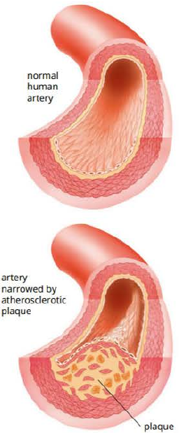
Both arteriosclerosis and atherosclerosis can affect arteries and arterioles throughout the body, restricting blood flow to tissues and organs. If atherosclerosis has developed, plaque can break away or blood clots can form around plaque. Both situations can cause stroke or heart attack.
Atherosclerosis can affect coronary blood vessels supplying blood to heart muscle. Plaque build-up restricts nutrient and oxygen supply to heart tissue. If coronary vessels become too narrow or completely blocked, a heart attack can result, possibly leading to heart tissue death.
Everyone will eventually develop some arteriosclerosis, but what causes more rapid development in some individuals and progression to life-threatening atherosclerosis is not fully understood. What is known is that high blood pressure, along with high cholesterol and triglyceride levels in blood, increase atherosclerosis risk. Smoking, poor diet, lack of exercise and obesity are risk factors. Some medications effectively lower cholesterol levels in the body.
Liver disease
People who drink alcohol excessively are prone to severe and often fatal liver disease. Medical evidence indicates that adding vitamins to alcoholic drinks, while good for nutrition, won't prevent chronic liver damage.
Alcohol is toxic. Special enzymes needed to break it down are found in the liver. Because biochemical pathways in heavy drinkers' liver cells are involved with removing alcohol, cells cannot carry out normal cellular respiration levels. Substances that should have been broken down for energy are converted to fats instead, and these fats accumulate in the liver.
Initially the situation is reversible, but then fat-filled cells start dying, causing alcoholic hepatitis. This is followed by cirrhosis (scar tissue formation in the liver). Finally, death may occur when the liver cannot carry out normal functions.
Cancer
Cancers are diseases commonly involving unregulated and abnormal cell growth and division. Cancer can be caused by genetic mutations increasing cell division rate and/or suppressing apoptosis (programmed cell death). Either case can lead to tumour growth.
Cancer can be caused by mutagens called carcinogens (cancer-causing agents). There are three carcinogen types: chemical, physical and biological. Fortunately, such mutations cannot pass from one generation to the next unless the mutation arises in parents' gametes (eggs and sperm).
Key Points to Remember:
-
Non-infectious diseases cannot be transmitted between individuals. Unlike infectious diseases caused by pathogens, they result from genetic factors, environmental exposure, nutritional deficiencies, or immune system malfunctions.
-
Genetic diseases result from inherited mutations that disrupt normal protein production. Examples include phenylketonuria (PKU), albinism and cystic fibrosis. Many can be managed through diet, medication or gene therapy approaches.
-
Hypersensitivity reactions occur when the immune system overreacts to normally harmless substances. Type I reactions (allergies) involve IgE antibodies and histamine release. Type IV reactions are mediated by T lymphocytes and cause delayed responses like contact dermatitis.
-
Autoimmune diseases develop when the body's immune system attacks its own cells. Examples include type 1 diabetes (attacking pancreatic beta cells), multiple sclerosis (attacking myelin sheaths) and rheumatoid arthritis (attacking joints). These conditions require ongoing management with immunosuppressant medications.
-
Nutritional diseases result from dietary deficiencies or imbalances. Vitamin C deficiency causes scurvy, vitamin D deficiency causes rickets, and poor diet combined with lifestyle factors contributes to coronary heart disease. Many nutritional diseases can be prevented through balanced diets and appropriate supplementation.