The Second Line of Defence (VCE SSCE Biology): Revision Notes
The Second Line of Defence
What is the second line of defence?
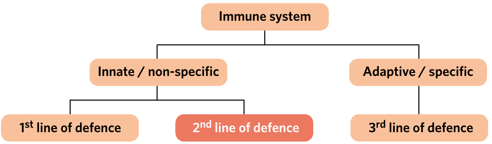
When pathogens manage to slip past or breach the first line of defence, your body has a backup plan ready to respond. The second line of defence is a component of the innate immune system characterised by the non-specific and immediate response to injury and pathogens by a variety of cells and molecules.
This defence system works quickly and doesn't need prior exposure to a pathogen to function. It responds the same way to all threats, which is why we call it "non-specific". The second line of defence consists of two main types of components: cellular components (various types of white blood cells) and non-cellular components (proteins and other molecules).
Key Features of the Second Line of Defence:
- Responds immediately without prior exposure to pathogens
- Non-specific - treats all threats the same way
- Consists of both cellular (white blood cells) and non-cellular (proteins and molecules) components
- Part of the innate immune system
Cellular components of the innate immune response
The cellular components of the second line of defence are all types of leukocytes, which is another name for white blood cells. Leukocytes are a group of blood cells responsible for protecting the body against pathogens and foreign material.
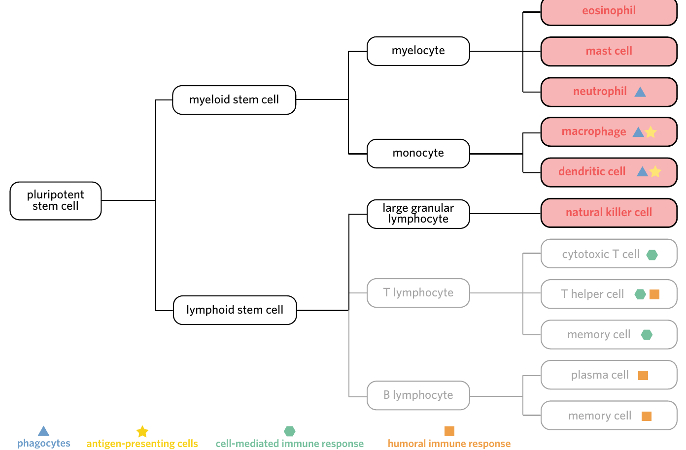
Phagocytes
Phagocytes are a group of leukocytes responsible for the endocytosis and destruction of pathogens, foreign material, and cell debris. These cells work by engulfing foreign material through a process called phagocytosis, which you may remember from previous studies on endocytosis.
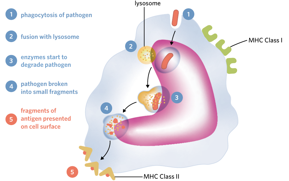
Once a phagocyte engulfs a pathogen or piece of debris, it traps it inside a vesicle. This vesicle then fuses with lysosomes (special compartments containing antimicrobial enzymes called lysozymes) which destroy the engulfed material. The three main types of phagocytes you need to know are neutrophils, macrophages, and dendritic cells.
How Phagocytosis Works:
Think of phagocytes as the "PAC-men" of your immune system! They:
- Engulf pathogens or debris, trapping them in a vesicle
- Fuse the vesicle with lysosomes containing antimicrobial enzymes
- Destroy the engulfed material using these powerful enzymes
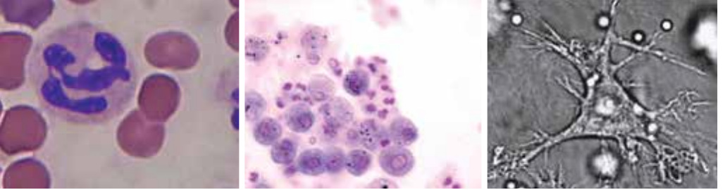
Neutrophils
Neutrophils are the most common type of leukocyte in the body. They engage in phagocytosis of pathogens and foreign material, as well as the release of cytokines. Neutrophils are typically the first immune cells to arrive at a site of infection or injury, and they work rapidly to destroy pathogens.
Macrophages
Macrophages are a type of leukocyte found throughout the body that engages in phagocytosis and antigen presentation. These cells are larger than neutrophils and can consume more material. Macrophages also play an important role in tissue repair and wound healing.
Dendritic cells
Dendritic cells are a type of leukocyte that engages in phagocytosis and antigen presentation. They have long, branching projections that help them sample their environment for potential threats.
Antigen-presenting cells
Both macrophages and dendritic cells belong to a special subgroup called antigen-presenting cells (also known as professional antigen-presenting cells). These cells are a subgroup of phagocytes that display antigens from consumed pathogens on their surface and interact with the adaptive immune system.
Critical Concept: MHC Class II Markers
You learned previously that all nucleated cells express MHC Class I markers on their surface. However, only specific cells express MHC Class II markers. Antigen-presenting cells are these special immune cells that express both MHC Class I and MHC Class II markers.
After consuming a pathogen through phagocytosis, antigen-presenting cells break down the pathogen into fragments. They then display these fragments (antigens) on their surface using MHC Class II markers. This process is crucial for activating the adaptive immune system, which you'll learn about in the next lesson.
Cytokines and immune communication
To communicate within the immune system, phagocytes release important signalling molecules called cytokines. Cytokines are signalling molecules released by cells (typically in the immune system) which aid in communication between immune cells and help protect against pathogens.
Think of cytokines as chemical messengers that help coordinate the immune response. They can guide other immune cells to the site of infection or injury, activate various defensive mechanisms, and help ensure all components of the immune system work together effectively.
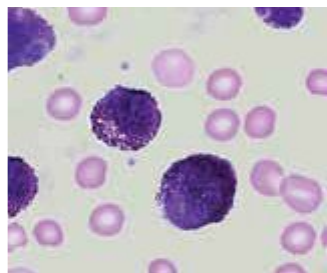
Natural killer (NK) cells
Natural killer (NK) cells are a type of leukocyte responsible for the recognition and destruction of damaged and/or infected host cells. These large granulated cells are particularly important for targeting abnormal cells (like cancer cells) and virally infected cells.
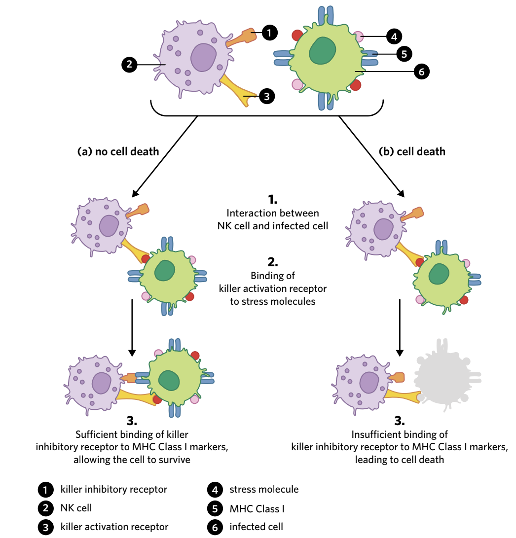
NK cells work using two types of receptors:
- Killer inhibitory receptor: This receptor examines the surface of cells looking for MHC Class I markers. When it finds sufficient MHC Class I markers, it sends an "inhibitory" signal that prevents the NK cell from killing the target cell.
- Killer activation receptor: This receptor binds to certain stress molecules that appear on cells undergoing cellular stress, such as infected or cancerous cells. When activated, it sends a signal to kill the target cell.
The "Missing Self" Response
The clever part of how NK cells work is the balance between these two signals. Under normal circumstances, healthy cells display MHC Class I markers, which keeps the killer inhibitory receptor satisfied and prevents cell death. However, many disease processes can alter MHC Class I expression:
- Viral infections can destroy or suppress the production of MHC Class I markers
- Cancer cells may have altered gene expression affecting MHC Class I production
When MHC Class I markers are absent or insufficient, the killer inhibitory receptor cannot override the killer activation signal. This leads to the destruction of the infected or abnormal cell - a process sometimes called the "missing self" response.
Think of it like checking for ID: no ID (MHC Class I) means the cell gets destroyed!
Mast cells
Mast cells are a type of leukocyte responsible for releasing histamine during allergic and inflammatory responses. These cells reside in connective tissues throughout your body, essentially acting as sentries watching for signs of trouble.
When mast cells detect injury to surrounding cells or are stimulated by antigens or allergens, they become activated and undergo degranulation - the release of granule contents from a cell. The main substance released during degranulation is histamine, a molecule that plays a key role in inflammation.
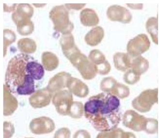
Eosinophils
Eosinophils are large granular leukocytes responsible for the release of toxic chemical mediators. These cells contain various toxic enzymes including DNases (which break down DNA), RNases (which break down RNA), and proteases (which break down proteins).
Eosinophils typically target pathogens that are too large to be phagocytosed, such as parasitic worms. When they encounter such pathogens, they degranulate on contact, releasing their toxic chemical mediators to destroy the invader.
Non-cellular components of the innate immune response
In addition to the various types of leukocytes, the second line of defence includes important non-cellular components. These molecular and systemic responses work alongside cellular components to provide comprehensive protection.
Interferons
Interferons are cytokines released by virally infected cells that increase the viral resistance of neighbouring uninfected cells. When a cell becomes infected with a virus, it releases interferons as a warning signal to nearby cells.
These interferons interact with receptors on neighbouring cells, triggering a series of changes that make them less susceptible to viral infection. This creates a protective barrier around the infected area, helping to prevent the virus from spreading between cells.
Complement proteins
Within your blood, there are numerous different complement proteins - a number of different types of proteins found in the blood that opsonise, cause lysis, and attract phagocytes to invading pathogens. Together, these proteins form the complement system.
When certain pathogens are detected, these complement proteins begin reacting with each other in a series of reactions called the complement cascade - a complex sequence of events which occurs after the activation of complement proteins.
The Complement Cascade: Three Major Outcomes
The complement cascade produces three major outcomes that work together to destroy pathogens. Remember them as "O-C-L" (or think of it as "Oh, Cell Lysis!"):
- Opsonisation - tagging pathogens for destruction
- Chemotaxis - attracting immune cells to the site
- Lysis - directly destroying pathogens
Opsonisation
Opsonisation is the mechanism by which complement proteins attach to the surface of pathogens, making them easier to phagocytose. The complement proteins essentially "tag" the pathogen, making it more recognisable to phagocytes as something that needs to be destroyed.
Think of opsonisation like putting a bright "eat me" sticker on a pathogen that makes it much easier for phagocytes to identify and consume it.
Chemotaxis
Chemotaxis is the attraction of phagocytes towards a pathogen. Complement proteins gather near a pathogen and act like a chemical beacon, attracting phagocytes to the site. This increases the likelihood that the pathogen will be quickly found and destroyed.
Lysis
Lysis is the disintegration or rupturing of a cell. Complement proteins can join together on the surface of pathogens to form a structure called a membrane attack complex (MAC) - a pore formed by complement proteins in the cell membranes of a pathogen, disrupting the membrane and leading to the pathogen's destruction.
The "MAC Attack"
The MAC creates holes in the pathogen's membrane, causing an influx of fluid into the pathogen. This causes the pathogen to swell and eventually burst, destroying it completely. Think of it as the complement system launching a "MAC attack" on invading pathogens!
Fever
A fever is a temporary increase in body temperature that occurs as an innate response to potential infection. During a fever, complex responses raise the body's set temperature point, and the body then initiates countermeasures to reach this new temperature, such as shivering and heat-conserving behaviours.
Fevers help fight infection in several ways:
- Many pathogens cannot survive at elevated temperatures, as the heat can denature their proteins and enzymes, causing loss of function
- Fever activates certain proteins in the body that strengthen immune defences
- The increased temperature can enhance the activity of some immune cells
However, it's important to note that prolonged fevers can be detrimental because they place additional stress on our own cells, which are no longer functioning at their optimal temperature.
Steps in the inflammatory response
The inflammatory response is a series of biochemical events that occur in the body as a result of infection and/or trauma, characterised by swelling, redness, pain, and heat in the affected tissue.
The inflammatory response serves several important purposes:
- Eliminates the effects of an injury
- Defends against potential pathogens
- Clears out damaged or destroyed cells
- Initiates tissue repair
This is a complex, non-specific process that always occurs in the same way regardless of the pathogen present or the type of injury. Let's examine the inflammatory response through the example of getting a splinter.
Process of inflammation
The inflammatory response can be divided into three main stages: initiation, vasodilation, and migration.
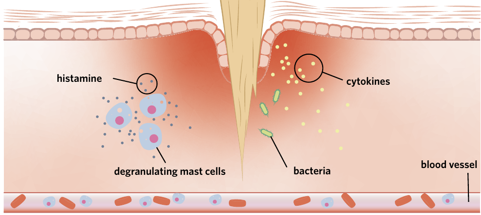
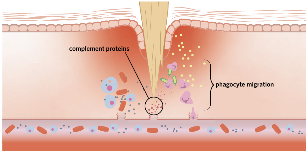
Worked Example: The Splinter Scenario
Let's follow what happens when you get a splinter in your finger while handling wood:
Step 1: Tissue Damage (Initiation) The splinter pierces your skin, damaging cells and introducing bacteria and other pathogens into your body. This tissue damage triggers the inflammatory response.
Step 2: Chemical Signalling In response to this injury, several things happen simultaneously:
- Macrophages already present in the tissue become activated
- Damaged cells release chemical signals
- Both macrophages and damaged cells release cytokines
- Mast cells detect the injury and degranulate, releasing histamine
Step 3: Blood Vessel Changes (Vasodilation) The histamine travels to nearby blood vessels, causing them to widen and become more permeable.
Step 4: Immune Cell Arrival (Migration) Phagocytes and complement proteins leave the bloodstream and enter the site of injury, where they destroy pathogens and clear debris.
Initiation
Imagine you get a splinter in your finger while handling wood. The splinter pierces your skin, damaging cells and introducing bacteria and other pathogens into your body. This tissue damage triggers the inflammatory response.
In response to this injury, several things happen simultaneously:
- Macrophages already present in the tissue become activated
- Damaged cells release chemical signals
- Both macrophages and damaged cells release cytokines
- Mast cells detect the injury and degranulate, releasing histamine
Vasodilation
Vasodilation is the widening of blood vessels. The histamine released from mast cells travels to nearby blood vessels and binds to specific receptors on the vessel walls. This binding causes two important changes:
- The blood vessels widen (dilate), increasing blood flow to the injury site
- Gaps form in the vessel wall, increasing its permeability to immune cells
Why Your Injury Looks Red and Feels Hot
These changes are responsible for several of the characteristic signs of inflammation:
- Redness: caused by increased blood flow to the area
- Heat: the additional warm blood makes the area feel hot
- Swelling: increased blood flow and leakage of fluid into tissues causes swelling

Migration
The widening and increased leakiness of blood vessels allows various components of the innate immune system to leave the bloodstream and enter the site of injury. This migration includes:
Phagocytes: Macrophages and neutrophils are guided by the cytokines released earlier. They follow this chemical trail to the injury site, where they phagocytose pathogens and digest them using antimicrobial enzymes such as lysozymes.
Complement proteins: These proteins are attracted to pathogens at the site of injury. They opsonise pathogens (making them easier to recognise), attract more phagocytes through chemotaxis, and can directly destroy pathogens through formation of membrane attack complexes.
The fourth cardinal sign of inflammation - pain - results from several factors including pressure from swelling, chemical mediators stimulating pain receptors, and tissue damage itself.
What is Pus?
An interesting side note: the pus that forms in an infected area is actually caused by this increase in blood flow and immune cell activity. Pus contains a large amount of dead immune cells (particularly neutrophils) and destroyed pathogens.
The inflammatory response continues until the site has been cleared of pathogens and debris, and the damaged tissue has been repaired. Eventually, the area will return to normal.
Summary of the inflammatory response
The inflammatory response can be summarised as a cascade of events:
- Tissue damage and infection occur
- Cytokines are released and mast cells degranulate, releasing histamine
- This leads to both increased vessel permeability and vasodilation
- Increased vessel permeability allows movement of phagocytes and complement proteins to the site
- Vasodilation causes increased blood flow
- These changes produce the cardinal signs: redness, heat, swelling, and pain
- The combined action of immune components leads to tissue repair and infection clearance
The Cardinal Signs of Inflammation
Remember "RED HOT" for the four cardinal signs:
- Redness - from increased blood flow
- hEat - from additional warm blood in the area
- Damaged/swelling - from fluid leakage into tissues
- hurT/pain - from pressure, chemical mediators, and tissue damage
All four signs result from the increased blood flow and immune activity at the site of injury or infection.
Summary of second line of defence components
Here is a comprehensive summary of all the cellular and non-cellular components of the second line of defence:
Cellular components
| Component | Role |
|---|---|
| Neutrophil | Phagocytosis of pathogens |
| Macrophage | Phagocytosis of pathogens and antigen presentation within the adaptive immune system |
| Dendritic cell | Phagocytosis of pathogens and antigen presentation within the adaptive immune system |
| Natural killer cell | Destroys infected or abnormal cells with insufficient MHC Class I markers |
| Mast cell | Causes inflammation through the release of histamine |
| Eosinophil | Releases toxic chemical mediators to destroy invading pathogens |
Non-cellular components
| Component | Role |
|---|---|
| Interferons | Released by virally-infected cells and causes changes to neighbouring cells that make them less susceptible to infection |
| Complement proteins | React with each other and aid in the destruction of pathogens via opsonisation, attraction of phagocytes to pathogens, and the formation of membrane attack complexes (MAC) |
| Fever | An abnormally high body temperature used by the body to kill pathogens |
This table provides a quick reference for all the key components of the second line of defence. Make sure you understand not just what each component is, but how they work together to provide comprehensive protection against pathogens.
Remember!
Key Points to Remember:
-
The second line of defence provides rapid, non-specific protection when pathogens breach the first line of defence through various cellular and non-cellular components.
-
Phagocytes (neutrophils, macrophages, and dendritic cells) engulf and destroy pathogens, while macrophages and dendritic cells also present antigens to activate the adaptive immune system.
-
The complement cascade produces three outcomes: opsonisation (tagging pathogens), chemotaxis (attracting phagocytes), and lysis (directly destroying pathogens through membrane attack complexes).
-
The inflammatory response occurs in three stages: initiation (cytokine release and mast cell degranulation), vasodilation (blood vessel widening and increased permeability), and migration (immune cells and proteins moving to the injury site).
-
The cardinal signs of inflammation - redness, heat, swelling, and pain - all result from the increased blood flow and immune activity at the site of injury or infection.