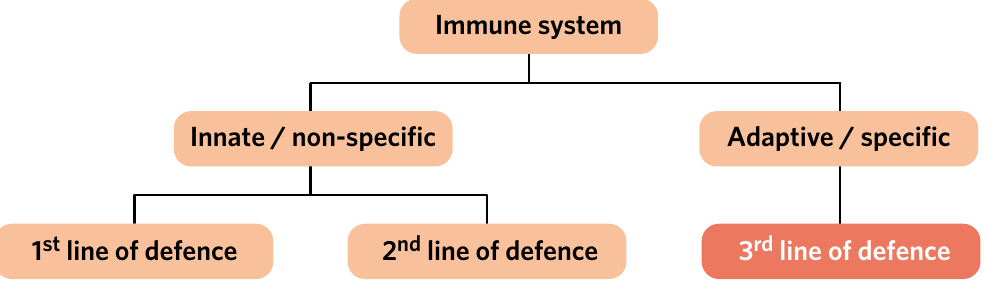
The Third Line of Defence (VCE SSCE Biology): Revision Notes
The Third Line of Defence
Introduction to adaptive immunity
The third line of defence, also called the adaptive immune system or specific immune response, is a sophisticated defence mechanism that activates when the innate immune system cannot control a pathogen. This system has two unique features that distinguish it from the second line of defence:
- Specificity – the adaptive immune system responds to each distinct pathogen in a unique and tailored manner
- Immunological memory – the adaptive immune system produces cells that enable the body to respond quickly and effectively to future re-infections by previously encountered pathogens
The adaptive immune system is like a specialized defense force that learns from each encounter with a pathogen. Unlike the innate immune system, which provides the same general response to all threats, the adaptive system creates a customized response for each specific pathogen it encounters.
The adaptive immune system consists of two main components:
- Humoral immunity (also called B cell immunity) – targets extracellular pathogens using antibodies
- Cell-mediated immunity (also called T cell immunity) – destroys infected or abnormal cells
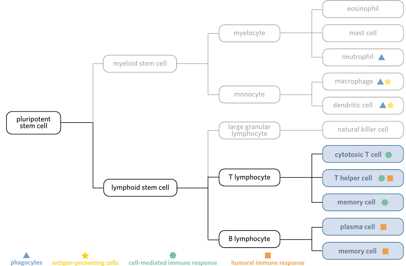
Cells of the adaptive immune system
The adaptive immune system uses specialised white blood cells called lymphocytes. These cells originate from pluripotent stem cells and differentiate into two main types:
B lymphocytes are white blood cells that play a central role in humoral immunity. They differentiate into:
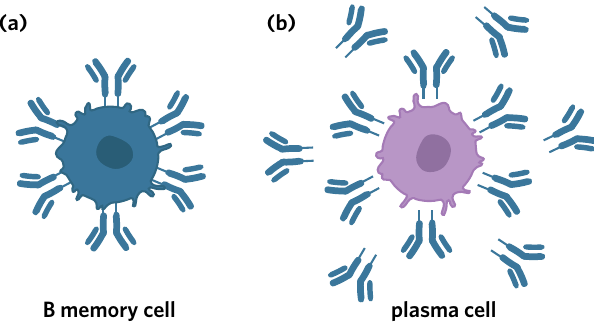
- Plasma cells (which secrete antibodies)
- B memory cells (which provide long-lasting immunity)
T lymphocytes are white blood cells that play important roles in both humoral and cell-mediated immunity. They differentiate into:
- T helper cells (which coordinate immune responses)
- Cytotoxic T cells (which destroy infected cells)
- T memory cells (which provide long-lasting immunity)
Think of lymphocytes as specialized soldiers in your immune army. B lymphocytes are like artillery units that fire antibodies at distant targets, while T lymphocytes are like special forces that either coordinate the battle (T helper cells) or engage in close combat to eliminate infected cells (cytotoxic T cells).
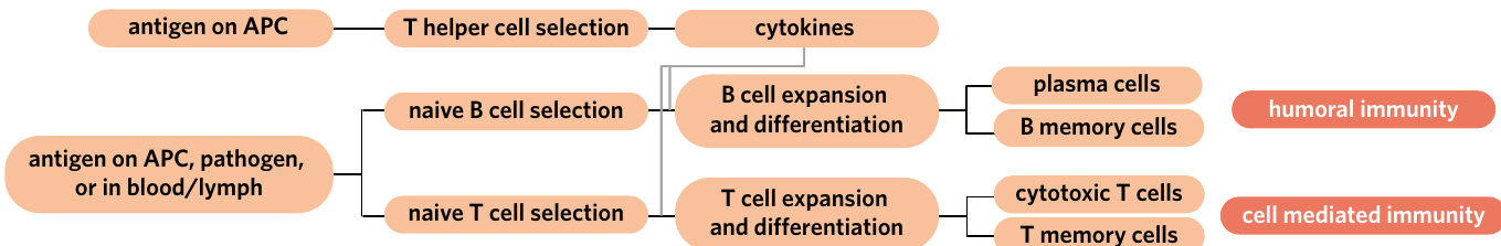
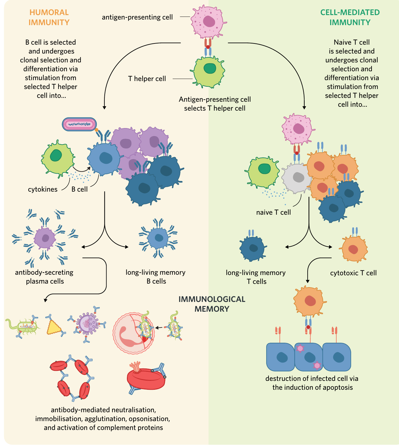
Initiation of the third line of defence
Antigen presentation
The adaptive immune response begins with a process called antigen presentation. This involves antigen-presenting cells (APCs) displaying foreign antigens to T helper cells.
Here's how the process works:
- Antigen-presenting cells (such as macrophages and dendritic cells) engulf pathogens through phagocytosis
- The APC digests the pathogen and displays its antigens on the cell surface using MHC Class II molecules
- The APC travels via the lymphatic system to lymph nodes
- In the lymph node, the APC presents the foreign antigen to T helper cells
- Each T helper cell has unique T cell receptors on its surface that are specific to a single antigen
- When a T helper cell encounters an APC displaying a complementary antigen, it becomes activated (this is called being 'selected')
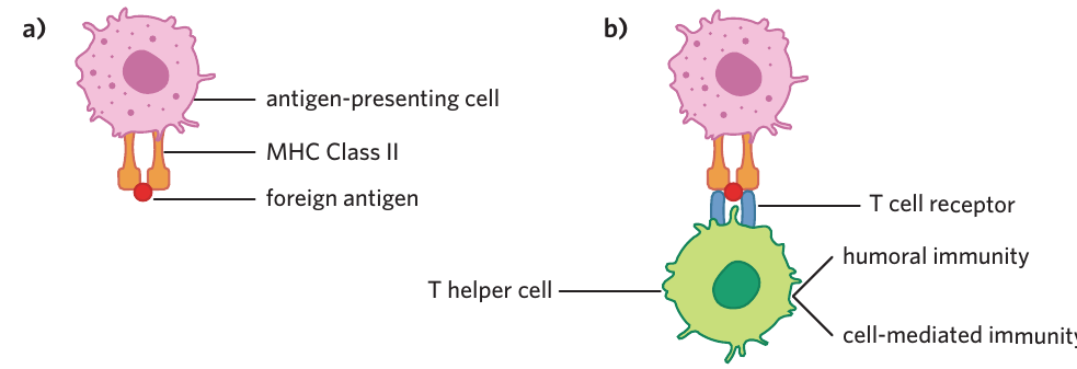
The activation of T helper cells through antigen presentation is the critical first step in launching the adaptive immune response. Without this step, neither humoral nor cell-mediated immunity can begin. This is why APCs are sometimes called the "bridge" between innate and adaptive immunity.
The activated T helper cell can then initiate either humoral immunity or cell-mediated immunity, depending on the type of pathogen encountered.
Key definitions:
- Antigen-presenting cell (APC): a type of phagocyte that displays antigens from consumed pathogens on its surface and interacts with the adaptive immune system
- T helper cell: a differentiated T lymphocyte that supports the functioning of various immune cells, including assisting in the cloning and differentiation of selected B and T cells
- Lymphatic system: a large network of vessels and tissues throughout the body that forms an important component of both the circulatory and immune systems
- Lymph node: a small secondary lymphoid tissue where antigen-presenting cells activate the adaptive immune system
Humoral immunity
Humoral immunity involves the neutralisation and destruction of extracellular pathogens through the production and secretion of antibodies.
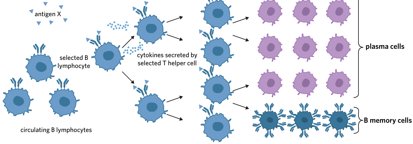
B lymphocyte activation
B lymphocytes are covered in B cell receptors (also known as antibodies). These cells travel through the bloodstream and are found in high numbers within lymph nodes. The activation of B lymphocytes occurs through four key stages:
1. Selection of a B cell A pathogen with an antigen that is complementary in shape to the B cell receptor binds to that B cell. When this occurs, the B cell is said to be 'selected'.
2. Stimulation by T helper cells Once a B cell is selected, a T helper cell (which was selected through antigen presentation and has a complementary receptor to the same antigen) recognises the selected B cell. The T helper cell secretes chemical messengers called cytokines, which cause the B cell to undergo clonal expansion – a process where many copies of the selected B cell are produced.
3. Differentiation The T helper cell also stimulates the selected B cell to undergo differentiation. This means the clones of the selected B cell develop into two specialised cell types:
- Plasma cells (effector cells)
- B memory cells
4. Antibody production Plasma cells are differentiated B cells that secrete large quantities of antibodies into the blood to defend against the pathogen. Each plasma cell can produce approximately 2,000 antibodies per second!
Worked Example: B Cell Activation Timeline
When you're first exposed to a pathogen like chickenpox virus:
Day 0-1: APCs engulf chickenpox virus and present antigens to T helper cells Day 2-3: Selected T helper cells encounter and activate B cells with complementary receptors Day 4-5: Selected B cells undergo clonal expansion, creating thousands of copies Day 5-7: B cells differentiate into plasma cells and B memory cells Day 7-14: Plasma cells produce millions of antibodies, clearing the infection
This entire process takes about 1-2 weeks for a primary response, which is why you feel sick for several days before recovering.
Key definitions:
- Clonal selection: the process in which B and T cells encounter an antigen that matches their antigen-binding site, then generate many copies of themselves
- Clonal expansion: the process in which many copies of a lymphocyte are generated
- Differentiation: the process in which cells develop specialised characteristics, typically transforming them from one cell type to another more specialised cell type
- Cytokine: a signalling molecule released by cells in the immune system which aids in communication between immune cells and helps protect against pathogens
- Plasma cell: a differentiated B lymphocyte responsible for the generation and secretion of antibodies during the humoral response
- B memory cell: a differentiated B lymphocyte responsible for providing long-lasting immunological memory of an antigen
Antibody structure
Antibodies (also called immunoglobulins) are proteins with a quaternary structure. They have a characteristic 'Y' shape and are composed of:
- Two heavy chains (the longer polypeptide chains)
- Two light chains (the shorter polypeptide chains)
- Disulphide bonds that join the heavy chains together
Each antibody has two regions:
- Variable region – the tops of the 'arms' that form two identical antigen-binding sites
- Constant region – the 'stem' of the antibody
Because antibodies have two antigen-binding sites, they can bind to two pathogens simultaneously.
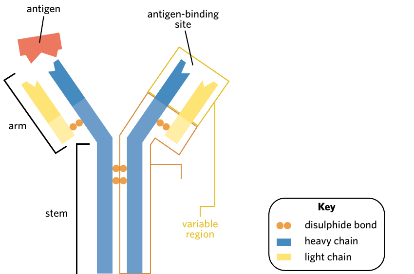
The Y-shape of antibodies is functional, not just decorative! The two arms (variable regions) act like precision-guided missiles, seeking out and binding to specific antigens. The stem (constant region) determines what happens next – whether the antibody triggers complement proteins, signals phagocytes, or performs another defensive function.
Types of antibodies
There are five types of antibodies, each serving slightly different functions:
| Type | Characteristic |
|---|---|
| IgA | Found in mucus, breast milk, and saliva |
| IgD | Important for the activation of other immune cells |
| IgE | Protects against parasitic worms; also responsible for allergic reactions |
| IgG | Most common antibody in the body; able to cross the placenta to the foetus |
| IgM | The first type of antibody produced by plasma cells in response to infection |
Mnemonic for antibody types: "All Dogs Eat Good Meat" (IgA, IgD, IgE, IgG, IgM)
Remember that IgM is always the first responder – it's the initial antibody type produced during a new infection, hence "M" for "first" in the alphabet of immune response!
Exam tip: You don't need to memorise detailed characteristics of each antibody type, but you should understand the general structure of antibodies.
Functions of antibodies
Antibodies combat pathogens through several mechanisms:
Neutralisation Antibodies block the sites on pathogens used to attack host cells (such as the site a virus uses to enter a cell) and can block the active sites of toxins, rendering them harmless.
Agglutination Antibodies bind to antigens on two separate pathogens, forming large antigen-antibody complexes. This makes it easier for phagocytes to recognise and destroy the pathogens.
Immobilisation Antibodies restrict the movement of pathogens around the body through the formation of large antigen-antibody complexes.
Opsonisation Antibodies bind directly to the surface of pathogens, making them easier to phagocytose (like coating them to make them more 'tasty' for phagocytes).
Activation of complement proteins Antibodies attached to pathogen surfaces facilitate the actions of complement proteins, including the formation of membrane attack complexes (MACs) – pores that disrupt the pathogen's membrane and lead to its death.
Mnemonic for antibody functions: "NAIAC"
- Neutralisation
- Agglutination
- Immobilisation
- Activation of complement
- Coating (opsonisation)
Think of antibodies as Swiss Army knives – they have multiple tools (functions) to combat pathogens in different ways!
Key definitions:
- Antibody: a protein produced by plasma cells during the adaptive immune response that is specific to an antigen and combats pathogens in various ways (also known as immunoglobulin)
- Antigen-antibody complex: a structure formed by the complementary binding between antigen and antibody molecules
- Agglutination: the clumping of particles together; in the immune system, antibodies help clump pathogens together
- Membrane attack complex (MAC): a pore formed by complement proteins in the cell membranes of a pathogen, disrupting the membrane and leading to the pathogen's death
Cell-mediated immunity
Cell-mediated immunity involves the destruction of infected or abnormal cells through the action of cytotoxic T cells.
Cytotoxic T cell activation
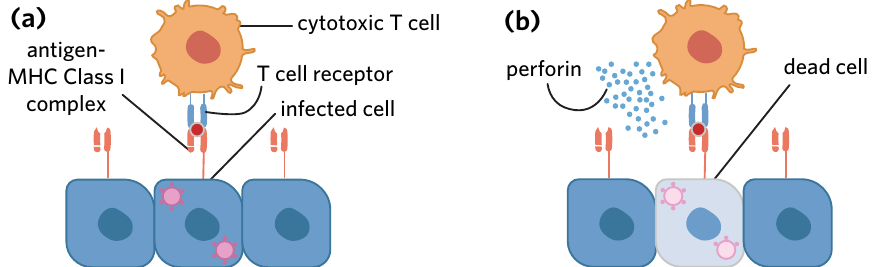
Cytotoxic T cells are a type of T lymphocyte that destroy infected cells by recognising foreign antigens displayed on MHC Class I markers. Remember that all nucleated cells display MHC Class I molecules on their surface. When a cell becomes infected, it displays foreign antigens on its MHC Class I molecules.
The cell-mediated immune response follows these steps:
1. Selection of naive T cells At the same time T helper cells are being selected, antigen-presenting cells interact with naive T cells. When an APC finds a naive T cell with a complementary T cell receptor, that T cell becomes selected.
2. Clonal expansion and differentiation The selected T helper cell secretes cytokines that stimulate the selected naive T cell to undergo clonal expansion and differentiation. This produces:
- Cytotoxic T cells (effector cells)
- T memory cells
3. Destruction of infected cells Cytotoxic T cells leave the lymph node and travel to the site of infection. Because of clonal selection, all cytotoxic T cells have T cell receptors specific to the foreign antigen. When a cytotoxic T cell finds an infected cell displaying the complementary antigen on its MHC Class I complex, it:
- Binds to the infected cell via interactions between the T cell receptor and the antigen-MHC Class I complex
- Secretes chemicals such as perforin to induce apoptosis (programmed cell death) in the infected cell
Critical distinctions between humoral and cell-mediated immunity:
- Humoral immunity primarily targets extracellular pathogens (bacteria and viruses floating in body fluids)
- Cell-mediated immunity primarily targets intracellular pathogens (viruses hiding inside cells, bacteria that can survive inside cells)
- Cell-mediated immunity also destroys abnormal cells (such as cancer cells)
- Cell-mediated immunity is the main cause of organ rejection after transplants
Understanding which immune response targets which type of threat is essential for exam success!
Key definitions:
- Cytotoxic T cell: a differentiated T lymphocyte responsible for the destruction of infected or abnormal cells
- T memory cell: a differentiated T lymphocyte responsible for providing long-lasting immunological memory
- Apoptosis: the controlled death of cells in the body (also known as programmed cell death)
Immunological memory
Immunological memory is the ability of the immune system to quickly and aggressively combat a previously encountered pathogen. This is made possible by B memory cells and T memory cells.
How memory cells work
B memory cells contribute to immunological memory by:
- Rapidly dividing and forming new antibody-producing plasma cells when they encounter a matching antigen
- Constantly secreting low amounts of their specific antibody, so a person immune to a pathogen always has trace amounts of that antibody in their blood
T memory cells contribute to immunological memory by:
- Rapidly proliferating into T helper cells and cytotoxic T cells when stimulated by an antigen-presenting cell presenting a previously encountered antigen
Advantages of immunological memory
Benefits of immunological memory:
Immunological memory provides several crucial advantages that protect us from re-infection:
- Creates a more rapid immune response upon re-infection (days instead of weeks)
- Produces antibodies at a greater rate during re-infection (higher concentrations faster)
- Generates cytotoxic T cells more quickly to destroy infected cells
- Can prevent disease development during re-exposure, as the pathogen cannot replicate fast enough to cause symptoms before being eliminated
This is the foundation of how vaccinations work – they create immunological memory without causing disease, preparing your immune system for future encounters with the real pathogen.
Summary of the third line of defence
The adaptive immune system consists of two interconnected responses:
Humoral immunity:
- B cells are selected when they encounter complementary antigens
- T helper cells stimulate B cells to undergo clonal expansion and differentiation
- Plasma cells secrete antibodies that neutralise, agglutinate, immobilise, opsonise pathogens, and activate complement proteins
- B memory cells provide long-lasting immunity
Cell-mediated immunity:
- Naive T cells are selected by antigen-presenting cells
- T helper cells stimulate naive T cells to undergo clonal expansion and differentiation
- Cytotoxic T cells destroy infected or abnormal cells by inducing apoptosis
- T memory cells provide long-lasting immunity
Both responses create immunological memory, allowing rapid and effective responses to previously encountered pathogens.
Remember!
Key Points to Remember:
-
The third line of defence (adaptive immune system) has two unique features: specificity and immunological memory
-
Humoral immunity uses B lymphocytes to produce antibodies that target extracellular pathogens through neutralisation, agglutination, immobilisation, opsonisation, and complement activation
-
Cell-mediated immunity uses cytotoxic T cells to destroy infected or abnormal cells by inducing apoptosis
-
T helper cells coordinate the adaptive immune response by secreting cytokines that stimulate B cells and naive T cells to undergo clonal expansion and differentiation
-
Memory cells (both B and T memory cells) remain in the body for extended periods, providing rapid and effective responses to previously encountered pathogens and forming the basis of vaccination