Proteins (VCE SSCE Chemistry): Revision Notes
Proteins
Introduction to proteins
Proteins are large biological molecules built from chains of amino acid units. They perform essential functions throughout the body, including:
- Acting as hormones that regulate biological processes
- Providing structural components in cell membranes, muscles, hair and feathers
- Functioning as antibody molecules in the immune system
- Transporting substances across cell membranes or around the body
- Serving as enzymes that speed up specific biochemical reactions
Understanding protein structure is crucial for comprehending how many medicinal substances work in the human body. The function of a protein depends on its shape, which is determined by its structure at four different levels: primary, secondary, tertiary, and quaternary.
Primary structure of proteins
The primary structure of a protein refers to the number, type, and sequence of amino acid units that make up the protein chain. This sequence is extremely important because it determines the protein's ultimate function.
Amino acids join together through peptide links (also called peptide bonds, represented as —CONH—) to form a polypeptide chain. These peptide links form when the carboxyl group () of one amino acid reacts with the amino group () of another amino acid, releasing a water molecule.
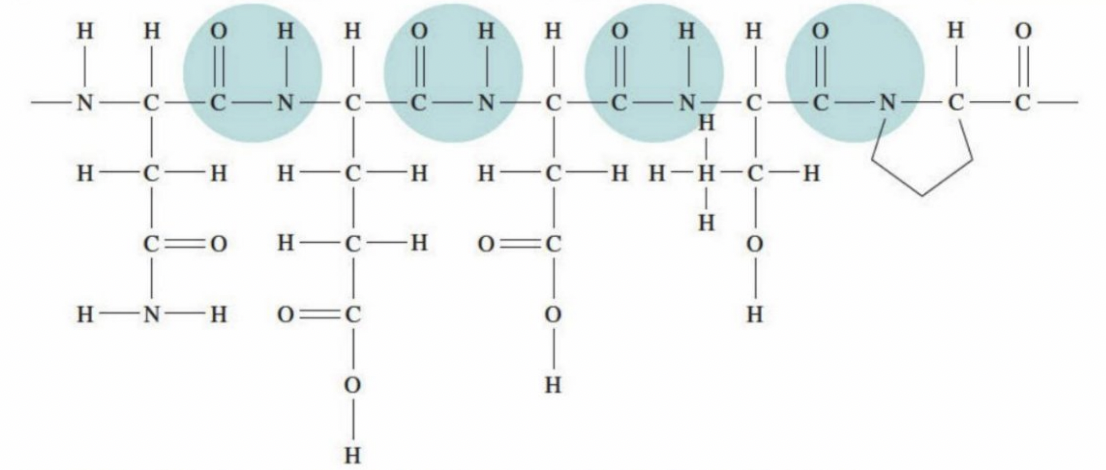
The diagram above shows part of the glycinin protein found in soybeans. Notice how the peptide links (highlighted in blue) connect each amino acid unit to the next. Each protein has a precise chemical composition and amino acid sequence, resulting in a unique three-dimensional shape.
The sequence of amino acids in the primary structure is critical – even a single amino acid change can dramatically alter the protein's shape and function. This is why genetic mutations that change amino acid sequences can have serious effects on protein function.
Secondary structure of proteins
The secondary structure of a protein describes the coiling and pleating of sections of the polypeptide chain. These regularly arranged sections are stabilised by hydrogen bonds that form between different parts of the molecule.
Hydrogen bonding in secondary structure
Hydrogen bonds form between the polar group in one peptide link and the polar group in another peptide link at regular intervals along the chain.
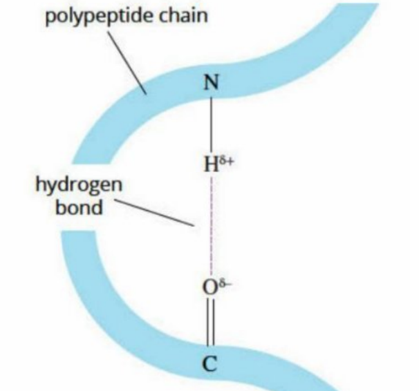
The hydrogen atom in the group carries a partial positive charge (), while the oxygen atom in the group carries a partial negative charge (). The attraction between these opposite charges creates the hydrogen bond.
α-helices
An α-helix is a spiral or spring-like structure formed when a polypeptide chain coils due to hydrogen bonding. Keratin, the protein found in hair and wool fibres, has an α-helical structure.
In keratin, hydrogen bonds form between the group of one peptide link and the group of a peptide link four amino acid units along the chain. This extensive hydrogen bonding throughout the chain causes it to coil into the characteristic helical shape.
β-pleated sheets
A β-pleated sheet forms when hydrogen bonds occur between peptide links in different sections of the polypeptide chain, causing these sections to line up parallel to each other.
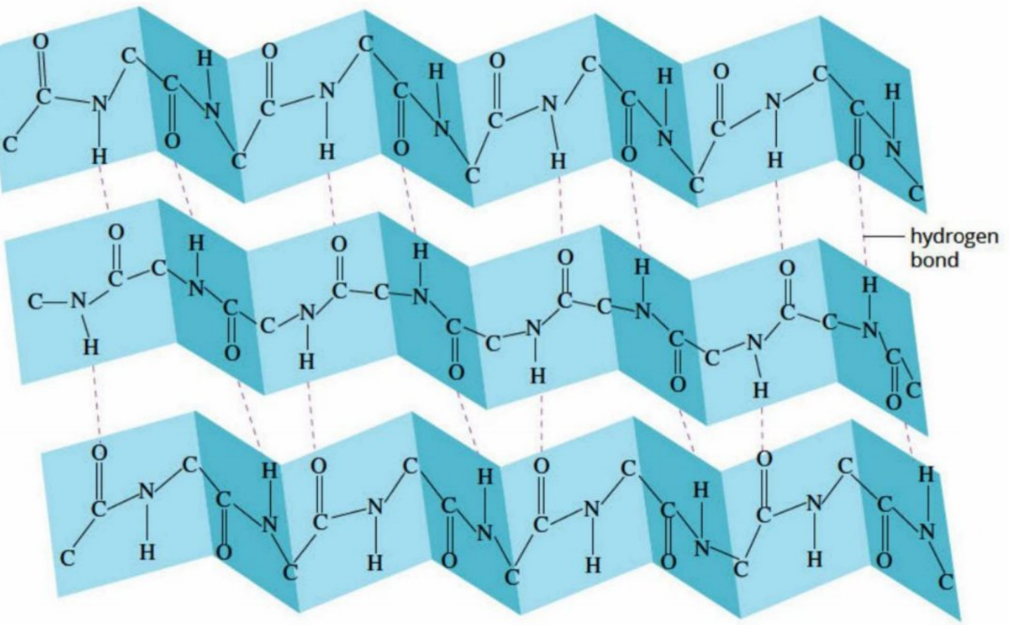
The repeating structure of the protein backbone () allows hydrogen bonds to form at regular intervals between adjacent sections. This creates a sheet-like structure with a pleated appearance.
Silk is an excellent example of a protein with a β-pleated sheet structure. The polypeptide chains in silk are mainly composed of the amino acids glycine, alanine, and serine.
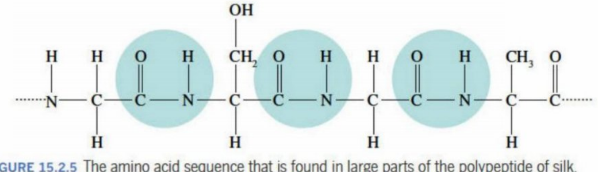
Notice that every second R group (side chain) in silk is simply a hydrogen atom (). These small side chains allow sections of the protein molecule to align closely together, enabling hydrogen bonds to form between adjacent sections and creating the β-pleated sheets that give silk its characteristic strength and texture.
Key point: The secondary structure of a protein results from hydrogen bonding within different regions of the amino acid sequence, leading to the formation of α-helices or β-pleated sheets.
Acid-base properties of amino acids
To understand how proteins fold into their final shapes and how pH affects protein structure, we need to understand the acid-base properties of amino acids.
Zwitterion formation
Amino acids contain both polar amine () and carboxyl () functional groups. This gives them special properties in solution:
- The group can act as a base, accepting a proton () to become
- The group can act as an acid, donating a proton to become
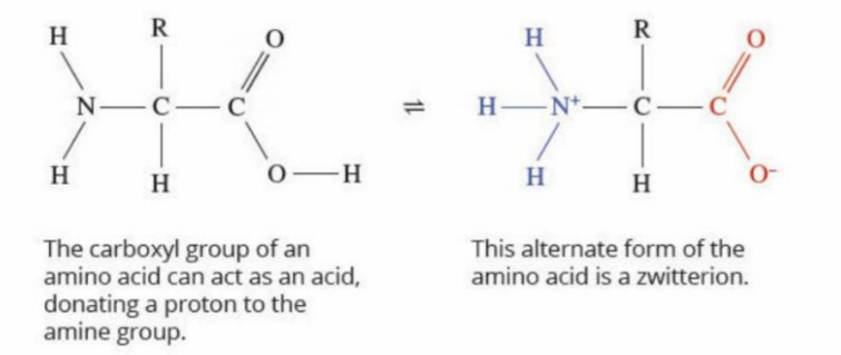
In aqueous solution, an amino acid can exist as a zwitterion – a dipolar ion that contains both a positive charge (on the amino group) and a negative charge (on the carboxyl group). Although the zwitterion has charged regions, its overall charge is neutral because the positive and negative charges balance each other.
The relatively high melting points of pure crystalline amino acids occur because zwitterions are present in the solid state, where the opposite charges create strong electrostatic attractions between molecules.
Effect of pH on amino acid structure
Because amino acids can act as both acids and bases, different chemical forms can exist in equilibrium in solution. The predominant form depends on the pH of the solution.
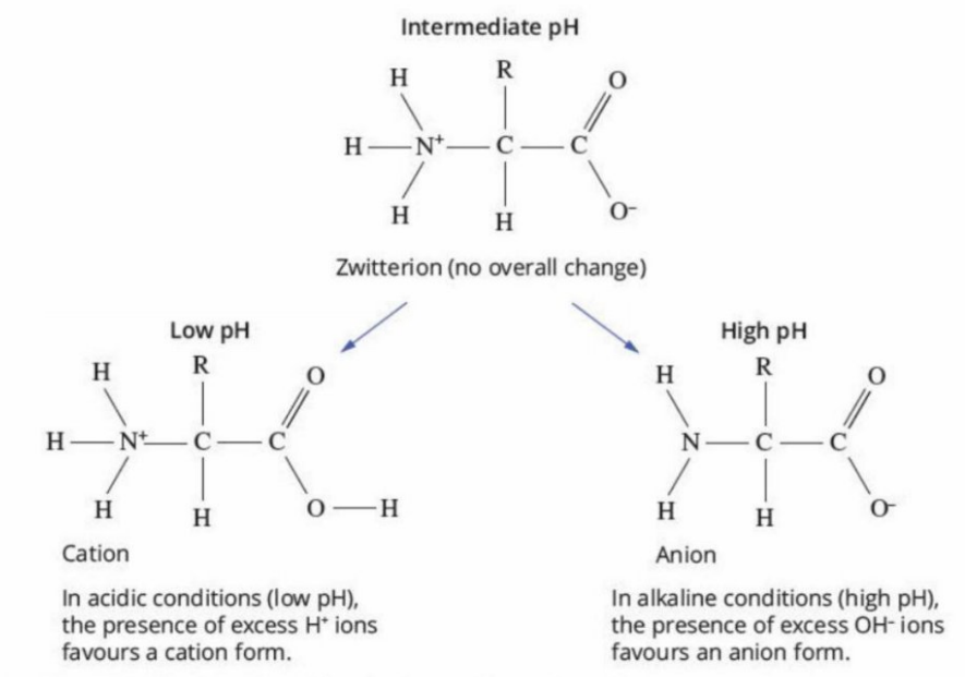
- At low pH (acidic conditions): The presence of excess ions favours the formation of a cation (positively charged ion). Both the amino and carboxyl groups are protonated.
- At intermediate pH: The zwitterion form predominates, with a protonated amino group () and a deprotonated carboxyl group (). The overall charge is neutral.
- At high pH (alkaline conditions): The presence of excess ions favours the formation of an anion (negatively charged ion). Both groups are deprotonated.
Effect of R groups on acid-base properties
The R group (side chain) of an amino acid can significantly influence its acid-base behaviour. If the R group contains functional groups with acid-base properties, additional charged forms can exist.
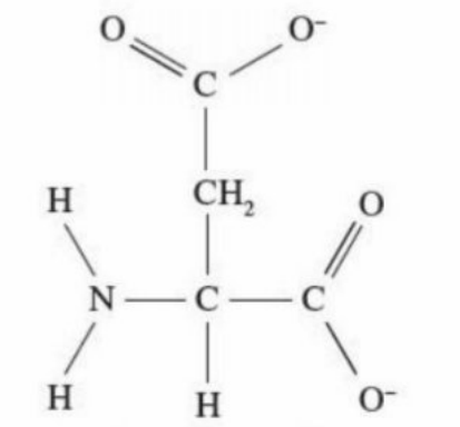
Acidic amino acids: Amino acids like aspartic acid have R groups containing carboxyl groups. At pH 12, aspartic acid exists predominantly as an ion with a charge of 2− because both carboxyl groups (the one in the backbone and the one in the R group) are deprotonated.
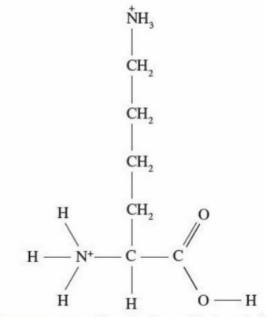
Basic amino acids: Amino acids like lysine have R groups containing amino groups. At pH 2, lysine exists predominantly as an ion with a charge of 2+ because both amino groups (the one in the backbone and the one in the R group) are protonated.
R groups can be classified into four main categories: polar, non-polar, acidic, or basic. These properties influence how the amino acid behaves in solution and how it contributes to protein structure.
Tertiary structure of proteins
The tertiary structure of a protein is its overall three-dimensional shape, formed by the folding of its secondary structures (α-helices and β-pleated sheets) in space. The protein can twist and fold back over itself to create a unique shape that is essential for its biological function.
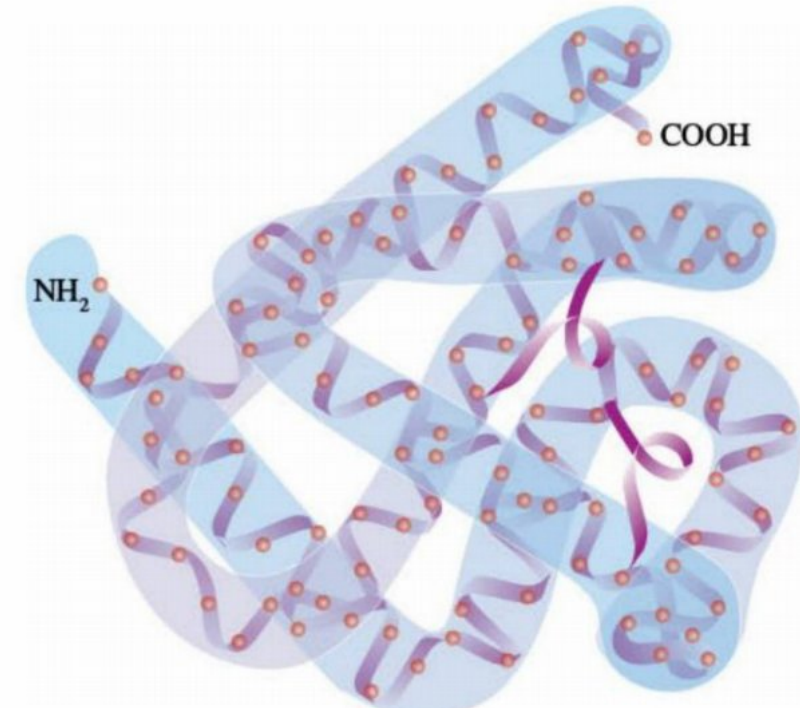
The diagram above shows myoglobin, a protein with a complex tertiary structure. Notice how the polypeptide chain folds into a compact, globular shape.
Factors influencing tertiary structure
The R groups (side chains) of amino acids play a crucial role in determining the final three-dimensional shape of a protein. Several factors contribute to this folding:
- Size of R groups: Some R groups, like that in phenylalanine, are relatively large and bulky
- Polarity: Some R groups contain polar functional groups
- Charge: Depending on pH, some R groups can become positively or negatively charged
- Hydrophobic nature: Non-polar R groups tend to fold towards the interior of protein molecules, away from water
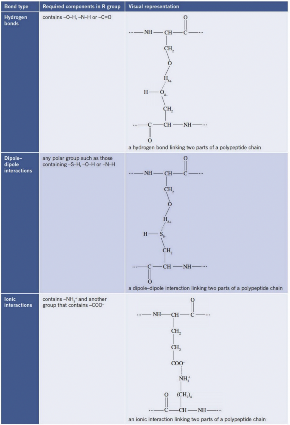
Types of bonding in tertiary structure
Five main types of interactions between R groups stabilise the tertiary structure of proteins:
1. Hydrogen bonds
Form between R groups containing , , or groups. These are similar to the hydrogen bonds in secondary structure but occur between different parts of the folded chain.
2. Dipole-dipole interactions
Occur between any polar R groups, such as those containing , , or groups. The partial charges on these groups attract each other.
3. Ionic interactions
Form between R groups with opposite charges, such as between an group on one amino acid and a group on another amino acid.
4. Covalent cross-links (disulfide bridges)
Form when two cysteine amino acids come close together. The thiol groups () on their R groups react to form a disulfide bridge (). These are the strongest interactions in protein tertiary structure.
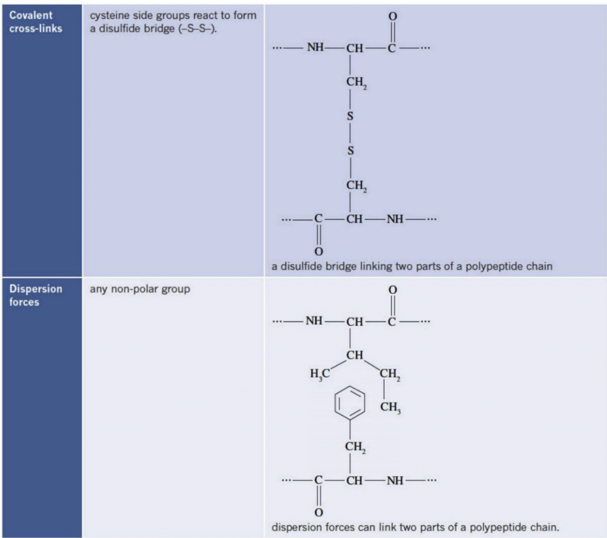
5. Dispersion forces
Occur between any non-polar R groups. Although individually weak, the cumulative effect of many dispersion forces can be significant, especially in proteins with many non-polar amino acids.
Key point: Interactions between the various R groups on amino acids lead to each protein having a unique three-dimensional structure.
Because of these different types of interactions, an enormous variety of protein shapes exist. Some proteins resemble flat sheets, others are long and helical, while others are compact and globular. The final shape is always determined by the sequence of amino acids in the primary structure.
Quaternary structure of proteins
Some proteins are composed of two or more polypeptide chains that interact together. When multiple chains and/or non-protein molecules combine to produce a larger, more complex functional unit, this is called the quaternary structure.
Haemoglobin: an example of quaternary structure
Haemoglobin is the oxygen-transporting protein found in red blood cells. It provides an excellent example of quaternary structure.
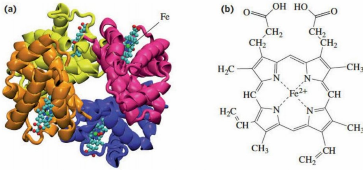
The haemoglobin molecule consists of four distinct subunits (shown in different colours above). Each subunit contains:
- A polypeptide chain (called globin) that is coiled into α-helices and folded into a tertiary structure
- An oxygen-binding site called a haem group (shown in light blue)
- One iron atom () within each haem group (shown in green)
Haemoglobin transports oxygen around the body by binding one oxygen molecule () to the iron atom in each haem group. Since there are four haem groups, one haemoglobin molecule can transport four oxygen molecules.
Interesting fact: Red blood cells are produced by bone marrow at approximately 2 million per second. Each red blood cell contains roughly 250 million molecules of haemoglobin and functions for about 3 months in the human body.
Summary of protein structure levels
The following table summarises the four levels of protein structure:
| Level of bonding | Effect of bonding | Types of bonding |
|---|---|---|
| Primary | Sequence of amino acids in the main chain | Covalent bonds (peptide links) |
| Secondary | Coiling or pleating of the protein chain | Hydrogen bonding between and on different parts of the molecule |
| Tertiary | Overall three-dimensional shape of the protein | Formed by a variety of bond types between the R groups on different parts of the protein molecule |
| Quaternary | Some proteins contain more than one peptide chain and form a more complex unit | Dispersion forces or other bonds between molecules |
Exam tip: In computer models and diagrams, protein structures are often represented using simplified visual conventions. An α-helix is typically shown as a twisted ribbon or coil, while β-pleated sheets appear as wide parallel ribbons with arrows. Many proteins contain several regions of both types of secondary structure.
Key Points to Remember:
-
Proteins have four levels of structure: Primary (amino acid sequence), secondary (α-helices and β-pleated sheets), tertiary (3D folding), and quaternary (multiple chains).
-
The function of a protein depends on its shape, which is determined by the order and type of amino acids in its primary structure.
-
Amino acids exist as zwitterions in solution, with both positive () and negative () charged groups. The predominant form changes with pH.
-
Five types of interactions stabilise tertiary structure: hydrogen bonds, dipole-dipole interactions, ionic interactions, disulfide bridges (covalent cross-links), and dispersion forces.
-
Haemoglobin demonstrates quaternary structure, consisting of four subunits, each containing a polypeptide chain and a haem group with an iron atom for oxygen binding.