Structure of Eukaryotic Cells (AQA A-Level Biology): Revision Notes
Structure of Eukaryotic Cells
Each cell functions as a metabolic compartment where specific chemical processes occur. Cells adapt to perform particular functions, and each cell type possesses an internal structure suited to its role. This internal organisation is called the ultrastructure of the cell.
Eukaryotic cells contain a distinct nucleus and possess membrane-bounded organelles. This distinguishes them from prokaryotic cells like bacteria. The membrane-bounded organelles can be observed using electron microscopy, revealing detailed structural arrangements within cells.
The presence of membrane-bounded organelles is the key distinguishing feature that separates eukaryotic cells from prokaryotic cells. This compartmentalisation allows for more complex cellular processes and specialised functions within different organelles.
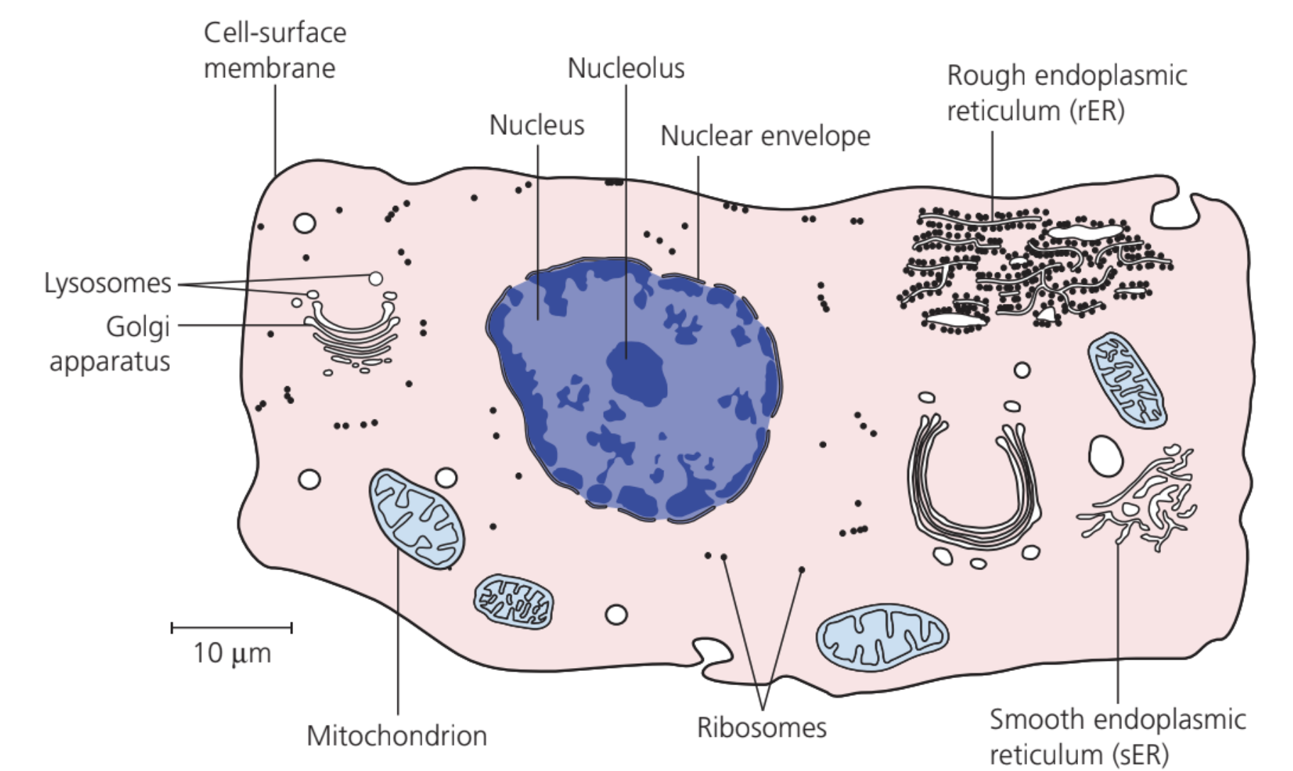
Structure of an animal cell:
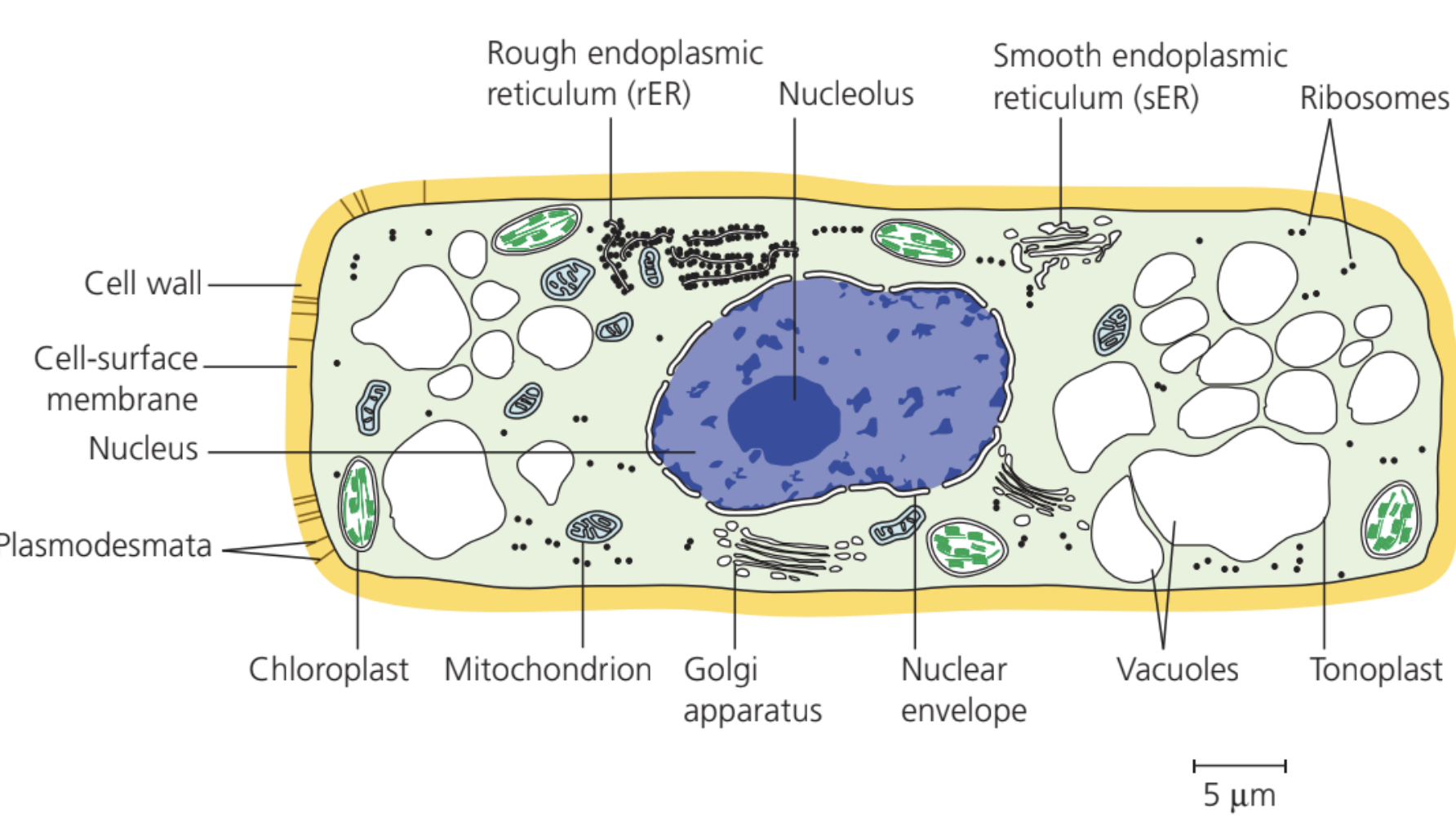
 Structure of a plant cell:
The nucleus
The nucleus represents the most noticeable structure in a eukaryotic cell, appearing as a large spherical organelle. Typically measuring 10-20 μm in diameter, the nucleus contains the organism's hereditary material and controls cellular activities.
Nuclear structure
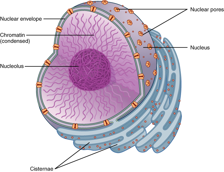
The nucleus comprises several key components that work together to maintain and regulate genetic information:
- Nuclear envelope - This double membrane surrounds the nucleus and connects with the endoplasmic reticulum of the cell. Ribosomes often attach to its surface. The envelope regulates the movement of materials into and out of the nucleus whilst containing the reactions occurring within it.
- Nuclear pores - These openings permit the passage of large molecules, particularly messenger RNA, out of the nucleus. Each nucleus typically contains around 3000 pores, with each pore measuring 40-100 nm in diameter.
- Nucleoplasm - The granular, jelly-like material forms the bulk of the nuclear interior.
- Chromosomes - These structures consist of protein-bound, linear DNA molecules.
- Nucleolus - This small spherical region within the nucleoplasm manufactures ribosomal RNA and assembles ribosomes. Multiple nucleoli may exist within a single nucleus.
Nuclear functions
The nucleus performs several essential roles:
- Acts as the control centre through production of mRNA and tRNA, enabling protein synthesis
- Retains genetic material in DNA and chromosome form
- Manufactures ribosomal RNA and ribosomes
The nucleus is often called the "control centre" of the cell because it contains all the genetic information needed to direct cellular activities and reproduction. Without a functional nucleus, eukaryotic cells cannot survive or reproduce.
The mitochondrion
Mitochondria appear as rod-shaped organelles, typically 1-10 μm in length. These structures are often called the powerhouses of the cell due to their role in energy production.
Mitochondrial structure
- Double membrane - A double membrane surrounds the organelle, controlling material entry and exit. The inner membrane folds to form extensions called cristae.
- Cristae - These extensions of the inner membrane span across the mitochondrion's width in some species. They create extensive surface area for enzyme attachment and other proteins involved in respiration.
- Matrix - This region comprises the remainder of the mitochondrion, containing proteins, lipids, ribosomes and DNA. This allows mitochondria to control production of some proteins independently. Many respiratory enzymes are located within the matrix.

The presence of DNA and ribosomes in mitochondria supports the endosymbiotic theory, which suggests that mitochondria were once independent bacteria that became incorporated into eukaryotic cells through evolution.
Mitochondrial function
Mitochondria serve as the sites for aerobic respiration stages (Krebs cycle and oxidative phosphorylation pathway). They produce the energy-carrier molecule ATP from respiratory substrates such as glucose.
Cells with high metabolic activity contain numerous large mitochondria with extensive cristae, as they require abundant ATP supply. Examples include muscle and epithelial cells. Epithelial cells in intestines need substantial ATP for absorbing substances through active transport.
Worked Example: Structure-Function Relationship in Mitochondria
Muscle cells contain hundreds of mitochondria with densely packed cristae because:
- Step 1: Muscle contraction requires enormous amounts of ATP
- Step 2: More cristae = greater surface area for ATP-producing enzymes
- Step 3: More mitochondria = greater total ATP production capacity
- Result: Muscle cells can sustain high-energy demanding contractions
Chloroplasts
Chloroplasts function as the organelles carrying out photosynthesis. They vary in shape and size but typically appear disc-shaped, measuring 2-10 μm long and 1 μm in diameter.
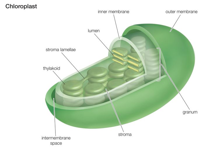
Chloroplast structure
- Chloroplast envelope - A double plasma membrane surrounds the organelle, showing high selectivity regarding substances entering and leaving the chloroplast.
- Grana - These represent stacks of up to 100 disc-like structures called thylakoids. Within thylakoids lies the photosynthetic pigment chlorophyll. Some thylakoids possess tubular extensions connecting with adjacent grana thylakoids. Grana locations host the first stage of photosynthesis (light absorption).
- Stroma - This fluid-filled matrix accommodates the second stage of photosynthesis (sugar synthesis). Within the stroma exist various structures, including starch grains.
Chloroplast adaptations
Chloroplasts show several adaptations for harvesting sunlight and conducting photosynthesis:
- Granal membranes create extensive surface area for chlorophyll, electron carriers and enzyme attachment needed for the first photosynthesis stage
- Stroma fluid contains all enzymes required for sugar production in the second photosynthesis stage
- Chloroplasts contain both DNA and ribosomes, enabling rapid manufacture of proteins needed for photosynthesis
Like mitochondria, chloroplasts also contain their own DNA and ribosomes, supporting the endosymbiotic theory. This suggests chloroplasts were once independent photosynthetic bacteria that became incorporated into plant cells.
Endoplasmic reticulum
The endoplasmic reticulum (ER) forms an elaborate, three-dimensional system of sheet-like membranes spreading throughout the cell's cytoplasm. It connects with the outer nuclear membrane. These membranes enclose networks of tubules and flattened sacs called cisternae. Two ER types exist:
Rough endoplasmic reticulum
Rough endoplasmic reticulum (RER) has ribosomes present on the membrane outer surfaces. Its functions include:
- Providing extensive surface area for protein and glycoprotein synthesis
- Creating pathways for material transport, especially proteins, throughout the cell
Smooth endoplasmic reticulum
Smooth endoplasmic reticulum (SER) lacks surface ribosomes and often appears more tubular. Its functions include:
- Synthesising, storing and transporting lipids
- Synthesising, storing and transporting carbohydrates
The distinction between RER and SER reflects their specialised functions. RER's ribosomes make it the protein synthesis factory, while SER specialises in lipid metabolism and detoxification processes.
Cells manufacturing and storing large quantities of carbohydrates, proteins and lipids possess very extensive ER. Such cells include liver and secretory cells, for example epithelial cells lining the intestines.
Golgi apparatus
The Golgi apparatus occurs in almost all eukaryotic cells and resembles SER in structure, though more compact. It consists of stacked membranes forming flattened sacs or cisternae, with small rounded hollow structures called vesicles..jpg)
Golgi structure and function
Proteins and lipids produced by the ER pass through the Golgi apparatus following a strict sequence. The Golgi modifies these proteins, often adding non-protein components such as carbohydrate groups. It also 'labels' proteins, enabling accurate sorting and direction to correct destinations.
Once sorted, modified proteins and lipids travel in Golgi vesicles, which regularly detach from Golgi cisternae ends. These vesicles may move to the cell surface, where they fuse with the membrane and release contents outside.
The Golgi apparatus functions include:
- Adding carbohydrate groups to proteins, forming glycoproteins
- Producing secretory enzymes, such as those secreted by the pancreas
- Secreting carbohydrates for plant cell wall construction
- Transporting, modifying and storing lipids
- Forming lysosomes
The Golgi apparatus acts like a post office for the cell, receiving proteins from the ER, modifying them, packaging them correctly, and shipping them to their final destinations within or outside the cell.
The Golgi apparatus shows particularly good development in secretory cells, such as epithelial cells lining the intestines.
Lysosomes
Lysosomes form when vesicles produced by the Golgi apparatus contain enzymes such as proteases and lipases. They also contain lysozymes - enzymes that hydrolyse bacterial cell walls. Up to 50 such enzymes may exist within a single lysosome.
Measuring up to 1.0 μm in diameter, lysosomes isolate these enzymes from the cell's remainder before releasing them, either to the outside or into phagocytic vesicles within the cell.
The isolation of digestive enzymes within lysosomes is crucial for cell survival. If these powerful enzymes were released freely into the cytoplasm, they would digest and destroy the cell's own components.
Lysosome functions
Lysosomes perform several important roles:
- Hydrolysing material ingested by phagocytic cells, such as white blood cells and bacteria
- Releasing enzymes to the cell exterior (exocytosis) to destroy material around the cell
- Digesting worn-out organelles, enabling useful chemical reuse
- Completely breaking down cells after death (autolysis)
Given these roles, lysosomes occur abundantly in secretory cells, such as epithelial cells, and in phagocytic cells.
Ribosomes
Ribosomes appear as small cytoplasmic granules present in all cells. They may occur in the cytoplasm or associate with the RER. Two types exist, depending on cell location:
80S ribosomes - Found in eukaryotic cells, approximately 25 nm in diameter 70S ribosomes - Found in prokaryotic cells, mitochondria and chloroplasts, slightly smaller
The presence of 70S ribosomes in mitochondria and chloroplasts (the same type found in bacteria) provides additional evidence for the endosymbiotic theory of organelle evolution.
Ribosomes contain two subunits - one large and one small - each containing ribosomal RNA and protein. Despite their small size, they exist in vast numbers, accounting for up to 25% of a cell's dry mass. Ribosomes represent the site of protein synthesis.
Cell wall
Characteristic of all plant cells, the cell wall consists of cellulose microfibrils embedded in a matrix. Cellulose microfibrils possess considerable strength, contributing to overall cell wall strength.
Cell wall features
Cell walls contain:
- Multiple polysaccharides, including cellulose
- A thin layer called the middle lamella, marking boundaries between adjacent cell walls and cementing adjacent cells together
Cell wall functions
The cellulose cell wall serves several purposes:
- Providing mechanical strength to prevent cell bursting under pressure created by osmotic water entry
- Giving mechanical strength to the plant as a whole
- Allowing water passage along it, contributing to water movement through the plant
Cell wall variations
Different organisms show cell wall variations:
- Algae cell walls comprise either cellulose or glycoproteins, or both mixtures
- Fungal cell walls lack cellulose but contain a mixture of nitrogen-containing polysaccharide called chitin, plus polysaccharide called glycan and glycoproteins
Vacuoles
A vacuole represents a fluid-filled sac bounded by a single membrane. Within mature plant cells exists usually one large central vacuole. The single membrane surrounding it is called the tonoplast.
Vacuole contents and functions
Plant vacuoles contain solutions of mineral salts, sugars, amino acids, wastes and sometimes pigments such as anthocyanins.
Plant vacuoles serve various functions:
- Supporting herbaceous plants and herbaceous parts of woody plants by making cells turgid
- Acting as temporary food stores for sugars and amino acids
- Colouring petals through pigments to attract pollinating insects
Worked Example: Vacuole Function in Plant Support
In herbaceous plants like sunflowers:
- Step 1: Vacuoles fill with water, creating turgor pressure
- Step 2: Turgor pressure pushes against the rigid cell wall
- Step 3: This creates structural support throughout the plant
- Result: Plants can stand upright without woody tissue
Relating cell ultrastructure to function
Each organelle possesses its specific function, making it possible to determine a cell's role by examining the number and size of organelles it contains. For example, since mitochondria produce ATP used as temporary energy storage, cells containing many mitochondria likely require substantial ATP and therefore demonstrate high metabolic rates.
Even within individual mitochondria, denser and more numerous cristae indicate greater metabolic rates for cells possessing these mitochondria.
Worked Example: Reading Cell Function from Structure
When examining different cell types under a microscope:
- Muscle cells: Packed with mitochondria with extensive cristae → High energy demand
- Pancreatic cells: Abundant RER and Golgi → Protein secretion specialists
- Leaf cells: Numerous chloroplasts → Photosynthesis specialists
- White blood cells: Many lysosomes → Pathogen destruction specialists
Key Points to Remember:
- Eukaryotic cells contain a membrane-bound nucleus and various membrane-bound organelles, each with specific functions
- The nucleus controls cell activities and contains genetic material, whilst mitochondria produce ATP through respiration
- Chloroplasts in plant cells conduct photosynthesis, containing grana for light reactions and stroma for sugar synthesis
- The endomembrane system (ER and Golgi apparatus) works together to synthesise, modify and transport proteins and lipids
- Cell structure directly relates to function - cells requiring high energy contain numerous mitochondria with extensive cristae
- Each organelle's structure is perfectly adapted to its specific role within the cell