The Cardiac Cycle (AQA A-Level Biology): Revision Notes
The Cardiac Cycle
The cardiac cycle refers to the sequence of events that occurs in the heart, repeating approximately 70 times per minute during rest. This cycle ensures continuous circulation of blood throughout the body.
The cardiac cycle consists of two main phases:
- Systole: when heart muscle contracts
- Diastole: when heart muscle relaxes
However, since the atria and ventricles don't always contract simultaneously, the cycle is better described in three distinct stages. Understanding these stages helps explain how the heart efficiently pumps blood to both the lungs and the rest of the body.
The cardiac cycle is fundamental to understanding cardiovascular function. Disruption of any stage can lead to serious health complications, making this one of the most critical biological processes to master.
Stages of the cardiac cycle
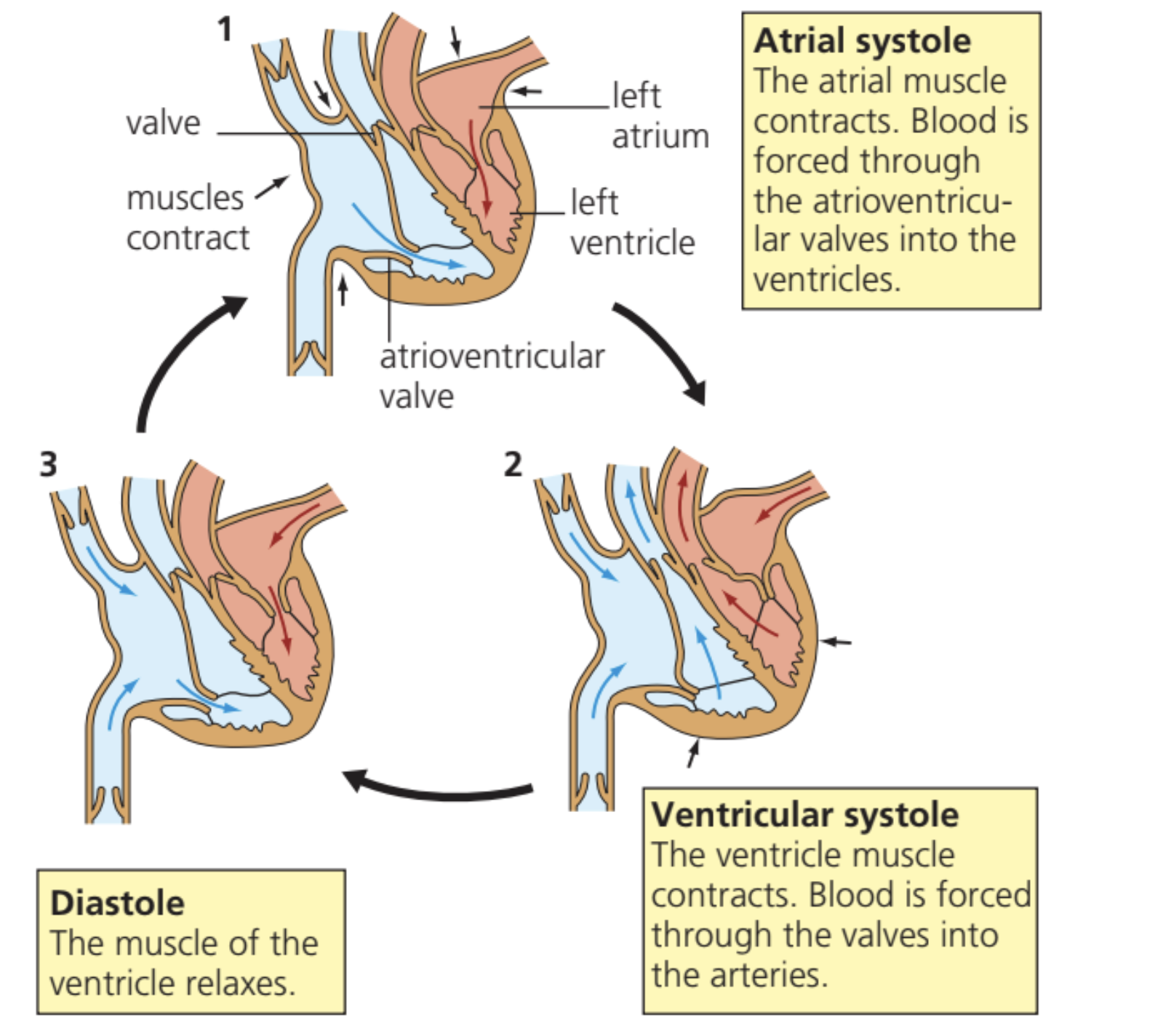
Diastole (relaxation of the heart)
During diastole, both the atria and ventricles are relaxed. Blood returns to the heart through the pulmonary veins (from the lungs) and the vena cava (from the body), causing the atrial pressure to rise gradually.
When atrial pressure exceeds ventricular pressure, the atrioventricular valves open, allowing blood to flow from the atria into the ventricles. This blood flow occurs passively due to the pressure difference, assisted by gravity.
The passive filling during diastole is crucial for cardiac efficiency. The heart doesn't need to actively "suck" blood in - the pressure differences created by relaxation naturally draw blood into the chambers.
As the ventricles relax, their walls recoil, which reduces the internal pressure. This creates a pressure difference that helps draw blood into the ventricles. The lower pressure in the ventricles compared to the major arteries causes the semi-lunar valves (in the aorta and pulmonary artery) to close, producing the characteristic 'dub' sound of the heartbeat.
Atrial systole (contraction of the atria)
During atrial systole, the atrial walls contract while the ventricular walls remain relaxed. This contraction forces any remaining blood from the atria into the ventricles, ensuring the ventricles are completely filled before they contract.
The ventricular muscles stay relaxed during this stage, allowing them to accommodate the additional blood being pushed in from the atria.
Atrial systole contributes approximately 10-15% of ventricular filling. While most filling occurs passively during diastole, this final "atrial kick" ensures maximum ventricular volume before contraction.
Ventricular systole (contraction of the ventricles)
In ventricular systole, the atria relax while the ventricles contract powerfully. This contraction dramatically increases the pressure within the ventricles.
The increased ventricular pressure forces the atrioventricular valves to close, preventing blood from flowing back into the atria. The closure of these valves creates the 'lub' sound of the heartbeat.
When ventricular pressure exceeds the pressure in the aorta and pulmonary artery, the semi-lunar valves open, allowing blood to be ejected from the heart. The thick muscular walls of the ventricles are essential for generating the high pressure needed to pump blood around the body, with the left ventricle being particularly muscular to pump blood to all body tissues.
The left ventricle must generate much higher pressures than the right ventricle because it needs to pump blood throughout the entire body, while the right ventricle only pumps to the nearby lungs. This is why left ventricular problems are often more serious than right ventricular issues.
Valve control of blood flow
How valves maintain blood flow direction
Blood naturally flows from regions of higher pressure to lower pressure. Valves throughout the cardiovascular system prevent unwanted backflow when pressure gradients would otherwise cause blood to flow in the wrong direction.
Valves are designed to open when the pressure difference across them favours the desired direction of blood flow, and to close when pressure differences would cause reverse flow.
Think of heart valves like one-way doors that only open in one direction. They respond automatically to pressure changes, requiring no conscious control or energy expenditure.
Types of valves
- Atrioventricular valves are located between the atria and ventricles (left and right sides). These valves prevent blood from flowing back into the atria when the ventricles contract. When ventricular pressure exceeds atrial pressure, these valves close, ensuring blood moves towards the arteries rather than backwards.
- Semi-lunar valves are found in the aorta and pulmonary artery. They prevent blood from flowing back into the ventricles when the pressure in these vessels exceeds ventricular pressure. This occurs when the elastic walls of the arteries recoil, maintaining pressure even when the ventricles relax.
- Pocket valves exist throughout the venous system (particularly in veins of the arms and legs). These ensure blood continues flowing towards the heart, especially when skeletal muscles contract and compress the veins.
Valve malfunction is a serious medical condition. When valves don't close properly (regurgitation) or don't open fully (stenosis), the heart must work harder to maintain adequate blood flow, potentially leading to heart failure over time.
Valve structure and function
All these valves share a similar basic design consisting of tough, flexible fibrous tissue flaps shaped like cups. When pressure is greater on the convex (curved outward) side, the flaps separate, allowing blood to pass through. When pressure is greater on the concave (curved inward) side, the flaps are pushed together, creating a tight seal that prevents blood flow.
Pressure and volume changes during the cardiac cycle
Cardiac output
Cardiac output represents the volume of blood pumped by one ventricle in one minute, typically measured in dm³ min⁻¹. This important measure depends on two factors:
- Heart rate: the number of heartbeats per minute
- Stroke volume: the volume of blood ejected from a ventricle with each heartbeat
The relationship is expressed as:
A typical resting cardiac output is about 5 dm³ min⁻¹ (5 litres per minute). This means your heart pumps your entire blood volume around your body approximately once every minute!
Pressure changes throughout the cycle
Ventricular pressure starts low during diastole but increases gradually as the ventricles fill with blood. During ventricular systole, pressure rises dramatically as the thick muscular walls contract. Once the semi-lunar valves open, pressure begins to fall as the ventricles empty, then drops rapidly during relaxation.
Atrial pressure remains relatively low throughout the cycle because the thin atrial walls cannot generate significant force. Pressure is highest during atrial contraction but falls when the atrioventricular valves open and blood moves into the ventricles.
Aortic pressure rises when the ventricles contract and blood is forced into the aorta. However, it never falls below approximately 12 kPa because the elastic arterial walls recoil, maintaining pressure even during ventricular relaxation.
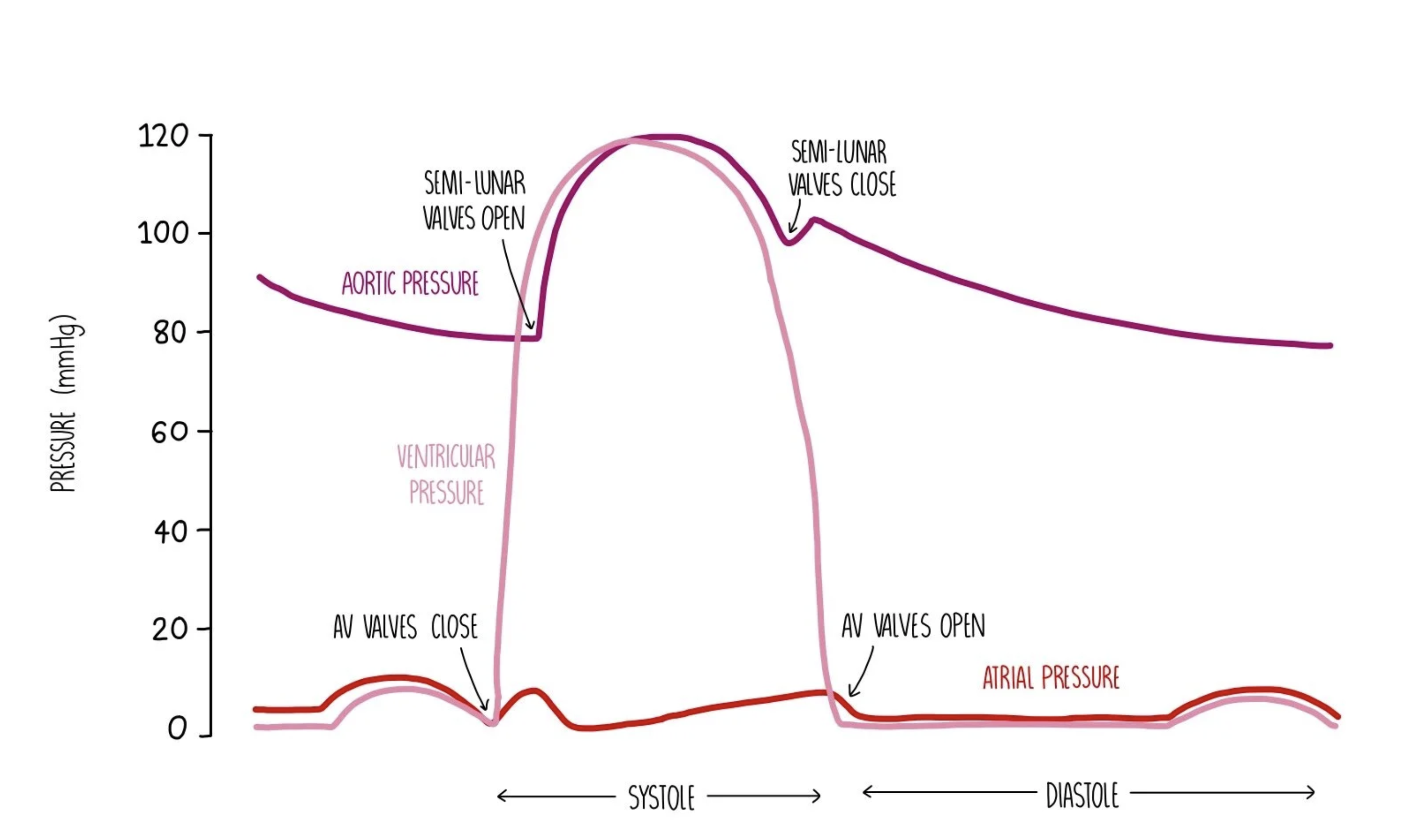
The fact that aortic pressure never falls to zero is crucial for continuous blood flow. The elastic recoil of arteries maintains pressure during diastole, ensuring organs receive blood even when the heart is relaxing.
Volume changes in the ventricles
Ventricular volume increases as the atria contract and the ventricles fill with blood. Volume then drops suddenly when blood is forced out through the aorta as the semi-lunar valves open. The volume continues to increase again as the ventricles refill with blood during the next cycle.
The closed circulatory system in mammals allows pressure to be maintained and regulated effectively, enabling precise control of blood flow throughout the body.
The closed circulatory system is much more efficient than open systems found in some invertebrates. It allows for higher pressures, faster circulation, and more precise control of blood distribution to different organs.
Key Points to Remember:
- The cardiac cycle consists of three main stages: diastole (relaxation), atrial systole (atrial contraction), and ventricular systole (ventricular contraction)
- Valves prevent backflow by opening when pressure differences favour forwards flow and closing when they would cause reverse flow
- Cardiac output equals heart rate multiplied by stroke volume and represents the volume of blood pumped per minute
- Ventricular pressure changes dramatically during the cycle, while atrial pressure remains relatively low due to thinner muscle walls
- The 'lub-dub' heart sounds correspond to the closure of atrioventricular valves (lub) and semi-lunar valves (dub)